Best Transducer and Positioning for Gallbladder Imaging
A 40-year-old man with right-sided epigastric pain is being evaluated for gallbladder issues. The best transducer for viewing the gallbladder is identified, along with proper positioning techniques for optimal imaging. The case includes details on cholelithiasis and management recommendations following an abdominal ultrasound.

Download Presentation

Please find below an Image/Link to download the presentation.
The content on the website is provided AS IS for your information and personal use only. It may not be sold, licensed, or shared on other websites without obtaining consent from the author. Download presentation by click this link. If you encounter any issues during the download, it is possible that the publisher has removed the file from their server.
E N D
Presentation Transcript
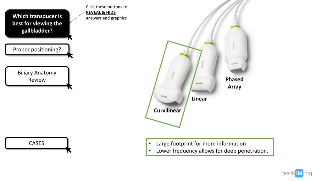
Click these buttons to REVEAL & HIDE answers and graphics Which transducer is best for viewing the Which transducer is best for viewing the gallbladder? gallbladder? Proper positioning? Proper positioning? Biliary Anatomy Review Biliary Anatomy Review Phased Array Linear Curvilinear CASES CASES Large footprint for more information Lower frequency allows for deep penetration.
Which transducer is best for viewing the gallbladder? Which transducer is best for viewing the gallbladder? Indicator cephalad Transducer position for Transducer position for long axis view? long axis view? Proper positioning? Biliary Anatomy Review Biliary Anatomy Review Maneuvers to improve visualization? visualization? Maneuvers to improve ~7 cm from xyphoid - - Inspiratory hold Left lateral decubitus Alternative window? Alternative window? Along inferior border of ribs CASES CASES
Which transducer is best for viewing the gallbladder? Which transducer is best for viewing the gallbladder? Proper position for long axis view? Proper position for long axis view? Proper positioning? Biliary Anatomy Review Biliary Anatomy Review Maneuvers to improve visualization? - - Inspiratory hold Left lateral decubitus Alternative window? Between Ribs CASES CASES - Useful if gas artifact in sub-costal view - Unable to perform sonographic murphy s
Which transducer is best for viewing gallbladder? Which transducer is best for viewing gallbladder? Proper positioning? Proper positioning? Biliary Anatomy Review Liver Add US image Add US image Common Bile Duct Hide Anatomy Hide Anatomy Hepatic Artery CASES Portal Vein CASES IVC Image courtesy of Brandon Fainstad, MD
Which transducer is best for viewing the gallbladder? 40-year-old man presents to clinic with two weeks of new intermittent right-sided epigastric pain following meals. He has had heart burn before, but not experiencing it during this time. His sister and mother both had their gallbladders removed. Which transducer is best for viewing the gallbladder? Proper positioning? Proper positioning? Biliary Anatomy Review Biliary Anatomy Review Multiple small gallstones in body of gallbladder US Image What is the diagnosis? Cholelithiasis with likely biliary colic What is your next step in management? CASES CASES Work up: Formal abdominal US Echogenic Shadow Case 1 Case 2 Treatment: Dietary restrictions. Consider general surgery consult Case 2 Image courtesy of Brandon Fainstad, MD
40-year-old man presents to the ED with two weeks of new abdominal pain that is now acutely worse with nausea and vomiting over the past two hours. T 38.5, HR 110, BP 110/75, RR 22. WBC 13, LFTs normal, Cr 1.3 (baseline 0.9), UA nml Which transducer is best for viewing the gallbladder? Which transducer is best for viewing the gallbladder? Proper positioning? Proper positioning? US Image Biliary Anatomy Review 7mm Biliary Anatomy Review What is the diagnosis? Acute cholecystitis What are normal & abnormal GB wall thicknesses? Normal <3mm Abnormal >4mm CASES CASES What is your next step in management? Case 1 Work up: Formal abdominal US Treatment: Admission with antibiotics, IVF, NPO, and general surgery consult. Case 2 Image courtesy of Brandon Fainstad, MD Courtesy of Brandon Fainstad, MD