Bacterial Cell Shapes and Simple Stain Procedure


Lab 7:
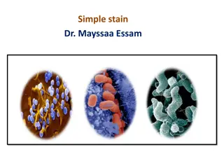
Simple stain
+ Gram stain

140 Micro


To recognize the three basic shapes
of bacterial cells.
1
-
C
o
c
c
u
s
S
p
i
r
a
l
3
-
B
a
c
i
l
l
u
s
2
-
having one of the following arrangements
:
Diplococcus: a pair of cocci
Streptococcus: a chain of cocci
Tetrad: a square of 4 cocci
Sarcina: a cube of 8 cocci
Staphylococcus:
cocci in irregular, often grape-like clusters
1
-
C
o
c
c
u
s
Bacillus: a single bacillus
Streptobacillus: bacilli in chains
Coccobacillus: oval and similar to a coccus
B
a
c
i
l
l
u
s
2
-
Vibrio: an incomplete spiral or comma-shaped
Spirillum: a thick, rigid spiral
Spirochete: a thin, flexible spiral
S
p
i
r
a
l
3
-
The simple stain is a very simple staining
procedure involving only one stain.
You may choose from
methylene blue
,
safranin
, and
crystal violet
.
1.
Prepare the smear.
- place a small drop of water on a clean
slide. Drag the sterile inoculating
needle tip through the edge of colony.
- Gently spread the mixture into a circle
to spread out.
2
.
L
e
t
t
h
e
s
m
e
a
r
a
i
r
d
r
y
c
o
m
p
l
e
t
e
l
y
.
3.
Heat-Fix
the smear.
Smears are heat-fixed by quickly passing the
slide through a flame two or three times.
This causes
the microbes to
stick
to the
slide and not get washed off during the
staining process.
4. Stain the smear.
Place the slide on a rack over the
sink. Flood the smear with stain
and let it for 60-90 seconds. Rinse
gently and blot dry.
5. Then, place a drop of
oil
directly on the
stained smear .Turn the oil lens into
position and fine focus to observe the
cells.
Coccus (cocci pl.)
Bacillus (Bacilli pl.)
Spirillum (Spirilli pl.)
(
(
ص
ب
غ
ة
ج
ر
ا
م
)
)
T
h
e
g
r
a
m
s
t
a
i
n
i
s
c
a
l
l
e
d
a
d
i
f
f
e
r
e
n
t
i
a
l
s
t
a
i
n
b
e
c
a
u
s
e
i
t
s
t
a
i
n
c
e
l
l
d
i
f
f
e
r
e
n
t
l
y
b
a
s
e
d
o
n
t
h
e
i
r
c
e
l
l
w
a
l
l
s
t
r
u
c
t
u
r
e
.
G
r
a
m
-
p
o
s
i
t
i
v
e
b
a
c
t
e
r
i
a
H
a
v
e
a
t
h
i
c
k
p
e
p
t
i
d
o
g
l
y
c
a
n
l
a
y
e
r
s
u
r
r
o
u
n
d
s
t
h
e
c
e
l
l
.
T
h
e
s
t
a
i
n
g
e
t
s
t
r
a
p
p
e
d
i
n
t
o
t
h
i
s
l
a
y
e
r
a
n
d
t
h
e
b
a
c
t
e
r
i
a
t
u
r
n
e
d
p
u
r
p
l
e
.
G
r
a
m
-
n
e
g
a
t
i
v
e
b
a
c
t
e
r
i
a
h
a
v
e
a
t
h
i
n
p
e
p
t
i
d
o
g
l
y
c
a
n
l
a
y
e
r
t
h
a
t
d
o
e
s
n
o
t
r
e
t
a
i
n
c
r
y
s
t
a
l
v
i
o
l
e
t
s
t
a
i
n
.
I
n
s
t
e
a
d
,
i
t
h
a
s
a
t
h
i
c
k
l
i
p
i
d
l
a
y
e
r
w
h
i
c
h
d
i
s
s
o
l
v
e
d
e
a
s
i
l
y
u
p
o
n
d
e
c
o
u
l
o
r
i
z
a
t
i
o
n
w
i
t
h
A
l
c
o
h
o
l
.
T
h
e
r
e
f
o
r
e
,
c
e
l
l
s
w
i
l
l
b
e
c
o
u
n
t
e
r
s
t
a
i
n
e
d
w
i
t
h
s
a
f
r
a
n
i
n
a
n
d
t
u
r
n
e
d
r
e
d
.
T
h
e
m
a
t
e
r
i
a
l
:
C
u
l
t
u
r
e
s
o
f
:
S
t
a
p
h
y
l
o
c
o
c
c
u
s
a
u
r
e
u
s
,
B
a
c
i
l
l
u
s
s
u
b
t
i
l
i
s
,
E
.
c
o
l
i
1.
Crystal violet .
2.
Iodine solution .
3.
Alcohol 95% .
4.
Safranin .
5.
Water .
G
r
a
m
–
v
e
G
r
a
m
+
v
e
S
S
h
h
a
a
p
p
e
e
:
:
C
C
o
o
c
c
c
c
i
i
A
A
r
r
r
r
a
a
n
n
g
g
m
m
e
e
n
n
t
t
:
:
i
i
r
r
r
r
e
e
g
g
u
u
l
l
a
a
r
r
c
c
l
l
u
u
s
s
t
t
e
e
r
r
s
s
C
C
o
o
l
l
o
o
u
u
r
r
:
:
V
V
i
i
o
o
l
l
e
e
t
t
G
G
r
r
a
a
m
m
’
’
s
s
r
r
e
e
a
a
c
c
t
t
i
i
o
o
n
n
:
:
G
G
r
r
a
a
m
m
’
’
s
s
+
+
v
v
e
e
N
N
a
a
m
m
e
e
o
o
f
f
m
m
i
i
c
c
r
r
o
o
o
o
r
r
g
g
a
a
n
n
i
i
s
s
m
m
:
:
S
S
t
t
a
a
p
p
h
h
y
y
l
l
o
o
c
c
o
o
c
c
c
c
i
i
S
S
h
h
a
a
p
p
e
e
:
:
B
B
a
a
c
c
i
i
l
l
l
l
i
i
A
A
r
r
r
r
a
a
n
n
g
g
m
m
e
e
n
n
t
t
:
:
C
C
h
h
a
a
i
i
n
n
s
s
C
C
o
o
l
l
o
o
u
u
r
r
:
:
V
V
i
i
o
o
l
l
e
e
t
t
G
G
r
r
a
a
m
m
’
’
s
s
r
r
e
e
a
a
c
c
t
t
i
i
o
o
n
n
:
:
G
G
r
r
a
a
m
m
’
’
s
s
+
+
v
v
e
e
N
N
a
a
m
m
e
e
o
o
f
f
m
m
i
i
c
c
r
r
o
o
o
o
r
r
g
g
a
a
n
n
i
i
s
s
m
m
:
:
B
B
a
a
c
c
i
i
l
l
l
l
u
u
s
s
ب
ا
ل
ت
و
ف
ي
ق
.
.
.
.
Explore the three basic shapes of bacterial cells - Coccus, Bacillus, and Spiral - and learn about different arrangements within each shape. Dive into the Simple Stain procedure, a straightforward staining technique using single stains like methylene blue and crystal violet, to prepare and visualize bacterial samples on slides effectively.

Download Presentation

Please find below an Image/Link to download the presentation.
The content on the website is provided AS IS for your information and personal use only. It may not be sold, licensed, or shared on other websites without obtaining consent from the author.If you encounter any issues during the download, it is possible that the publisher has removed the file from their server.
You are allowed to download the files provided on this website for personal or commercial use, subject to the condition that they are used lawfully. All files are the property of their respective owners.
The content on the website is provided AS IS for your information and personal use only. It may not be sold, licensed, or shared on other websites without obtaining consent from the author.
E N D
Presentation Transcript
Purpose To recognize the three basic shapes of bacterial cells.
Thethree common shapes of bacteria: 1-Coccus 2- Bacillus 3- Spiral
1-Coccus having one of the following arrangements : Diplococcus: a pair of cocci Streptococcus: a chain of cocci Tetrad: a square of 4 cocci Sarcina: a cube of 8 cocci Staphylococcus: cocci in irregular, often grape-like clusters
2- Bacillus Bacillus: a single bacillus Streptobacillus: bacilli in chains Coccobacillus: oval and similar to a coccus
3- Spiral Vibrio: an incomplete spiral or comma-shaped Spirillum: a thick, rigid spiral Spirochete: a thin, flexible spiral
Simple stain : The simple stain is a very simple staining procedure involving only one stain. You may choose from methylene blue, safranin, and crystal violet.
Simple stain : 1. Prepare the smear. - place a small drop of water on a clean slide. Drag the sterile inoculating needle tip through the edge of colony. - Gently spread the mixture into a circle to spread out.
Simple stain : 2. Let the smear air dry completely.
Simple stain : 3. Heat-Fix the smear. Smears are heat-fixed by quickly passing the slide through a flame two or three times. This causes the microbes to stick to the slide and not get washed off during the staining process.
Simple stain : 4. Stain the smear. Place the slide on a rack over the sink. Flood the smear with stain and let it for 60-90 seconds. Rinse gently and blot dry.
Simple stain : 5. Then, place a drop of oil directly on the stained smear .Turn the oil lens into position and fine focus to observe the cells.
Result Result
Gram stain )) ((
It is used to differentiate between gram-positive and gram-negative bacteria, which have distinct and consistent differences in their cell walls.
The gram stain is called a differential stain because it stain cell differently based on their cell wall structure .
Gram-positive bacteria Have a thick peptidoglycan layersurrounds the cell. The stain gets trapped into this layer and the bacteria turned purple. Gram-negative bacteria have a thin peptidoglycan layer that does not retain crystal violet stain. Instead, it has a thick lipid layerwhich dissolved easily upon decoulorization with Alcohol. Therefore, cells will be counterstained with safranin and turned red.
The material : Cultures of : Staphylococcus aureus, Bacillus subtilis, E.coli Crystal violet . 1. Iodine solution . 2. Alcohol 95% . 3. Safranin . 4. Water . 5.
The bacteria under the microscope
Gram ve Gram +ve
Results: Shape: Cocci Arrangment: irregular clusters Colour: Violet Gram s reaction: Gram s +ve Name of microorganism: Staphylococci
Results: Shape: Bacilli Arrangment: Chains Colour: Violet Gram s reaction: Gram s +ve Name of microorganism: Bacillus
Thank you Thank you ....


")
")
")