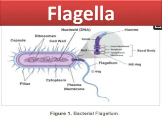
Bacterial Flagella: Structure and Function


FLAGELLA

•
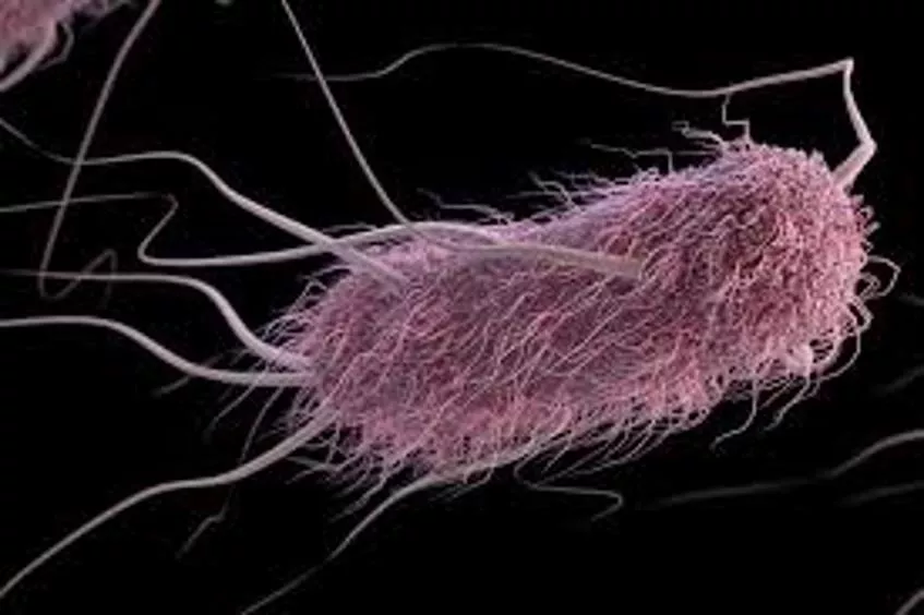
Bacterial flagella are long, thin appendages free at one end and
attached to the cell at the other end.
•
Bacterial flagella are so thin (15–20 nm, depending on the species)
that a single flagellum cannot be seen by light microscopy unless it is
stained to increase its
diameter
Flagellar Structure
•
The bacterial flagellum is composed of three parts.
(1) The longest and most obvious portion is the filament, which
extends from the cell surface to the tip.
(2) The basal body is embedded in the cell envelope.
(3) A short, curved segment, the hook, links the filament to
its basal
body.
•
The filament is
a hollow, rigid cylinder constructed of subunits of the
protein
flagellin.
•
The base of the flagellum is structurally different from the filament. There is
a wider region at the base of the filament called the
hook
.
•
The hook consists of a single type of protein and connects the filament to
the flagellum motor
in the base
.
•
The flagellum motor is anchored in the cytoplasmic membrane and
cell wall.
•
The motor consists of a central rod that passes through a series of
rings. In gram-negative bacteria, an outer ring, called the
L ring
, is
anchored in the lipopolysaccharide layer.
•
A second ring, called the
P ring
, is anchored in the peptidoglycan layer
of the cell wall. A third set of rings, called the
MS
and
C rings
, are
located within the cytoplasmic membrane and the
cytoplasm,
respectively.
•
In gram-positive bacteria,
which lack an outer membrane, only the
inner pair of rings is present.
•
Surrounding the inner ring and anchored in the cytoplasmic membrane
are a series of proteins called
Mot proteins
.
•
A final set of proteins, called
Fli proteins
, function as the motor switch,
reversing the direction of rotation of the flagella
in response to
intracellular signals.
Flagella in Gram negative bacteria
Flagella in Gram positive bacteria
Bacterial flagella are long, thin appendages crucial for motility in bacteria. Composed of filament, basal body, and hook, these structures play a vital role in bacterial movement. This article dives into the detailed structure and functionality of bacterial flagella, highlighting their importance in different types of bacteria.

Download Presentation

Please find below an Image/Link to download the presentation.
The content on the website is provided AS IS for your information and personal use only. It may not be sold, licensed, or shared on other websites without obtaining consent from the author.If you encounter any issues during the download, it is possible that the publisher has removed the file from their server.
You are allowed to download the files provided on this website for personal or commercial use, subject to the condition that they are used lawfully. All files are the property of their respective owners.
The content on the website is provided AS IS for your information and personal use only. It may not be sold, licensed, or shared on other websites without obtaining consent from the author.
E N D
Presentation Transcript
Bacterial flagella are long, thin appendages free at one end and attached to the cell at the other end. Bacterial flagella are so thin (15 20 nm, depending on the species) that a single flagellum cannot be seen by light microscopy unless it is stained to increase its diameter
Flagellar Structure The bacterial flagellum is composed of three parts. (1) The longest and most obvious portion is the filament, which extends from the cell surface to the tip. (2) The basal body is embedded in the cell envelope. (3) A short, curved segment, the hook, links the filament to its basal body.
The filament is a hollow, rigid cylinder constructed of subunits of the protein flagellin. The base of the flagellum is structurally different from the filament. There is a wider region at the base of the filament called the hook. The hook consists of a single type of protein and connects the filament to the flagellum motor in the base.
The flagellum motor is anchored in the cytoplasmic membrane and cell wall. The motor consists of a central rod that passes through a series of rings. In gram-negative bacteria, an outer ring, called the L ring, is anchored in the lipopolysaccharide layer. A second ring, called the P ring, is anchored in the peptidoglycan layer of the cell wall. A third set of rings, called the MS and C rings, are located within the cytoplasmic membrane and the cytoplasm, respectively.
In gram-positive bacteria, which lack an outer membrane, only the inner pair of rings is present. Surrounding the inner ring and anchored in the cytoplasmic membrane are a series of proteins called Mot proteins. A final set of proteins, called Fli proteins, function as the motor switch, reversing the direction of rotation of the flagella in response to intracellular signals.
Flagella in Gram negative bacteria Flagella in Gram positive bacteria