Fluorescence Microscopy: Principles and Applications
Fluorescence microscopy, pioneered by British scientist Sir George G. Stokes, reveals hidden details in specimens using fluorescent dyes that emit light of longer wavelengths. This innovative technique allows for visualization of cellular components that are otherwise colorless under conventional microscopy. By exciting fluorophores with high-intensity light, fluorescence microscopy provides sensitive and specific imaging, distinguishing bright objects on dark backgrounds. Explore the fascinating world of fluorescence microscopy to uncover intricate details about specimens.

Download Presentation

Please find below an Image/Link to download the presentation.
The content on the website is provided AS IS for your information and personal use only. It may not be sold, licensed, or shared on other websites without obtaining consent from the author.If you encounter any issues during the download, it is possible that the publisher has removed the file from their server.
You are allowed to download the files provided on this website for personal or commercial use, subject to the condition that they are used lawfully. All files are the property of their respective owners.
The content on the website is provided AS IS for your information and personal use only. It may not be sold, licensed, or shared on other websites without obtaining consent from the author.
E N D
Presentation Transcript
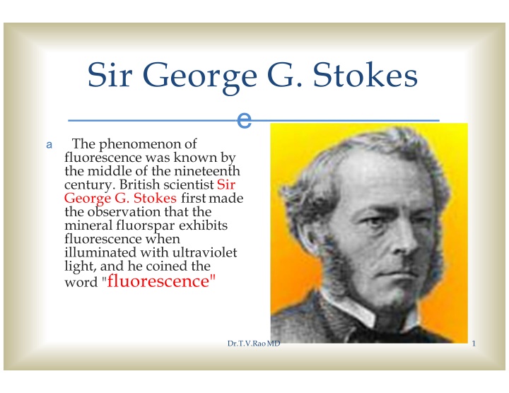
Sir George G. Stokes e a The phenomenon of fluorescence was known by the middle of the nineteenth century. British scientist Sir George G. Stokes firstmade the observation that the mineral fluorspar exhibits fluorescence when illuminated with ultraviolet light, and he coined the word "fluorescence" Dr.T.V.RaoMD 1
Differences between Conventional and Fluorescent Microscope e aThe Conventional microscope uses visible light (400-700 nanometers) to illuminate and produce a magnified image of a sample. a A fluorescence microscope, uses a much higher intensity light source which excites a fluorescent species in a sample of interest. This fluorescent species in turn emits a lower energy light of a longer wavelength that produces the magnified image instead of the original light source. Dr.T.V.RaoMD 2
What is Fluorescence? e aFluorescence islight produced by a substance when it is stimulated by another light. Fluorescence is called "cold light" because it does not come from a hot source like an incandescent light bulb. Dr.T.V.RaoMD 3
What is Fluorescence Microscopy? e a Fluorescence microscopy is a unique way of using a microscope to discover facts about specimens that often are not shown by standard bright field microscopy. In bright field microscopy, specimens are illuminated from outside, below or above, and dark objects are seen against a light background. In fluorescence microscopy, specimens are self-illuminated by internal light, so bright objects are seen in vivid color against a dark background. Bright objects against dark backgrounds are more easily seen. This characteristic of fluorescence microscopy makes it very sensitive and specific. Dr.T.V.RaoMD 4
Principle of Fluorescent Microscopy e a Most cellular components are colorless and cannot be clearly distinguished under a microscope. The basic premise of fluorescence microscopy is to stain the components with dyes. Fluorescent dyes, also known as fluorophores of fluorochromes, are molecules that absorb excitation light at a given wavelength (generally UV), and after a short delay emit light at a longer wavelength. The delay between absorption and emission is negligible, generally on the order of nanoseconds. The emission light can then be filtered from the excitation light to reveal the location of the fluorophores. Dr.T.V.RaoMD 5
Principle of Fluorescent Microscopy e Fluorescence microscopy uses a much higher intensity light to illuminate the sample. This light excites fluorescence species in the sample, which then emit light of a longer wavelength. The image produced is based on the second light source or the emission wavelength of the fluorescent species -- rather than from the light originally used to illuminate, and excite, the sample. a Dr.T.V.RaoMD 6
Works on Principles of Light Pathways e a Specifically, a dichroic mirror is used to separate the excitation and emission light paths. Within the objective, the excitation/emission share the same optics. In a fluorescence microscope, the dichroic mirror separates the light paths. Dr.T.V.RaoMD 7
Advantages of Fluorescent Microscopy e a Fluorescence microscopy is the most popular method for studying the dynamic behavior exhibited in live cell imaging. This stems from its ability to isolate individual proteins with a high degree of specificity amidst non-fluorescing material. a The sensitivity is high enough to detect as few as 50 molecules per cubic micrometer. a Different molecules can now be stained with different colors, allowing multiple types of molecule to be tracked simultaneously. These factors combine to give fluorescence microscopy a clear advantage over other optical imaging techniques, for both in vitro and in vivo imaging. Dr.T.V.RaoMD 8
Epifluorescence Microscopy Epifluorescence microscopy is a e method of fluorescence microscopy that is widely used in life sciences The excitatory light is passed from above (or, for inverted microscopes, from below), through the objective lens and then onto the specimen instead of passing it first through the specimen. The fluorescence in the specimen gives rise to emitted light which is focused to the detector by the same objective that is used for the excitation a Dr.T.V.RaoMD 9
The Specimens to be Stained e a Most specimens for fluorescence microscopy must be stained. Fluorescent stains are called "fluorochromes." Acridine orange, auramine O, and fluorescent antibody (FA) are the fluorochromes used most. Dr.T.V.RaoMD 10
How to Use a Fluorescence Microscope Dr.T.V.RaoMD 11
How to Use a Fluorescence Microscope a The object to be studied ise marked with a molecule called a fluorophore (a dye). When the florescent light is activated, the light used for illumination is separated from the florescent molecule (the fluorophore), which is much weaker. This is done through an emission filter. Dr.T.V.RaoMD 12
Step 1 e a Locate the light switch on the side of the microscope that turns on the light. Turn the microscope on. a Write down the exact time you turn on the light. The florescent light is mercury-based, and a time log must be kept for exposure and use of the light. Dr.T.V.RaoMD 13
Step 2 e aLocate the toggle switch on the right side of the microscope between the oculars and objectives. This switch controls the shutter for the mercury light to the objective lens. Dr.T.V.RaoMD 14
Step 3 e a Select the appropriate dye for your object (this will depend entirely on what you are going to be studying). The most common dyes include I3 (for use with CTC, DTAF and fluorescein), A (for use with DAPI and f420), N21 (for use with Rhoda mine) and L3 (for use Dr.T.V.Rao MDwith fluorescein). 20
Step 4 e aPut the filter (dye) into the tray operated by the silver sliding knob. To remove the tray, simply pull the silver knob out. Dr.T.V.RaoMD 16
Step 5 e a Select the lens you would like to use. The 63x objective lens will have the highest numerical aperture. The 100x objective lens will have the highest magnitude that can be used with the mercury-based florescent light source. Dr.T.V.RaoMD 17
Step 6 e a Turn the light off when finished, and mark the time. Wait 30 minutes before turning the light back on, or the lamp could explode. It is a good idea to keep track of how many hours the lamp is in use and replace it according to the manufacture's guidelines. Dr.T.V.RaoMD 18
Step 7 e aClean off the microscope lens with lens paper, or if really dirty, use a cotton swap and glass cleaner. Dr.T.V.RaoMD 19