Types of Blastulae and Their Formation in Frog and Chick Embryos
The development of blastula, an important stage in embryonic development, gives rise to various types such as coeloblastula, discoblastula, and blastocyst in different animal species. In frogs and birds, varied forms of blastulae are observed based on the amount of yolk present, leading to distinct organ-forming areas. Understanding the characteristics and formation of these blastula types provides insights into early embryonic development processes in vertebrates.
Download Presentation

Please find below an Image/Link to download the presentation.
The content on the website is provided AS IS for your information and personal use only. It may not be sold, licensed, or shared on other websites without obtaining consent from the author. Download presentation by click this link. If you encounter any issues during the download, it is possible that the publisher has removed the file from their server.
E N D
Presentation Transcript
BLASTULA.TYPE BLASTULAE.FATE MAPS. PRESUMPTIVE ORGAN FORMING AREAS IN FROG AND CHICK. B.Bindu Bennet Assistant Professor, PG Department of Zoology Sarah Tucker College Tirunelveli
BLASTULA The term blastula may be defined as a stage of embryonic development intervening between the morula stage and end of cleavage. Blastula as a hollow sphere of blastomeres surrounding a cavity, the blastocoel. or .A spherical or a disc-shaped embroyonic stage marking the end of cleavage.
Types of Blastulae 1. Coeloblastula. 2. Discoblastula. 3. Blastolcyst. 4. Superfical blastula. 5. Steroblastula.
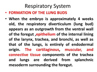
1. COELOBLASTULA: A coeloblastula is formed as a result of holoblastic cleavage. If forms with scantly amount of yolk, such as branchiostoma, the blastula has a hollow spherical cavity is referred to as segmentation (or cleavage) cavity (or blastocoels). In Branchiostoma, the coeloblastula result from equal holoblastic cleavage. In frogs and toads, and other amphibians, with a moderate amount of yolk, the blastocel is ecentrically displaced toward the animal pole. This modified coeloblastula is formed as a result of unequal holoblastic cleavage.
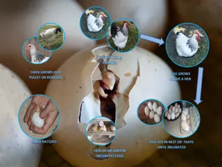
2. DISCOBLASTULA: In the eggs of birds, reptiles and egg laying mammals, the cleavage divisions are confined to a thin discoidal area at the animal pole; for this reason the blastula is called discoblastula. In other words, the discoblastula is formed as a result of meroblastic cleavage. Here, the blastocoel is highly modified, and appears like a mere slit like cavity. It is roofed over by blastomeres; but as the segmentation cavity combines with the yolk cavity, the bottom of the bastocoel consists of undivided yolk mass. The discoidal cap of cells overlying the blastocoel consists the blestoderm
3. BLASTOCYST: The term blastocyst is used for the mammalian blastula. In mammals the morula stage progresses rapidly into a blastula during blastocyst formation, the solid morula develops a cavity, the blastocoels; and the blastomeres rearrange themselves in the form of a hollow sphere. Except one region, the wall of the hollow sphere is single- layered in thickness, and is called the trophoblast. At one region of the blastocyst, there is a cluster of cells, which from the inner cell mass.
4. SUPERFICAL BLASTULA: In centrolecithal eggs of insects, the cleavage divisions are confined to the surface area of the cytoplasm and the blastoderm surrounds the central mass of uncleaved yolk such a blastula is called superficial blastula. 5. STEROBLASTULA: The solid blastula of become there is no blastocoel cavity called steroblastula.
FATE MAPS. DEFINATION: A topographical surface mapping of ovum or an early blastula, showing the ultimate fate of the various areas, is called fate map. CONSTRUCTION OF FATE MAPS: The fate maps can be constructed either by using 1. natural markings or by employing 2. artificial marking methods
NATURAL MARKINGS: In developing amphibian eggs, particularly in frog, three different areas can be marked soon after the fertilization of the ovum. The animal hemisphere is clearly marked out by the black pigment distribution in the cytoplasm. In fact, upper 2/3 of the surface areas of the ovum is black in colour. The lower 1/3 of the surface areas in the vegetal hemisphere appears crescentic area, called gray crescent, can be located just below the equator, on one side of the ovum as well as in early blastula. The black pigmented area presumptive endoderm, which will give rise to the lining of the developing embryo. However, this kind of natural marking does not occur in the all animal eggs, and the is such cases different artificial marking methods are used to construct the fate maps.
ARTIFICALMARKINGS: In artificial marking, the following methods have been used: (i) VITAL DYE STAINING METHOD: Vital staining technique was devised by w. Vogt (1925) for the construction of maps of amphibians by using vital dye such as Nile blue sulphate, neutral red, Bismarck brown, janus green etc. with a particular dye and placing a small piece of it on the surface of the embryo in arequired position for a short duration. The dye immediately diffuses from the piece of ager or cellophane into the blastimeres. Different areas of the blastula can be marked simultaneously by specific dyes and the movements of blastomeres can be followed during gastrulation. Since the dye persists in the persists in the ultimate areas in the embryo formed after gastrulation, by locating the marked materials it is possible to construct a map of prospective organ forming areas.
CARBON PARTICLE MARKING METHOD: This method was devised by spratt (1946), in which he marked the surface of the embryo with insoluble carbon particles. These carbon particles stick to the surface of the cells and their movement was observed. By conducting a series of the such marking experiments, it is possible to construct the fate maps of the vertebrate embryos. (iii) RADIOACTIVE SUBSTANCES AS MARKERS: Radioactive substances like tritiated thymidine, c14, p22etc, are o now used as markers for determining presumptive organ forming areas of the embryos. This technique involves labeling of the blastomeres with radioactive markers and subsequently fate maps are constructed.
ORGAN FORMING AREAS (OR FATE MAPS) in chick. BLASTULA o By the time the area of the central cells is clearly established, latitudinal cleavages also take place. These latitudinal cleavages intersect the furrows visible from the surface and thus separate a single superficial layer of central cells from the protoplasm below. Further meridional and latitudinal divisions continue to take place and as a result the area of o central soon becomes several layered thick. The area of the central and marginal cells is also increased. There is still an unregimented area at the marginal cells at the margin of the marginal cell of the blastoderm; This region is slightly darker in colour than the central area and is about 0.5 mm wide. This unsegmented area is called marginal periblast . The nuclei of the marginal cells near the periblast divide rapidly, but this nuclear division is o not followed by cytoplasmic division. Gradually the blastoderm becomes larger. The original segmentation cavity also becomes enlarged and this enlarged space is also called by the name subgerminal cavity.
As the blastoderm expands, the yolk below to from the zone of overgrowth. The cells, which lie inside the zone of overgrouth are in contact with the yolk below and constitute the zone of junction. The cell, which are located inside the zone of junction and are recently incorporated to the central cells, constitute the germ wall. Usually the egg is the blastula stage, in which the central mass of the cells over the segment cavity, and the marginal periblast are clearly therefore this central blastoderm is trabsparent since it is free from yolk and therefore this central region is called areas pellucid. On the other hand the area of marginal periblast is opaque because of the adhering yolk below and as such this opaque region is called area opaca. The organ forming areas in chick are differentiated only during later cleavages. Therefore the egg of chick is considered as indeterminate .
Thank you Thank you


