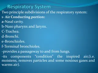
Differentiating between Viral and Bacterial Infections in Upper Respiratory Tract


•
Done by:
•
Saleh Alrashed
432102163
•
Abdullah Alsebti
•
432100932
•
Fahad Alturki
•
432102724
•
Objectives
:
How can we differentiate between viral and bacterial infections?
Sore throat (clinical features, differential diagnosis, complications, management)
Sinusitis including allergic rhinitis (Clinical features and management)
Otitis media in children (AOM and Secretory OM, Features, management)
How can we modify help seeking behavior of patients with flu illness?
Q 1 : A 55 year old patient came to the primary complaining of sore throat. The
patients reports having a low grade fever, which has resolved. On physical examination
there is no swelling on the neck or exudate in the mouth.
From the previous findings, what is your next step in management. ?
a.
Send the patient home with some analgesics.
b.
Start the patient on penicillin.
c.
Preform RDT for the patient.
d.
Throat culture.
Q 2: A patient presents to your primary care clinic complaining of recurrent
sore throat. On physical examination you notice that the patient has enlarged
tonsils. For the previous case, when will you advise the patient to have
tonsillectomy ?
1.
More than six episodes of streptococcal pharyngitis (confirmed by positive culture) in 1
year.
2.
Nine episodes of streptococcal pharyngitis in 2 consecutive years
3.
Ten or more infections of the tonsils and/or adenoids per year for 3 years in a row despite
adequate medical therapy.
4.
acute or recurrent tonsillitis associated with the streptococcal carrier state
Q 3: 3 year old child diagnosed with acute otitis media , what is the drug of choice to
treat him :
a.
Amoxicillin
b.
Cephalosporin
c.
Macrolides
d.
Doxycycline
Q 4: A Nine-month child brought to by his mother to the primary clinic, with the mother
complaining that “he won’t stop crying”. You notice the child keeps touching his ear. You
suspect otitis media. You decide to do otoscopy. However, every time you touch the
child’s ear he screams more. If the previous case was left untreated, what would the
Intratemporal complications that the patient might have?
a.
Facial paralysis
a.
Meningitis
b.
Lateral sinus thrombosis
c.
Cavernous sinus thrombosis
Q 5: Ahmad is 30 year old gentleman complaining of headache increase on leaning
forward during praying and mucopurulent post nasal discharge ,For the last 2 weeks .
On examination, there was nasal discharge in both nasal fossae. What is the most likely?
a.
Acute Bacterial Rhinosinusitis .
b.
Acute Viral Rhinosinusitis
c.
Common Cold .
d.
Chronic Bacterial Rhino Sinusitis
Allergic Rhinitis and
Sinusitis
•
Allergic rhinitis
, or allergic rhinosinusitis, is characterized by paroxysms of sneezing,
rhinorrhea, and nasal obstruction, often accompanied by itching of the eyes, nose, and
palate. Postnasal drip, cough, irritability, and fatigue are other common symptoms.
•
Epidemiology
— Allergic rhinitis is common, affecting 10 to 30 percent of children and adults
in the United States and other industrialized countries. It may be less common in some parts
of the world, although even developing countries report significant rates
Risk factors
—
The following are proposed or identified risk factors for allergic rhinitis:
•
Family history of atopy (ie, the genetic predisposition to develop allergic diseases)
•
Male sex
•
Birth during the pollen season
•
Firstborn status
•
Maternal smoking exposure in the first year of life
•
Exposure to indoor allergens, such as dust mite allergen
•
Serum IgE >100 int. units/mL before age six
•
Presence of allergen-specific immunoglobulin E (IgE)
Signs and symptoms
— Allergic rhinitis presents with paroxysms of sneezing, rhinorrhea,
nasal obstruction, and nasal itching. Postnasal drip, cough, irritability, and fatigue are other
common symptoms.
Diagnosis
— The diagnosis of
allergic rhinitis can be made
on
clinical grounds
based upon
the presence of characteristic
symptoms (ie, paroxysms of
sneezing, rhinorrhea, nasal
obstruction, nasal itching,
postnasal drip, cough, irritability,
and fatigue), a suggestive clinical
history (including the presence of
risk factors), and supportive
findings on physical examination.
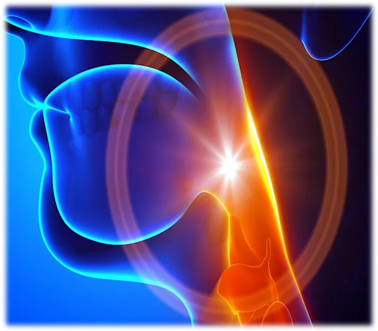
Sinusitis
:
Is inflammation of the sinuses resulting in symptoms. Common signs and
symptoms include thick nasal mucous, a plugged nose, and pain in the face.
The maxillary sinuses are the most common site (85%), followed by
ethmoidal (65%), sphenoidal (39%), and frontal (32%) involvement
.
Etiology
Infection
•
Viral:
the vast majority of rhinosinusitis episodes are caused by viral infection. Most viral upper
respiratory tract infections are caused by rhinovirus, but coronavirus, influenza A and B,
parainfluenza, respiratory syncytial virus, adenovirus, and enterovirus are also causative agents.
•
Bacterial
: the most common pathogens isolated from maxillary sinus cultures in patients with
acute bacterial rhinosinusitis include
Streptococcus pneumoniae, Haemophilus
influenzae,
and
Moraxella catarrhalis.
•
Fungal
( Rare )
RF:
•
Cilia in the sinuses do not work properly due to some medical conditions (
kartegner syndrome
).
•
Colds and allergies
may cause too much mucus to be made or block the opening of the sinuses.
•
A deviated nasal septum
,
nasal bone spur
, or
nasal polyps
may block the opening of the sinuses.
Clinical Presentation
•
Purulent nasal discharge (v. imp) (diagnostic )
•
Pain over cheek and radiating to frontal region or teeth, increasing with straining or bending down
•
Redness of nose, cheeks, or eyelids
•
Tenderness to pressure over the floor of the frontal sinus immediately above the inner canthus
•
Referred pain to the vertex, temple, or occiput
•
Postnasal discharge
•
A blocked nose
•
Persistent coughing or pharyngeal irritation
•
Facial pain
Physical Examination
●
Press over the air sinuses to check for:
o
Tenderness
o
Yellow
to
yellow
-
green
nasal discharge
.
●
Check the inside of the nasal passages by torch to check the mucus
and look for any structural abnormalities.
If signs and symptoms are not typical of sinusitis,
rule out an alternative diagnosis .
Differential diagnosis :
Allergic rhinitis.
Nasal foreign body.
Adenoiditis and tonsillitis
.
Sinonasal tumour.
Other causes of facial pain or headache
Investigations
•
Usually not necessary:
•
Diagnosis- Sinus Aspiration
•
Mucus culture
•
Nasal endoscopy
•
X-ray
•
Allergy testing
•
CT
•
Blood work
Management
Antibiotics
•
Amoxicillin/potassium clavunate (Augmentin)
•
Erythromycin-sulfisoxazole
Other Medications (facilitate drinage):
• Antihistamines if there is allergy
• Decongestants
• Anti-inflammatory agents ex. Steroids which will decrease the edema.
Non-pharmacological
•
Humidifier to relieve the drying of mucous membranes associated with mouth breathing
•
Increase oral fluid intake
•
Saline irrigation of the nostrils
•
Moist heat over affected sinus
Intracranial Complications
•
Meningitis (the most imp)
•
Epidural abscess
•
Subdural abscess
•
Intracerebral abscess
•
Cavernous sinus, venous sinus thrombosis
When to advise the patient to follow-up?
•
If symptoms rapidly deteriorate.
•
If they develop a high temperature.
•
Marked local pain that is predominately unilateral.
4 year old boy complaining of right earache, fever and
decrease of hearing on the same ear. He had a history of URTI
one week ago.
What is the most likely diagnosis ?
Otitis Media
https://www.youtube.com/watch?v=m-1UsNAoNyE
•
First-line
therapy — We suggest amoxicillin as the first-line therapy for children with
AOM who are treated with antibiotics and at low-risk for amoxicillin resistance.
•
amoxicillin-clavulanate
as the first-line therapy IN CASE OF Increased risk of beta-
lactam resistance.
•
Penicillin allergy: delayed reaction: cefdinir, cefpodoxime, cefuroxime, cefuroxime
and ceftriaxone
•
Immediate reaction — macrolide or lincosamide antibiotics.
Mr. Ali a 20 year old student came to the clinic complaining
of pain in his throat for 4 days which is worse upon
swallowing and talking. He also have runny congested nose
and cough.
What is the most likely dignosis?
How can we modify help seeking behavior of
patients with flu illness ?
MCQs
Q 1 : A 55 year old patient came to the primary complaining of sore throat. The
patients reports having a low grade fever, which has resolved. On physical examination
there is no swelling on the neck or exudate in the mouth.
From the previous findings, what is your next step in management. ?
a.
Send the patient home with some analgesics.
b.
Start the patient on penicillin.
c.
Preform RDT for the patient.
d.
Throat culture.
Q 2: A patient presents to your primary care clinic complaining of recurrent
sore throat. On physical examination you notice that the patient has enlarged
tonsils. For the previous case, when will you advise the patient to have
tonsillectomy ?
1.
More than six episodes of streptococcal pharyngitis (confirmed by positive culture) in 1 year.
2.
Nine episodes of streptococcal pharyngitis in 2 consecutive years
3.
Ten or more infections of the tonsils and/or adenoids per year for 3 years in a row despite adequate
medical therapy.
4.
acute or recurrent tonsillitis associated with the streptococcal carrier state
Q 3: 3 year old child diagnosed with acute otitis media , what is the drug of choice to
treat him :
a.
Amoxicillin
b.
Cephalosporin
c.
Macrolides
d.
Doxycycline
Q 4: A Nine-month child brought to by his mother to the primary clinic, with the mother
complaining that “he won’t stop crying”. You notice the child keeps touching his ear. You
suspect otitis media. You decide to do otoscopy. However, every time you touch the
child’s ear he screams more. If the previous case was left untreated, what would the
Intratemporal complications that the patient might have?
a.
Facial paralysis
a.
Meningitis
b.
Lateral sinus thrombosis
c.
Cavernous sinus thrombosis
Q 5: Ahmad is 30 year old gentleman complaining of headache increase on leaning
forward during praying and mucopurulent post nasal discharge ,For the last 2 weeks .
On examination, there was nasal discharge in both nasal fossae. What is the most likely?
a.
Acute Bacterial Rhinosinusitis .
b.
Acute Viral Rhinosinusitis
c.
Common Cold .
d.
Chronic Bacterial Rhino Sinusitis
References
:
NICE Guidelines
UpToDate
http://www.nhs.uk/Conditions/Otitis-media/Pages/Introduction.aspx
http://emedicine.medscape.com/article/994656-overview#a2
http://www.aafp.org/afp/2013/1001/p435.html
Thank you
Understanding the distinction between viral and bacterial upper respiratory tract infections is crucial for effective management and treatment. This article discusses key differences in symptoms, diagnostic methods, and treatment approaches to aid in accurate differentiation between the two types of infections.

Download Presentation

Please find below an Image/Link to download the presentation.
The content on the website is provided AS IS for your information and personal use only. It may not be sold, licensed, or shared on other websites without obtaining consent from the author. Download presentation by click this link. If you encounter any issues during the download, it is possible that the publisher has removed the file from their server.
E N D
Presentation Transcript
Upper Respiratory Tract Infection Done by: Saleh Alrashed 432102163 Abdullah Alsebti 432100932 Fahad Alturki 432102724
Objectives: How can we differentiate between viral and bacterial infections? Sore throat (clinical features, differential diagnosis, complications, management) Sinusitis including allergic rhinitis (Clinical features and management) Otitis media in children (AOM and Secretory OM, Features, management) How can we modify help seeking behavior of patients with flu illness?
Q 1 : A 55 year old patient came to the primary complaining of sore throat. The patients reports having a low grade fever, which has resolved. On physical examination there is no swelling on the neck or exudate in the mouth. From the previous findings, what is your next step in management. ? a. Send the patient home with some analgesics. b. Start the patient on penicillin. c. Preform RDT for the patient. d. Throat culture.
Q 2: A patient presents to your primary care clinic complaining of recurrent sore throat. On physical examination you notice that the patient has enlarged tonsils. For the previous case, when will you advise the patient to have tonsillectomy ? 1. More than six episodes of streptococcal pharyngitis (confirmed by positive culture) in 1 year. 2. Nine episodes of streptococcal pharyngitis in 2 consecutive years 3. Ten or more infections of the tonsils and/or adenoids per year for 3 years in a row despite adequate medical therapy. 4. acute or recurrent tonsillitis associated with the streptococcal carrier state
Q 3: 3 year old child diagnosed with acute otitis media , what is the drug of choice to treat him : a. Amoxicillin b. Cephalosporin c. Macrolides d. Doxycycline
Q 4: A Nine-month child brought to by his mother to the primary clinic, with the mother complaining that he won t stop crying . You notice the child keeps touching his ear. You suspect otitis media. You decide to do otoscopy. However, every time you touch the child s ear he screams more. If the previous case was left untreated, what would the Intratemporal complications that the patient might have? a. Facial paralysis a. Meningitis b. Lateral sinus thrombosis c. Cavernous sinus thrombosis
Q 5: Ahmad is 30 year old gentleman complaining of headache increase on leaning forward during praying and mucopurulent post nasal discharge ,For the last 2 weeks . On examination, there was nasal discharge in both nasal fossae. What is the most likely? a. Acute Bacterial Rhinosinusitis . b. Acute Viral Rhinosinusitis c. Common Cold . d. Chronic Bacterial Rhino Sinusitis
18 year male came to PHC complaining of several episodes of headache associated with fever, nasal congestion and discharge, The headache concentrated in front of the head aggravated when he is praying. What is the most likely diagnosis ?
Allergic Rhinitis and Sinusitis
Allergic rhinitis, or allergic rhinosinusitis, is characterized by paroxysms of sneezing, rhinorrhea, and nasal obstruction, often accompanied by itching of the eyes, nose, and palate. Postnasal drip, cough, irritability, and fatigue are other common symptoms. Epidemiology Allergic rhinitis is common, affecting 10 to 30 percent of children and adults in the United States and other industrialized countries. It may be less common in some parts of the world, although even developing countries report significant rates
Risk factors The following are proposed or identified risk factors for allergic rhinitis: Family history of atopy (ie, the genetic predisposition to develop allergic diseases) Male sex Birth during the pollen season Firstborn status Maternal smoking exposure in the first year of life Exposure to indoor allergens, such as dust mite allergen Serum IgE >100 int. units/mL before age six Presence of allergen-specific immunoglobulin E (IgE)
Signs and symptoms Allergic rhinitis presents with paroxysms of sneezing, rhinorrhea, nasal obstruction, and nasal itching. Postnasal drip, cough, irritability, and fatigue are other common symptoms.
Diagnosis The diagnosis of allergic rhinitis can be made on clinical grounds based upon the presence of characteristic symptoms (ie, paroxysms of sneezing, rhinorrhea, nasal obstruction, nasal itching, postnasal drip, cough, irritability, and fatigue), a suggestive clinical history (including the presence of risk factors), and supportive findings on physical examination.
Sinusitis: Is inflammation of the sinuses resulting in symptoms. Common signs and symptoms include thick nasal mucous, a plugged nose, and pain in the face.
The maxillary sinuses are the most common site (85%), followed by ethmoidal (65%), sphenoidal (39%), and frontal (32%) involvement.
Etiology Infection Viral: the vast majority of rhinosinusitis episodes are caused by viral infection. Most viral upper respiratory tract infections are caused by rhinovirus, but coronavirus, influenza A and B, parainfluenza, respiratory syncytial virus, adenovirus, and enterovirus are also causative agents. Bacterial: the most common pathogens isolated from maxillary sinus cultures in patients with acute bacterial rhinosinusitis include Streptococcus pneumoniae, Haemophilus influenzae, and Moraxella catarrhalis. Fungal ( Rare ) RF: Cilia in the sinuses do not work properly due to some medical conditions (kartegner syndrome). Colds and allergies may cause too much mucus to be made or block the opening of the sinuses. A deviated nasal septum, nasal bone spur, or nasal polyps may block the opening of the sinuses.
Clinical Presentation Purulent nasal discharge (v. imp) (diagnostic ) Pain over cheek and radiating to frontal region or teeth, increasing with straining or bending down Redness of nose, cheeks, or eyelids Tenderness to pressure over the floor of the frontal sinus immediately above the inner canthus Referred pain to the vertex, temple, or occiput Postnasal discharge A blocked nose Persistent coughing or pharyngeal irritation Facial pain
Physical Examination Press over the air sinuses to check for: Tenderness o Yellow to yellow-green nasal discharge. o Check the inside of the nasal passages by torch to check the mucus and look for any structural abnormalities.
If signs and symptoms are not typical of sinusitis, rule out an alternative diagnosis . Differential diagnosis : Allergic rhinitis. Nasal foreign body. Adenoiditis and tonsillitis. Sinonasal tumour. Other causes of facial pain or headache
http://www.drpaulose.com/wp-content/uploads/antral-lavage1.jpghttp://www.drpaulose.com/wp-content/uploads/antral-lavage1.jpg Investigations Usually not necessary: Diagnosis- Sinus Aspiration http://www.corbisimages.com/images/Corbis-IH056424.jpg?size=67uid=f704c2f3-85e8-4748-b1ff-538e846f84d5 Mucus culture Nasal endoscopy X-ray Allergy testing CT Blood work
Management Antibiotics Amoxicillin/potassium clavunate (Augmentin) Erythromycin-sulfisoxazole Other Medications (facilitate drinage): Antihistamines if there is allergy Decongestants Anti-inflammatory agents ex. Steroids which will decrease the edema.
Non-pharmacological Humidifier to relieve the drying of mucous membranes associated with mouth breathing Increase oral fluid intake Saline irrigation of the nostrils Moist heat over affected sinus
Intracranial Complications Meningitis (the most imp) Epidural abscess Subdural abscess Intracerebral abscess Cavernous sinus, venous sinus thrombosis
When to advise the patient to follow-up? If symptoms rapidly deteriorate. If they develop a high temperature. Marked local pain that is predominately unilateral.
4 year old boy complaining of right earache, fever and decrease of hearing on the same ear. He had a history of URTI one week ago. What is the most likely diagnosis ?
Otitis Media (OM) is any inflammation of the middle ear, without reference to etiology or pathogenesis. It can be classified into many variants on the basis of etiology, duration, symptomatology, and physical findings. Peak prevalence of OM in both sexes occurs in children aged 6-18 months. Some studies show bimodal prevalence peaks; a second, lower peak occurs at age 4-5 years and corresponds with school entry.
Classifications of OM symptom, and physical findings Duration Etiology Recurrent OM Sub acute OM 3weeks up to 3months Acute OM Chronic OM AOM Bacterial 3 episodes within 6 months <3weeks >3 months OME viral
Risk Factors: Age The age-specific attack rate for acute OM peaks between 6 and 18 months of age. After that, the incidence declines with age. Family history Day care The transmission of bacterial and viral pathogens is common in day care centers. Lack of breastfeeding. Tobacco smoke and air pollution. Developing areas. Social and economic conditions (poverty and household crowding increase the risk). Season (increased incidence during the fall and winter months). Altered host defenses and underlying disease (eg, cleft palate, Down syndrome, allergic rhinitis).
Route Of Infection: Eustachian tube External auditory canal Blood borne Etiology: Bacteria: Viruses: Respiratory syncytial virus. Picornaviruses (eg, rhinovirus, enterovirus). Coronaviruses. Influenza viruses Adenoviruses. Human metapneumovirus.
Acute Otitis Media: Acute otitis media (AOM) is defined by moderate to severe bulging of the tympanic membrane (TM) accompanied by acute signs of illness and signs or symptoms of middle ear inflammation. Pathogensis:
Signs: bulging of the tympanic membrane. Erythema of the tympanic membrane. Opacity. Loss of landmarks: handle and long process of malleus not visible. Symptoms: Ear pain (otalgia). Fever. Irritability. Headache. Apathy. anorexia. Vomiting. Diarrhea. Otorrhea. hearing loss.
Otitis media with effusion: Otitis media with effusion (OME), also called serous otitis media, is defined as middle-ear effusion without acute signs of infection. OME often occurs after acute otitis media (AOM), but it also may occur with eustachian tube dysfunction in the absence of AOM. Signs & Symptoms: Hearing loss Conductive hearing loss occurs whenever fluid fills the middle ear. Other symptoms Other symptoms that may occur in children with OME include ear pain, sleep disturbance, a feeling of fullness in the ear, tinnitus, or balance problems. Clinical course Otitis media with effusion usually resolves spontaneously. Approximately 30 to 40 percent of children have recurrent episodes.
Diagnosis and examination: Otoscopy: The key to distinguishing acute otitis media (AOM) from otitis media with effusion (OME) is the performance of otoscopy using appropriate tools and an adequate light source.
Position The position of the tympanic membrane is the most critical characteristic in distinguishing AOM from OME. Position may be described as neutral, retracted, full, or bulging
Color Assessment of the color of the tympanic membrane is another important aspect of the otoscopic examination. Under normal conditions, the color of the tympanic membrane is pearly gray or pink . When there is uninfected fluid in the middle ear (ie, OME), the color is usually amber, gray, or blue. A white or pale yellow tympanic membrane usually indicates pus in the middle ear cavity, which is a sign of AOM Translucency. Mobility.
Investigations: Tympanometry and Acoustic reflectometry Tympanometry and acoustic reflectometry are techniques to predict the presence or absence of middle ear effusion. Audiology Audiology evaluation can facilitate the diagnosis of OME if the middle ear fluid is difficult to see Conductive hearing loss in conjunction with a flat tympanogram is suggestive of OME. Tympanocentesis (aspiration of the middle ear fluid) for culture is required for etiologic diagnosis. Scans On the very rare occasions where there is a possibility the infection has spread out of the middle ear and into the surrounding area, CT or MRI may be carried out.
Management: AOM: Most middle ear infections (otitis media) will clear up within three days and don't need any specific treatment. Symptomatic therapy: We suggest oral ibuprofen or acetaminophen for pain control in children with AOM. Antibiotic therapy: Child has a serious health condition that makes them more vulnerable to complications, such as cystic fibrosis or congenital heart disease. Child is less than three months old, or they are less than two years old and have an infection in both ears. Child's symptoms are severe. Child has discharge coming from their ear. Child's symptoms show no signs of improvement after four days.
First-line therapy We suggest amoxicillin as the first-line therapy for children with AOM who are treated with antibiotics and at low-risk for amoxicillin resistance. amoxicillin-clavulanate as the first-line therapy IN CASE OF Increased risk of beta- lactam resistance. Penicillin allergy: delayed reaction: cefdinir, cefpodoxime, cefuroxime, cefuroxime and ceftriaxone Immediate reaction macrolide or lincosamide antibiotics.
OME: Watchful waiting "Watchful waiting" for three months from the onset of the effusion (or the diagnosis of effusion if the date of onset is not known) is recommended for children who have OME, are not at risk for speech, language, or learning problems, and otherwise have normal hearing (ie, hearing loss 20 dB). Tympanostomy tubes. Potential indications: OME in children who are at risk of speech, language, or learning problems, regardless of hearing status. Structural damage to the tympanic membrane. Persistent OME-associated hearing loss. Bilateral OME for 3 months; unilateral OME for 6 months ecurrent episodes of OME. Signs of eustachian tube dysfunction.
Complications: Intratemporal complications: Hearing loss. Balance and motor problems. Tympanic membrane perforation, tympanosclerosis. Middle ear atelectasis (retraction or collapse of the tympanic membrane due to chronic or recurrent decreased pressure in the middle ear). Cholesteatoma. Adhesive otitis media. Extension of the suppurative process to adjacent structures (mastoiditis, petrositis, labyrinthitis). Facial paralysis.
Intracranial complications: Meningitis. Epidural abscess. Brain abscess. Lateral sinus thrombosis. Cavernous sinus thrombosis. Subdural empyema. Carotid artery thrombosis.
Mr. Ali a 20 year old student came to the clinic complaining of pain in his throat for 4 days which is worse upon swallowing and talking. He also have runny congested nose and cough. What is the most likely dignosis?


,")
,")


 is any inflammation of the middle ear,")