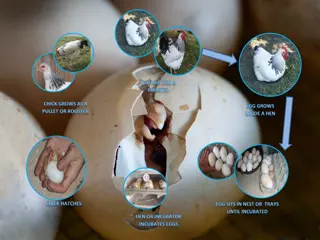
Extra Embryonic Membrane Formation in Chick and Their Functions
The development of extra embryonic membranes in chick embryos plays a crucial role in their growth and protection. These membranes, including the amnion and chorion, serve various functions such as providing protection, facilitating gas exchange, and supporting the formation of the placenta. Understanding the types and functions of these membranes is essential for comprehending the early stages of chick embryo development.

Download Presentation

Please find below an Image/Link to download the presentation.
The content on the website is provided AS IS for your information and personal use only. It may not be sold, licensed, or shared on other websites without obtaining consent from the author. Download presentation by click this link. If you encounter any issues during the download, it is possible that the publisher has removed the file from their server.
E N D
Presentation Transcript
Extra embryonic membrane formation in Chick and their functions By Dr. R. C. Nath; Associate Professor Department of Zoology Govt. Degree College, DMR.
Definition of Extra Embryonic Membrane: The blastoderm in birds , reptiles and mammals not only gives rise to embryo, but also to certain structures that lie outside the embryo. These extra embryonic structures are called Extra Embryonic Membrane.
Types of extra embryonic membranes There are basically four types of extra- embryonic membranes in Chick- Amnion Chorion Allantois and Yolk sac
1.Amnion: ectoderm and outer mesoderm) above the embryo. Between the amnion and embryo, there is amniotic cavity filled with amniotic fluid secreted by both embryo and amnion. Amnion protects the embryo while amniotic fluid acts as shock absorber and also prevents desiccation of embryo. It is innermost fold of somatopleur (inner
Functions of the Amnion : Amnion protects the embryo from shock and injury. Amniotic fluid prevents its desiccation.
2.Chorion: surrounds the embryo. In reptiles, birds and prototherians, allantochorion acts as extra embryonic lung and helps in exchange of gases. But in primates including human beings, only chorion forms the placenta (chorionic placenta) while in other eutherian, allantochorion forms allantoic placenta. It is outermost fold of somatopleur and
Functions of the Chorion : It protects the foetus. Provides place for the growth of allantois. Helps in the formation of the placenta.
3.Allantois: from the hind gut of the embryo. It is well developed in amniotes with polylecithal egg (e.g., reptiles, birds and prototherians) and stores the nitrogenous wastes of the embryo so acts as extra embryonic kidney. In most of eutherian, it combines with chorion to form allantochorion which takes part in placenta formation (Allantoic placenta). It isreduced in human beings. It is a fold of splanchnopleur developed
Functions of the Allantois : Store insoluble nitrogenous waste matter, uric acid. Functions as extra embryonic lung . Gaseous exchange taking place between blood and external air through it. Carries on excretion,respiration and nutrition. Allantois function as a soft, elastic cushion for protecting the embryo from shock . Allantois helps in a formation of umbilical chord.
4.Yolksac: endoderm and outer mesoderm) and is well developed in reptiles, birds and prototherians having poly lecithal egg. It is mainly digestive in function so acts as extra embryonic gut. It also absorbs the dissolved yolk and passes it to developing embryo. In human beings, it is vestigial. It is formed of splanchnopleur (inner
Functions of the Yolk Sac : Digest the yolk . Transfer the products of digestion to the embryo. Digestive surface increased by force off the walls of the yolk sacs called yolk sac septa . In Mammals yolk sac is less nutritive organ then Reptiles and Aves .
and chorion are closely associated, Amnion is bag like covering over the embryo, it separates the embryo from internal environment, Amnion is developed from somato-pleuric amniotic folds. These folds are head fold, late- ral folds and tail folds. a) At about 30 hours of incubation, in front of the head of embryo a head fold is developed, it is called amniotic head fold. In the development of embryo, amnion
b) At about third day of incubation amniotic tail fold is developed. It grows opposite to head fold. c) Mean while lateral folds will develop, they grow dorso-medially. d) After some time head fold, lateral folds, and tail fold will fuse near posterior end of a embryo. e) At 72 of incubation they are still not fused. They show an opening called amniotic umblicus, afterwards they unite.
f) After their union at the point of union "sero- amniotic raphae" is present. It is a fold. g) Because of this union outer chorion inner amnion will form, because it is developed from somatopleure. In chorion ectoderm is present out side and mesoderm is present inside. In amnion ectoderm is inside, mesoderm is out side. Hence the space between amnion and chorion is called exocoel or extraembryonic coelome.
THANKS TO ALL THANKS TO ALL


 At about third day of incubation amniotic tail")
 After their union at the point of union \"sero-")
 After their union at the point of union \"sero-")