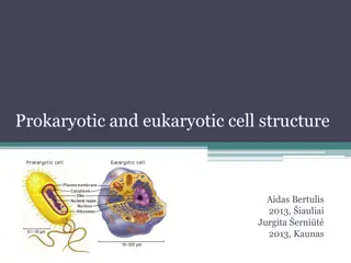
Evolution of Membrane Models: From Early Observations to Fluid-Mosaic Structure
Researchers in the early 20th century noted the permeability of lipid-soluble molecules into cells, paving the way for membrane studies. The development of membrane models progressed from the sandwich model to the widely accepted fluid-mosaic model. The complexity of the plasma membrane, comprising lipids, proteins, glycolipids, and cholesterol, plays a crucial role in cell function. The fluidity of the phospholipid bilayer allows for membrane flexibility, essential for cellular processes.

Download Presentation

Please find below an Image/Link to download the presentation.
The content on the website is provided AS IS for your information and personal use only. It may not be sold, licensed, or shared on other websites without obtaining consent from the author. Download presentation by click this link. If you encounter any issues during the download, it is possible that the publisher has removed the file from their server.
E N D
Presentation Transcript
Chapter 5 Outline and Terms Membrane Models Have Changed
A.Early Observations : 1. At turn of the century, researchers noted lipid-soluble molecules entered cells more rapidly than water-soluble suggesting lipids are component of plasma membrane. 2. Later chemical analysis revealed membrane contains phospholipids. 3. In 1925, Garter and Grendel found amount of phospholipid extracted from a red blood cell was just enough to form one bilayer; suggested nonpolar tails directed inward, polar heads outward. molecules,
To account for permeability of membrane to 4. nonlipid substances, Danielli and Davson proposed sandwich model (later phospholipid bilayer between layers of protein. 5. With electron microscope available, Robertson proposed proteins were membrane and all membranes in cells had similar compositions 3/4 the unit membrane model. B. In 1972, Singer and Nicolson introduced the currently accepted fluid-mosaic model of membrane structure. 1. Plasma membrane is phospholipid bilayer in which protein molecules are partially or wholly embedded. 2. Embedded proteins are scattered throughout membrane in irregular pattern; varies among membranes. proved wrong) with embedded in outer
The Plasma Membrane Is Complex A. Fluid-mosaic Model 1. Membrane structure has two components, lipids and proteins. . 2. Lipids are arranged into a bilayer. a. Most plasma membrane lipids are phospholipids, which spontaneously arrange themselves into a bilayer. . b. Nonpolar tails are hydrophobic and directed inward; polar heads are hydrophilic and are directed outward to face extracellular and intracellular fluids. c. Plasma membranes contain glycolipids with a structure similar to phospholipids except the hydrophilic head is a variety of sugar; they are protective and assist in various functions. d. Cholesterol is a lipid found in animal plasma membranes; reduces the permeability of the membrane.
B-The Membrane Is Fluid 1. At body temperature, the phospholipid bilayer has consistency of olive oil. 2. The greater the concentration of unsaturated fatty acid residues, the more fluid the bilayer. 3. In each monolayer, the fatty acid tails wiggle, and entire phospholipid molecules can move sideways at a rate of about 2 ---the length of a prokaryotic cell---per second. 4. However, phospholipid molecules rarely flip-flop from one layer to the other. 5. The fluidity of the phospholipid bilayer allows cells to be pliable. 6. Some membrane proteins are held in place by cytoskeletal filaments; most drift in fluid bilayer.
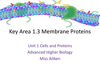
C. Proteins in the Plasma Membrane 1. Transmembrane proteins extend through both sides of a cell membrane; they have hydrophobic regions embedded within the membrane and hydrophilic regions that project from both surfaces of the bilayer. 2. Many trans membrane proteins are glycoproteins with a carbohydrate chain that projects externally. a. Some are anchored to membrane by covalently attached lipid or covalently bonded to carbohydrate chain of a glycolipid. b. Others held in place by noncovalent interactions; are disrupted by gentle shaking or change in pH. 3. Plasma membrane is asymmetrical; lipid and protein composition of inside half differs from outside half. . 4. Carbohydrate chains of glycolipids and glycoproteins form carbohydrate coat enveloping outer surface of plasma membrane. 5. Some proteins of inside surface serve as links to cytoskeletal filaments; on outer surface some serve as links to extracellular matrix.
D. Cell-Cell Recognition: 1. Carbohydrate chains of glycolipids and glycoproteins identify cell; diversity of the chains is enormous. a. Chains vary by number of sugars (from 15 to several hundred). b. Chains vary in branching. c. Sequence of sugars in chains varies, and by isomers as well. 2. Glycolipids and glycoproteins vary from species to species, from individual to individual of same species, and even from cell to cell in same individual. 3. In development, different type cells in embryo develop their own carbohydrate chains; these chains allow tissues and cells of the embryo to sort themselves out. 4. Immune system rejection of transplanted tissues is due to recognition of unique glycolipids and glycoproteins; blood types are due to unique glycoproteins on the membranes of red blood cells (RBC).
E. The Membrane Is a Mosaic : 1. Plasma membrane and organelle membranes have unique proteins; RBC plasma membrane contains 50+ types of proteins. 2. Membrane proteins determine most of the membrane's functions. 3. Channel proteins allow a particular molecule to cross the membrane freely (e.g., Cl- channels). 4. Carrier proteins selectively interact with a specific molecule so it can cross the plasma membrane (e.g., Na+-K+ pump). 5. Cell recognition proteins histocompatibility complex) glycoproteins that are different for each person; allows immune system to recognize foreign tissues. 6. Receptor proteins are shaped so a specific molecule (e.g., hormone or another molecule) can bind to it. 7. Enzymatic proteins catalyze specific metabolic reactions; membrane protein, adenylate cyclase, is involved in ATP metabolism. include MHC (major
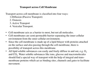
5.3. How Molecules Cross the Plasma Membrane : A. Types of Membranes and Transport:- 1. A permeable membrane allows all molecules to pass through. 2. an impermeable membrane allows no molecules to pass through. 3. a semipermeable membrane allows some molecules to pass through. a. Small noncharged lipid molecules pass through the membrane freely. b. Macromolecules cannot freely cross a plasma membrane. c. Ions and charged molecules have difficulty crossing the membrane. = The plasma membrane is differentially permeable; only certain molecules can pass through freely. = Both passive and active mechanisms are involved in the movement of molecules across the membrane. a. Passive transport moves molecules across the membrane without expenditure of energy by cell; includes diffusion and facilitated transport. b. Active transport uses energy (ATP) to move molecules across a plasma membrane; includes exocytosis, endocytosis, and pinocytosis.
B. Use of Diffusion and Osmosis: 1. In diffusion, molecules move from higher to lower concentration concentration gradient). a. A solution contains a solute, usually a solid, and a solvent, usually a liquid. b. In the case of a dye diffusing in water, dye is a solute and water is the solvent. 2. Membrane chemical and physical properties allow only a few types of molecules to cross by diffusion (i.e., down their
3. Osmosis is the diffusion of water across a differentially permeable membrane. a. Osmotic pressure is hydrostatic pressure, on side of membrane with higher solute concentration, produced by water diffusing to that side of membrane; thistle tube example: b.Although sugars and salts pass through membranes, differences in permeability between water and these solutes is so great that cells in sugar and salt solutions must deal with osmotic movement of water. c. Osmosis is constant process in life: for example, water is absorbed in large intestine, retained by kidneys, and taken up by blood.
4. Tonicity is strength of a solution in relationship to osmosis; determines movement of water into or out of cells. a. Isotonic is where the relative solute concentration of two solutions are equal. B- Hypotonic is where a relative solute concentration of one solution is less than another solution. . c. Hypertonic is where relative solute concentration of one solution is greater than another solution. . d. Swelling of cell in hypotonic solution creates turgor pressure; how plants maintain erect position. e. Solutions that cause cells to shrink are hypertonic solutions; red blood cells placed in salt solutions above 0.9% shrink and wrinkle, a condition called crenation.
C. Transport by Carrier Proteins 1. Plasma membrane impedes passage of most substances but many molecules enter or leave at rapid rates. 2. Carrier proteins are membrane proteins that combine with and transport only one type of molecule; are believed to undergo a change in shape to move molecule across in active and facilitated transport. 3. Facilitated transport is passive transport of specific solutes down their concentration gradient, facilitated by a carrier protein. 4. Active transport is transport of specific solutes across plasma membranes against the concentration gradient through use of cellular energy (ATP).
5. Use of membrane-assisted transport a. In exocytosis, a vesicle often formed by Golgi apparatus fuses with the plasma membrane as secretion occurs; method by which insulin leaves insulin-secreting cells. b. During endocytosis, cells take in substances by vesicle formation as plasma c. In phagocytosis, cells engulf large particles forming an endocytic vesicle. .(e.g., WBC engulfed Bacteria ). d. Pinocytosis occurs when vesicles form around a liquid or very small particles. e. Receptor-mediated endocytosis occurs when specific macromolecules bind to plasma membrane receptors. membrane pinches off.
The Cell Surface Is Modified : A. Plasma Membrane: 1. The plasma membrane is outer living boundary of a cell. 2. Many cells have an extracellular component formed outside of membrane; plant, fungi, algae and bacteria form cell walls, while animal cells have an extracellular matrix. B. Plant Cells Have a Cell Wall . 1. Plant cells are surrounded by a porous cell wall that varies in thickness, depending on function of cell. 2. Plant cells have primary cell wall composed of cellulose polymers united into threadlike micro fibrils that form fibrils. 3. Cellulose fibrils form a framework whose spaces are filled by noncellulose molecules: a. Pectins allow the cell wall to stretch and are abundant in the middle lamella that holds cells together. b. Noncellulose harden the wall of mature cells.
4. Lignin, a substance that adds strength, is a common ingredient of secondary cell walls in woody plants. 5. Plasmodesmata are narrow channels that pass through cell walls of neighboring cells and connect their cytoplasms, C. Animal Cells Have an Extracellular Matrix . 1. Extracellular matrix is meshwork of insoluble proteins with carbohydrate chains that are produced and secreted by animal cells; fills spaces between animal cells. 2. This matrix most likely influences the development, migration, shape and function of cells. 3. Collagen gives the matrix strength and elastin gives it resilience. 4. Fibronectins and laminins bind to membrane receptors; permit communication between matrix and cytoplasm. 5. Fibronectins and laminins form pathways that direct the migration of cells during development. 6. Proteoglycans are glycoproteins that provide a packing gel that joins the various proteins in matrix and most likely regulate signaling proteins that bind to receptors in the plasma protei
D. Animal Cells Have Junctions: 1. Cell junctions are points of contact that physically link neighboring cells or provide functional links; three types exist between animal cells: adhesion junctions, tight junctions, and gap junctions. 2. In adhesion junctions (desmosomes), internal cytoplasmic plaques, firmly attached to cytoskeleton within each cell are joined by intercellular filaments; hold cells together where tissues stretch (e.g., in heart, stomach, bladder). 3. In tight junctions, plasma membrane proteins attach to each other, producing zipperlike fastenings; hold cells together so tightly that the tissues (e.g., epithelial lining of stomach and kidney tubules) are barriers. 4. A gap junction allows cells to communicate. Gap junctions are important to function of heart muscle and smooth muscle because they permit diffusion of ions required for cells to contract.
. . .