Understanding Platelet Structure and Function in Physiology
Platelets play a crucial role in hemostasis and bleeding disorders. This lecture by Dr. Mohammed Alotaibi covers the ultrastructure of platelets, functions of organelles and surface receptors, mechanisms of platelet functions, and the relationship of membrane receptors and granule content in normal hemostasis. It also delves into platelet formation, regulation by thrombopoietin, and the intricate ultrastructure involving microtubules, granules, Von Willebrand factor, and more.

Download Presentation

Please find below an Image/Link to download the presentation.
The content on the website is provided AS IS for your information and personal use only. It may not be sold, licensed, or shared on other websites without obtaining consent from the author. Download presentation by click this link. If you encounter any issues during the download, it is possible that the publisher has removed the file from their server.
E N D
Presentation Transcript
Platelet Structure & Function DR.MOHAMMED ALOTAIBI MRes, PhD (Liverpool, England) Assist. Professor of Physiology, College of Medicine King Khalid University Hospital King Saud University
Objectives At the end of this lecture the student is expected to: - Understand plateletnormal ultrastructure - Understand the functions of different platelets organelles and surface receptors - Understand the mechanisms of platelet functions - Relate membrane receptors and granule content to normal function in hemostasis and bleeding (platelet) disorders
What are platelets? BLOOD Plasma Cells RBC WBC Platelet
What are platelets? Mature platelets Bone marrow Megakaryocyte
Platelets cont. Site of formation: Bone marrow Steps: Stem cell Megakaryoblast Megakaryocyte Platelets
Platelets Formation (Thrombopoiesis) Regulation of thrombopoiesis by Thrombopoietin
Platelet ultra-structure (Electron-microscope -EM)
Platelets (Thrombocytes) - Discs ~ 1-4 M in diameter - Their normal concentration in the blood is between 150,000 and 300,000 / l
(Thrombocytes) Anuclear and discoid cell activated Life span: 7 10 days Sequestered in the spleen; hypersplenism may lead to low platelet counts. spherical when
Platelet Ultrastructure Microtubules Mitochondria Granules von Willebrand Factor Fibrinogen Chemokines (PF4,etc.) Thrombospondin P-selectin Open canalicular system Dense Granules ADP/ATP Calcium Serotonin
Platelet Receptors Platelet (GP Ia, GP VI) Collagen GP Ib-IX-V (vW Factor) (TP ) TXA2 GP IIb-IIIa (Fibrinogen, vWF) (P2Y12) ADP
General functions of the platelets
HEMOSTASIS 1. VASCULAR PHASE 2. PLATELET PHASE 3. COAGULATION PHASE 4. FIBRINOLYTIC PHASE
Platelet Activation Adhesion Shape change Aggregation Secretion (Release reaction) Clot Retraction
Platelet function Adhesion GP Ib-IX-V (vW Factor) (GP Ia, GP VI) Collagen (vWF) Platelet GP IIb-IIIa (Fibrinogen, vWF)
Platelet function Activation
Resting platelet Activated platelet
Convolutions Spread to psuedopods Smooth surface psuedopods Convolutions In body Early Further Spread of Convolutions to psuedopods Spread platelet
Platelet function (GP Ia, GP VI) Collagen GP Ib-IX-V (vW Factor) Platelet (TP ) TXA2 GP IIb-IIIa (Fibrinogen, vWF) Aggregation (P2Y12) ADP Aggregation: Fibrinogen is needed to join platelets to each other via platelet fibrinogen receptors
Platelet Aggregation Activated platelet Resting platelet Fibrinogen Agonist GP IIb/IIIa receptors (unreceptive state) Aggregating platelets
von Willebrand factor (vWF) and Platelet Adhesion Blood Flow Fibrinogen vWF Platelet A1 domains GPIb- IX-V Plug Recruitment Adhesion Rolling Activation Formation
Platelet function Secretion
Activated Platelets Secrete: 1. 5HT (serotonin) 2. Platelet phospholipid (PF3) formation 3. Thromboxane A2 (TXA2) is a prostaglandin formed from arachidonic acid Function: Vasoconstriction Platelet aggregation (TXA2 inhibited by aspirin) vasoconstriction clot
Clot Retraction Myosin, actin filaments and thrombosthenin in platelets are stimulated to contract during aggregation further reinforcing the plug and help release of granule contents
Platelet haemostatic plug formation Platelets activated by adhesion Extend projections to make contact with each other Release: thromboxane A2, serotonin & ADP >>> activating other platelets Serotonin & thromboxane A2 are vasoconstrictors decreasing blood flow through the injured vessel. ADP causes stickiness and enhances aggregation
Platelet Activation- summary Platelets are activated when brought into contact with collagen exposed when the endothelial blood vessel lining is damaged Activated platelets release a number of different coagulation and platelet activating factors Transport of negatively charged phospholipids to the platelet surface; provide a catalytic surface for coagulation cascade to occur Platelets adhesion receptors (integrins): Platelets adhere to each other via adhesion receptors forming a hemostatic plug with fibrin Myosin and actin filaments in platelets are stimulated to contract during aggregation further reinforcing the plug and help release of granule contents GPIIb/IIIa: the most common platelet adhesion receptor for fibrinogen and von Willebrand factor (vWF)
General functions of the platelets: Platelet plug formation Platelets and blood coagulation
Maintenance of vascular integrity Adequate number and function of platelet is essential to participate optimally in hemostasis Stabilization of hemostatic plug by contributing to fibrin formation Initial arrest of bleeding by platelet plug formation
Bleeding Disorders Bleeding can result from: Platelet defects: Deficiency in number (thrombocytopenia) Defeciency in function (acquired or congenital)
Platelet function Bleeding disorders abnormal number or function of platelet
Congenital Platelet Disorders Disorders of Adhesion: . Bernard-Soulier Syndrome Disorder of Aggregation: . Glanzmann thrombosthenia Disorders of Granules: . Grey Platelet Syndrome . Storage Pool deficiency . Hermansky-Pudlak syndrome .Chediak-Higashi syndrome
Bernard-Soulier Syndrome Platelet GP Ib-IX-V Collagen (GP Ia, GP VI) (vW Factor) TXA2 GP IIb-IIIa (Fibrinogen, vWF) ADP
Bernard-Soulier Syndrome Platelet Collagen (GP Ia, GP VI) (vW Factor) TXA2 GP IIb-IIIa (Fibrinogen, vWF) ADP
Glanzmann Thromasthenia Platelet GP Ib-IX-V Collagen (GP Ia, GP VI) (vW Factor) TXA2 GP IIb-IIIa (Fibrinogen, vWF) ADP
Glanzmann Thromasthenia Platelet GP Ib-IX-V (vW Factor) Collagen (GP Ia, GP VI) TXA2 ADP
Glanzmann Thrombasthenia Fibrinogen IIb-IIIa Normal No Gp IIb-IIIa Receptors
How to investigate for platelet disorders? Platelet function tests
Laboratory Testing of Platelet Functions Platelet count (& shape) Bleeding time Platelet Aggregation Platelet Function Analyzer (PFA-100) Flow-cytometry Electron-microscopy Granule release products White GC et al. Approach to the Bleeding Patient. In Colman, RW et al. Hemostasis and thrombosis 2nd ed. 1987
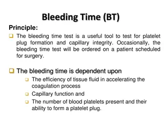
Bleeding Time Bleeding time 1 Bleeding time 2 Bleeding time 3
Laboratory Testing of Platelet Functions Platelet Aggregometer (in PRP): Provides information on time course of plat. activation. Agonists: ADP Adrenaline Collagen Arachidonic acid Ristocetin Thrombin Reference ranges need to be determined for each agonist (+ Dose responses)
Platelet Aggregation Agonists: ADP Adrenaline Collagen Arachidonic acid Ristocetin Thrombin Whole blood RBC PRP
Summary Platelets are cell fragments derived from megakaryocyte in the bone marrow Platelets play a pivotal role in hemostasis by arresting bleeding from injured blood Vessels Bleeding can result from: Platelet defects acquired or congenital


")


")