Differential Diagnosis of Ascites in a 50-Year-Old Man with Liver Disease
Ascites, jaundice, and confusion in a 50-year-old man raise concerns about liver disease. Differential diagnosis includes cirrhosis, malignancy, alcoholic liver disease, and biliary obstruction. Physical examination findings such as spider naevi, hepatomegaly, and asterixis help narrow down the possibilities, with consideration for underlying causes and complications.

Download Presentation

Please find below an Image/Link to download the presentation.
The content on the website is provided AS IS for your information and personal use only. It may not be sold, licensed, or shared on other websites without obtaining consent from the author. Download presentation by click this link. If you encounter any issues during the download, it is possible that the publisher has removed the file from their server.
E N D
Presentation Transcript
Ascites Anas Alahmad Mohammad Alkhateeb Abd Alrahman saadah
Contents Clinical case Definition Types and Causes Clinical features Complications History Physical examination Investigations Management
Clincal Cases Presenting problem : A 50-year-old man is referred to the medical admissions unit with 4week history of increasing abdominal distension and anorexia, followed by 2 weeks of jaundice. There has been no change in bowel habit or vomiting and he denies any weight loss. His wife has noticed him to be confused over the last hours. He takes no regular medication. He has drunk 8 pints of beer a day for about 10 years, but stopped 10 days ago when he started to feel unwell. The general practitioner has checked some blood tests, which are shown in Box 65.1.
What would your differential diagnosis include before examining the patient? Abdominal distension in the absence of symptoms of bowel obstruction suggests either an abdominal mass or ascites, of which the most common causes in a previously fit man are cirrhosis and malignancy. Cirrhosis is usually asympto- matic until portal hypertension develops and synthetic liver function worsens; then symptoms of chronic liver failure, such as ascites, appear quite quickly. The presence of jaundice indicates either liver disease, which may be acute or chronic, or obstruction to the biliary tree, which is usually due to malignancy Acute liver disease can be excluded, as this is not associated with ascites However, the presence of jaundice with ascites does not differentiate carcinoma of the head of the pancreas with peritoneal metastases from chronic liver failure. Investigations in this case do narrow down the differential diagnosis, as the presence of pancytopenia suggests chronic liver disease. Platelets are often disproportionately lower than the rest of the blood count in cirrhosis due to a combination of splenomegaly and reduced hepatic thrombopoietic production. The prolonged prothrombin time and low albumin can be seen in both chronic liver failure and biliary obstruction, but in the latter the prolonged clotting is reversible with vitamin K. It is common for individuals to stop drinking about 2 weeks before jaundice develops in alcoholic liver disease because they feel so unwell; alcohol detoxifi- cation will not, therefore, be needed in this man
on examination The patent have 5-6 spider naevi on his upper chest wall . He has a 6 cm firm non tender palpable liver . Ascitis is confirmed on abdominal examination . Asterixis ( flapping tremor ) is demonstrated Does this narrow down your differential diagnosis? The finding of stigmata of chronic liver disease makes the diagnosis of cirrhosis most likely. However, it is important to remember that not evervone with decompensated chronic liver disease will have these cutaneous stio mata. A large liver is also frequently seen in individuals with cirrhosis due to alcohol. Alcohol excess increases the risk of progression to cirrhosis in chronic liver disease due to other causes such as chronic viral infection, hepatitis B and C., and haemochromatosis. These conditions must also be excluded before concluding that the liver disease is due to alcohol alone Jaundice in an alcoholic with cirrhosis may either reflect end-stage liver disease or be due to a superimposed alcoholic hepatitis The patient's confusion in the presence of a hepatic flap is likely to be due to hepatic encephalopathy
On further investigation On further investigation : U/S show that the liver is enlarge and has a coarse texture but no bile duct dilatation the spleen is also enlarged no focal lesions are seen in the liver the the main portal vein is patent The result of ascitic tap was : Result analysis : 1. Ultrasound is very good at excluding an obstructive cause for jaundice 2. Splenomegaly suggests portal hypertension and would not be expected with the other causes of ascites. A transudative ascites is also consistent with cirrhosis 3. This has been complicated by spontaneous bacterial peritonitis, as shown by the elevated ascitic white cell count.
Definition From Greek origin (askites) which means bag or sac like. Pathological accumulation of fluid in the peritoneal cavity(>25 ml , symptomatic if > 100 ml ) Note that Healthymenhavelittleornointra-peritoneal fluid,butwomenmaynormallyhaveasmuchas20mL, dependingonthephaseoftheirmenstrualcycle.
Mnemonic Ascites is one of the causes of abdominal distension. Other causes includes (6Fs): 1. Fat (obesity). 2. Flatus (pseudo-obstruction ,obstruction) 3. Faeces (subacute obstruction,constipation) 4. Fetus (pregnancy). 5. Fluids (Ascites) 6. Functional (bloating as in irritable bowel disease).
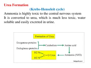
pathogenesis According to starling s hypothesis the exchange of fluids between the blood and tissue spaces is controlled by the balance between two factors; 1. Capillary blood pressure 2. Osmotic pressure of plasma proteins (plasma colloid osmotic pressure) Increase capillary blood pressure / decrease in Plasma colloid osmotic pressure = Ascites
Thefactorsthat are involvedinitspathogenesis include: 1.portalhypertension (75%) exerts a local hydrostatic pressure causing transudation of fluid to peritoneal cavity 2.sodiumandwaterretention(decreasealdosteronesecretion). 3.lowserumalbuminasaconsequenceofpoorsyntheticliver function,thisleadto reductioninplasmaoncoticpressure. 4.lymphaticobstruction (TB , malignancy )
Types according to protein concentration 1.Transudate:totalproteinconcentrationlessthan2.5 g/dL,itindicatessystemicdisease. Like:livercirrhosis,renalfailure,hypoalbuminemia(nephrosis), cardiac(RHF,pericarditis,valvedisease) 2.Exudate:totalproteinconcentrationmorethan2.5g/dL ,itindicateslocaldisease. Like:malignancy,pancreatitis,infection(TB)lymphatic obstruction,venousobstruction.
Causes of ascites the causes of ascites was classified to several categories in order to make it easy to identify the cause and reach the diagnosis according to : Serum-ascitesalbumingradient(SAAG) 1. 2. Both albumin ( SAAG ) and total protein 3. Type of ascitic fluid
Serum-ascitesalbumingradient(SAAG): SAAG=serumalbumin ascitesalbumin Ifitsmorethan1.1mg/dlitsduetoportal hypertension.(impliesTransudate) Lessthan1.1mg/dlnon-portalhypertensive etiology.(impliesexudate) It s the most useful measure for fluid protein.
according to Serum-ascites albumin gradient (SAAG)
according to Both albumin ( SAAG ) and total protein
according toType of ascitic fluid
Clinical features Ascites may be asymptomatic especially if it is mild As more fluid accumulates : abdominal distension rapid weight gain Abdominal discomfort andpain Nausea appetitesuppression SOB leg swelling : liver cirrhosis and heart failure Constitutional symptoms
Stria Scrotal edema Hernia Divarication of recti ( straight muscles, as of the abdomen, eye, neck, and thigh). Gynecomastia Dilated superficial veins
GRADING OF ASCITES
Minimum amount of fluid required For physical examination Puddle sign 120 Shifting dullness 500 Fluid thrill 1000-1500 Diagnostic tap 10-20 Ultrasound scan 100 CT scan 100
Complications Infection spontaneous bacterial peritonitis -itisacommoncomplicationofcirrhosis,andoccurin10%to30%of patientadmittedtohospital. -whichwillusuallycauseabdominalpain,tenderness,feverornausea. -Thesourceofinfection:cantusuallybedeterminedbutmostorganism isolated areofentericoriginandesherichiacoliisfrequentlyfound. - ThediagnosisofSBPrequiresparacentesisfromtheabdominalcavity. - Ifthefluidcontainsbacteriaor largenumberofneutrophilgranulocytes, infectionisconfirmedandantibioticsrequiredtoavoidcomplications.
Complications 2. Hydrothorax 3. Gastro-oesophageal reflux 4. Respiratory distress and atelectasis due to elevation of diaphragm 5. Inguinal / umbilical / femoral hernia 6. Scrotal oedema 7. Collection of fluid in the pleural sac 8. Mesenteric venous thrombosis 9. Functional renal failure.
History Ask about hx of gastrointestinal carcinoma,weight loss,painful ascites. Ask about voice and skin changes and cold intolerance. All come with hypothyroidism. History of cancer,heart failure,renal disease/hemodialysis, pancreatitis,tuberculosis
History Askaboutriskfactorsofchronicliverdisease(Alcoholintake,hx ofviralhepatitis,IVdrugs,multiplesexualpartners,bloodtransfusion) Askaboutcomplicationsofchronicliverdiseaseincludingjaundice, pedaledema,gastrointestinalhemorrhage,orhepaticencephalopathy. Alsonon-alcoholicliverdiseasefactors;obesity, hypercholesrolemia,andtype2diabetesmellituswhichmayleadto steatohepatitisandeventuallylivercirrhosis.
General examinations Enlarged lymph nodes : Suggestive of TB , leukaemia , malignancy , and lymphomas . Associated jaundice : Cirrhosis of liver . Dyspnoea , PND , orthopnoea , and oedema : congestive cardiac failure . Periorbital oedema , puffiness of face and oedema associated with ascites : acute nephritis , nephrotic synd. Severe anaemia : Ascites of haematologic origin . Other signs of malnutrition with ascites : Kwashiorkor .
Abdominal Examination Inspection : Abdomen is distended . Umbilicus is everted and slit transversely(laughing umbilicus) The distance between umbilicus and xiphisternum is more than the distance between umbilicus and pubic symphysis . Flanks are full. Nearly 1500 mL of fluid is required to make the flanks full . Veins are dilated over the abdomen . Scrotal oedema indicates nephrotic synd.
Palpation of abdomen
percussion Ascitesshouldbedistinguishedfrompanniculus (Fat Upper Pelvic Area),massive hepatomegaly,gaseousoverdistention, intra-abdominalmasses,andpregnancy. Bulgingflanks Shiftingdullness Fluidthrill Puddlesign
Puddle sign Patientinpronefor3-5minutes Putthestethoscopeonthemost dependentarea Flickafingeruntilsounddetected, soundincreasesasyougotoward theoppositeflank 120mloffluidisrequired
Ausculation It is not of much use in ascites
Investigations 1) Investigations to detect the presence of ascites: Abdominal ultrasonography & Abdominal CT: -Detect the presence of even minimal ascites (high sensitivity and specificity). -Differentiate free ascites from encysted ascites. -Detect the cause, e.g. liver cirrhosis.
II. Investigations to detect the type of ascites: Aspiration of ascitic fluid:(Paracentesis) Analysis for: physical, chemical, cytological, bacteriological characters: 1. Serum-Ascites Albumin Gradient (SAAG) 2. Cell count : An ascitic fluid with high RBCs suggests: Malignancy TB Pancreatitis An ascitic fluid with high WBCs : PMN > 250/mm3 suggests SBP Lymphocytes > 70% suggests TB peritonitis 3. Culture & Sensitivity test: Ziehl-Neelsen stain (ZN) for TB (detects acid-fast organisms) Lowenstein-Jensen medium or BACTEC : selective medium for Mycobacteria 4. Amylase to exclude pancreatic ascites. 5. Cytology for malignant ascites.
a) Features of exudate & transudate effusions: Transudate < 3 gm% < 1016 < 200 IU/L < 1000/cmm >= 1.1 Exudate > 3 gm% > 1016 > 200 IU/L >1000/cmm < 1.1 Proteins Specific Gravity LDH Cells (WBCs) SAAG
b) Features of Spontaneous Bacterial Peritonitis: The ascitic polymorph count exceeds 250 cells / cmm. The ascitic fluid culture is positive for organisms. The infecting organisms are usually: gram negative organisms. c) Features of Hemorrhagic ascites: - The fluid is bloody & contains many RBCs. d) Features of Chylous ascites: - The fluid is milky white & contains many fat, clears on addition of ether & stains orange with: Sudan III. e) Features of malignant ascites: - Features of exudate: massive, hemorrhagic, may contain malignant cells. - Rapidly reaccumulating after tapping.
III. Investigations to detect the cause of ascites: For liver cirrhosis e.g. liver function tests For heart failure e.g. echocardiography. For TB & malignancy e.g. laparoscopy & biopsy.
Treatment I. Treatment of the cause of ascites. II. Treatment of ascites in cases of liver cirrhosis: N.B. Always keep the patient Wet & Wise and not Dry & Drowsy. N.B. Treat ascites only if: mild liver failure & no encephalopathy. A) GENERAL MEASURES: (Conservative) 1. Bed rest: lying in recumbent position improves venous return and CO and thus better renal perfusion which will lead to diuresis ( RAAS). 2. Diet: Salt: restriction (<2 g/d ; cornerstone of therapy) Fluid: restriction in severe cases of hyponatremia. Protein: high protein diet (protein is restricted if there is encephalopathy or severe liver cell failure). 3. Follow up: Daily measurement of: urine volume & body weight. Daily measurement of: electrolytes (Na & K) & renal functions.
B) MEASURES TO REMOVE THE ASCITIC FLUID: 1. Diuretics: Indication: If weight loss is less than 1 Kg after 4 days on diet control. Drugs: a) At first: K-sparing diuretics, e.g. spironolactone (100-400 mg/day). b) If there is no improvement: Frusemide (40-160 mg/day)+ K supplement. c) In resistant cases: IV Mannitol or IV infusion of Dopamine. 2. Albumin (IV): May be given to correct hypoalbuminemia. 3. Tapping of ascites: . Indication: [Tense ascites] - Respiratory distress. - Impending rupture of umbilical hernia. Contra-indication: Severe liver failure, encephalopathy, renal failure. Volume: 4-5 litres at one time combined with IV albumin.
C) TREATMENT OF REFRACTORY ASCITES: - Definition: . Ascites unresponsive to 400 mg of spironolactone plus 120 mg of frusemide daily for at least one weeks. Resistance to treatment may be due to: Lack of salt restriction: treated by adequate salt restriction. Severe hypoalbuminemia: treated by IV albumin. Dilutional hyponatremia: treated by fluid restriction & IV mannitol. Serious problems as SBP, TB peritonitis or malignant ascites: treat the cause - Severe terminal cases may be treated by: 1) Le Veen shunt: ( peritoneo-venous shunt/peritoneo-caval shunt) Technique: - A catheter with one way valve is placed between the peritoneal cavity and the SVC to drain the ascitic fluid into the circulation - Complications: Hypervolemia, pulmonary edema, infection, DIC.
2) Transjugular intrahepatic porto - systemic shunt: (TIPS) Technique: It is an artificial channel in the liver from the portal vein to a hepatic vein to reduce the portal pressure (shunting). The catheter is introduced percutaneously via the IJV. The shunt is maintained open by a metal stent. Complications: Hepatic encephalopathy,TIPS stenosis. 3) Ascites ultrafiltration & reinfusion: - Ultrafiltration: removes the ascitic fluid & concentrates it. - Reinfusion: returns the fluid to the patient IV. 4) HEPATIC TRANSPLANTATION.
Treatment of Tuberculous peritonitis : Add specific treatment : 1) Anti-tuberculous drugs 2) Corticosteroids: prednisone 1 mg/Kg/day; to decrease inflammation & fibrosis. + TREAT COMPLICATIONS (intestinal obstruction, tense ascites...)
Treatment of Malignant ascites: Palliative: - Intraperitoneal injection of cytotoxic drugs. - Tapping of ascites a severe cases (tense ascites).
Reference 1. Kumar and Clarks clinical medicine, Ninth edition, Page 472 - 473 Medstudy, internal medicine core, 18th edition, Page 14/64 -14/66 Davidsons clinical cases 2008, Page 225- 227 Macleods clinical examination, 14th edition, Page 99, 109 InCapsule Series, Internal Medicine, Gastroenterology, Page (26/89-92) 2. 3. 4. 5. 6. Approach to ascites (slideshare.net)


:")


 Features of exudate & transudate effusions:")
 Features of Spontaneous Bacterial Peritonitis:")


 MEASURES TO REMOVE THE ASCITIC FLUID:")
 TREATMENT OF REFRACTORY ASCITES:")
 Transjugular intrahepatic porto - systemic shunt: (TIPS)")