A Rare Case of Ascites in a 22-Year-Old Female: Clinical Presentation and Investigations
A 22-year-old female presented with abdominal distension and pain. Her medical history, family history, and personal habits were reviewed. Examination revealed mild hepatomegaly and shifting dullness. Lab investigations showed normal values with no signs of infection. Ascitic fluid analysis indicated a transudative nature. Radiological investigations also supported the diagnosis. Overall, the case highlights a complex presentation of ascites in a young female.
Download Presentation

Please find below an Image/Link to download the presentation.
The content on the website is provided AS IS for your information and personal use only. It may not be sold, licensed, or shared on other websites without obtaining consent from the author. Download presentation by click this link. If you encounter any issues during the download, it is possible that the publisher has removed the file from their server.
E N D
Presentation Transcript
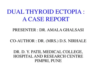
A RARE CASE OF ASCITES Dr. Siva Krishna DEPARTMENT OF MEDICINE
CHIEF COMPLAINTS 22 yr/Female, Resident of Lonavala, came with chief complaints of: Abdominal distension and Pain in abdomen since 3 days No h/o loose stool, vomiting, fever No h/o blood in stools No h/o breathlessness
Family history Past history No H/O HTN/DM Not significant No similar history in the No H/O TB/Asthma family members No H/O Thyroid/Epilepsy No H/O similar complaints in past No H/O Jaundice H/O Normal delivery 6 months back No H/O OC Pills usage.
Personal history Sleep normal Appetite decreased Bladder normal Bowel normal Mixed diet No any addictions
On examination Afebrile Pulse: 92/min BP: 110/70mmhg RR: 18/min
General examination No Pallor No Icterus No Edema No Cyanosis, Clubbing, Lymphadenopathy. No signs of Liver cell failure
Systemic examination PA : Soft and distended, mild hepatomegaly Shifting dullness present No fluid thrill CVS : Normal RS : Normal vesicular breath sounds CNS : Conscious oriented to time place and person
Lab investigations Hb TLC DLC Platelet Count Hematocrit Blood urea/ Creatinine Total Bilirubin/Direct Bilirubin SGPT/SGOT Na+/ K+ Total Protein Albumin/Globulin Pt-Inr/aPtt Amylase/Lipase HIV/HbsAg/HCV 11.8 gm% 7800 N:46 L:37 E:05 M:12 2.10 lakh 35.3 25/0.7 0.59/0.28 28/24 134/3.7 5.34 3.01/2.31 1.0/39.8 58/30 Negative
Ascitic fluid Physical Appearance Clear, pale yellow. No clots, coagulum or cobweb R/M Total Proteins/ Albumin Sugars ADA LDH Amylase/ Lipase Zero cells 2.5/1.4 128 3.8 65 28/64 Ascitic fluid suggestive of transudative nature SAAG: 1.61
Radiological investigations CXR: Normal USG Abdomen & Pelvis: Free fluid noted in pouch of Liver 16cms enlarged, with Douglas, pelvis, perihepatic normal echogenicity. and perisplenic region. Portal veins-normal Right sided minimal pleural Pseudo gall bladder wall effusion. thickening Spleen, Pancreas, Kidney: Normal
Color Doppler of Portal System Liver 18cms, relative atrophy of left lobe of liver with enlargement of caudate lobe of liver. Hepatic veins- Right hepatic vein appears thrombosed / thick hyper echoic. Middle hepatic vein appears patent. Left hepatic vein- thrombosed.
IVC normal, no thrombosis Portal vein 7mm with reversal of flow Collaterals- intrahepatic venovenous collaterals seen with no flow s/o thrombosis. Conclusion: Budd-Chiari Syndrome with portal hypertension.
CECT A/P A B
CECT A/P c D
On Upper GI endoscopy Mild portal hypertensive gastropathy Nodular pangastritis Pro thrombotic profile was NORMAL Diagnosis Budd-chiari syndrome most probable post partum etiology.
Treatment Inj. Enoxaparin 0.6cc BD for 5 days & overlapped with Tab. Acenocoumaral 2mg OD on day 3 of enoxaparin Tab. Furosemide/Spironolactone 20/50mg BD Tab. Propranolol 40mg OD
Follow up after one month Patient had symptomatically improved. Patient was continued on anticoagulation medication and INR was maintained between 2-3. On USG: No evidence of ascites.
DISCUSSION BUDD-CHIARI SYNDROME is a very rare condition affecting one in a million. Presents with classical triad of Abdominal pain, Ascites, liver enlargement. Etiology : Idiopathic in approximately 1/3rd of patients, Thrombotic diathesis, pregnancy, post partum, OCP, Chronic infection, Chronic inflammatory diseases, hematological disorders like Polycythemia rubra vera, Paroxysmal nocturnal hemoglobinuria, antiphospholipid antibody syndrome.
Clinical variants of Budd-chiari syndrome: Acute and sub acute form: Characterized by rapid development of abdominal pain, ascites, hepatomegaly, jaundice and renal failure Chronic form: Progressive ascites, 50% pt have renal impairment, Jaundice is absent. Fulminant form: Hepatic failure present, ascites, tender hepatomegaly, jaundice, and renal failure.
References 1. Rajani R, Melin T, Bj rnsson E, Broom U, Sangfelt P, Danielsson A, Gustavsson A, Grip O, Svensson H, L f L, Wallerstedt S, Almer SH (Feb 2009). "Budd Chiari syndrome in Sweden: epidemiology, clinical characteristics and survival - an 18- year experience". Liver International. 29 (2): 253 9. doi:10.1111/j.1478- 3231.2008.01838.x. PMID 18694401. 2. "Etiology, management, and outcome of the Budd Chiari syndrome". 3. "Hepatic vein thrombosis (Budd Chiari syndrome)". 4. "The Budd Chiari syndrome: a review". 5. "Budd Chiari syndrome: long-term survival and factors affecting mortality". 6. "Budd Chiari Syndrome: clinical patterns and therapy". 7. "Budd Chiari syndrome: etiology, diagnosis and management". 8. "Case records of the Massachusetts General Hospital. Weekly clinicopathological exercises. Case 51-1987. Progressive abdominal distention in a 51-year-old woman with polycythemia vera"


