Methods of Imaging the Brain: X-ray, CT Scan, and MRI

Different methods of imaging the brain, including X-ray, CT scan, and MRI, offer non-invasive ways to study brain structure and function. X-rays measure tissue density, CT scans provide detailed cross-sectional images, and MRI produces 3D anatomical images. MRI, in particular, has become a vital tool in neuroscience due to its safety and superior visualization capabilities.

Download Presentation

Please find below an Image/Link to download the presentation.
The content on the website is provided AS IS for your information and personal use only. It may not be sold, licensed, or shared on other websites without obtaining consent from the author. Download presentation by click this link. If you encounter any issues during the download, it is possible that the publisher has removed the file from their server.
E N D
Presentation Transcript
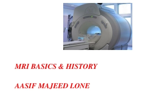
Lecture 1&2 Method of imaging the Head
Method of imaging the brain Human brain imaging methods noninvasive, though they may include exposure to ionizing radiation or contrast agents. These methods can be equally used to noninvasively study brain structure and function. New technologies allow non-invasive spatial mapping, (morphology), and observations of processes within the brain during set tasks. There are a range of scanning techniques, their purpose are described below
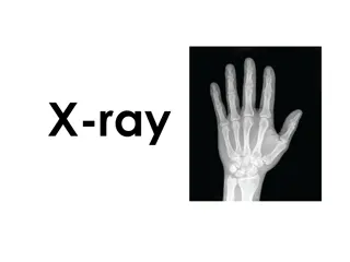
1. X-ray the x-ray, early structural imaging developed in 1895. X- rays measure the density of tissues. X-rays use photons, a quantum of visible light that possesses energy; the photons are passed through the body and deflected and absorbed to different degrees by the person s tissues. The photons were initially recorded onto a silver halide film as they passed out of the body. Dense structures such as bone, which block most of the photons, appear white; structures containing air appear black; and muscle, fat, and fluids appear in various shades of gray.
2. CT Acomputerized tomography (CT) scan is a series of X-ray images converted into cross-sectional images of your brain. These X-rays are combined to form cross-sectional slices or even a 3-D model of your brain. The results of a CT scan can also provide more detail than a standard X-ray. CT scans can: find certain types of brain injuries (hemorrhagic strokes) identify cancer locate brain swelling or bleeding reveal structural brain changes from schizophrenia
3.MagneticResonanceImagingMagnetic This imaging method is a painless, non-invasive imaging technology that produces 3D detailed anatomical images of our brain. Resonance Imaging (MRI) is based on the principle of nuclear magnetic resonance (NMR) and uses radiofrequency waves the scanner uses magnetic fields that emit radio waves to manipulate the magnetic position of the hydrogen protons of the body. The rotation and energy release of the protons are detected by a powerful antenna which sends the information to a computer. The computer analyzes the information, and through complex mathematical calculations, creates a clear, cross-sectional black-and-white image of the body.These images can be converted into three-dimensional pictures of the scanned area, with the results looking something like the images below.
Clinically, MRI has become the most important diagnostic imaging modality in neuroscience. One of the many benefits of MRI in 1. The central nervous system is that the radiofrequency signals readily penetrate the skull and spinal column, allowing the tissue within it to be imaged with no interference. 2. Radiofrequency energy is also non-ionizing, so MRI is safer than CT scanning. 3. MRI provides the best visualization of parenchymal (tissue) abnormalities in the brain and spinal cord including tumors, demyelinating lesions, infections, vascular lesions (such as stroke), developmental abnormalities, and traumatic injuries.
4.fMRI Functional magnetic resonance imaging (fMRI) can detect changes in blood flow and oxygen levels that result from your brain s activity. It uses the magnetic field of the scanner to affect the magnetic nuclei of hydrogen atoms, so they can be measured and converted into images. MRIs display anatomic structure and fMRIs measure metabolic function. fMRIs have many uses, such as: assessing brain activity finding brain abnormalities creating pre-surgicalbrain maps
5.PET A positron emission tomography (PET) scan uses a radioactive tracer that attaches to the glucose in your bloodstream. Since your brain uses glucose as its primary fuel source, the tracer accumulates in areas of higher brain activity. A PET scan is able to see these tracers and observe how they move and accumulate in your brain.This allows doctors to see trouble spots where glucose isn t moving correctly. PET scans can evaluate: seizures Alzheimer s tumors
6.EEG An electroencephalography (EEG) test measures your brain waves. Before the scan, clinicians will attach small electrodes to your scalp that are attached to wires. These electrodes detect electrical activity in your brain and send it to a computer where it creates a graph-like image. Each type of frequency appears on its own line and gives your doctor information about your brain activity. EEG can detect issues such as: anxiety head injuries epilepsy sleep disruption
7.MEG Magnetoencephalography (MEG) measures the magnetic field from neuron electrical activity. This type of scan can locate and identify malfunctioning neurons in your brain. Doctors use MEG to evaluate both spontaneous brain activity, as well as neuronal responses triggered by stimuli. MEG allows doctors to assess areas such as: epilepsysources motor areas sensory areas language and vision
8.NIRS Near-infrared spectroscopy (NIRS) monitors your brain s oxygen saturation. It uses infrared light to detect variations in hemoglobin oxygen levels in your blood. Since oxygen is critical for your brain to function properly, NIRS can assist doctors in any clinical setting where brain oxygen levels may fluctuate. NIRS is used to monitor: brain oxygen levels during cardiac surgery brain function and oxygenation levels in preterm infants in a neonatal intensive care unit (NICU) setting
IndicationsforMRIbrain Ataxia Transient ischaemic atta(TIA), cerebrovascular attack (CVA) Infection, inflammation, meningitisck, encephalitis, HIV, AIDS, TB Cognitive decline, neurodegenerative disorders, dementia Demyelinating disease, multiple sclerosis Loss of consciousness, seizures, epilepsy Brain tumour, metastases, abscess Cerebellar lesion, brainstem lesion Congenital abnormalities Post-operative follow-up Vascularpathologies Headaches Haemorrhage Trauma
Contraindications Any electrically, magnetically or mechanically activated implant (e.g. cardiac pacemaker, insulin pump biostimulator, neurostimulator, cochlear implant, and hearing aids) Intracranial aneurysm clips (unless made of titanium) Pregnancy (risk vs benefit ratio to be assessed) Ferromagnetic surgical clips or staples Metallic foreign body in the eye Metal shrapnel or bullet
Patient preparationfor MRI brain Ask the patient to remove all metal objects including keys, coins, wallet, cards with magnetic strips, jewellery, hearing aid and hairpins If possible provide a chaperone for claustrophobic patients (e.g. relative or staff) The exam must be explained to the patient before the scan Before injecting Gadolinium make sure that the patient has normal kidney function tests (Blood urea and serum creatinine levels) (normal GFR)
Positioning forMRI brain Head first supine Position the head in the head coil and immobilise with cushions Give cushions under the legs for extra comfort Centre the laser beam localiser over the glabella
Suggested protocols,parameters and planning Localiser Athree plane localizer must be taken in the beginning to localise and plan the sequences. Localizers are usually less than 25sec. T1 weighted low resolution scans.
T1SEcoronal Plan the coronal slices on the sagittal plane; angle the position block perpendicular to the corpus callosum. Check the positioning block in the other two planes. An appropriate angle must be given in the axial plane on a tilted head (perpendicular to mid line of the brain). Slices must be sufficient to cover the whole brain from the frontal sinus to the line of the occipital protuberance.
T2axial Plan the axial slices on the sagittal plane; angle the position block parallel to the genu and splenium of the corpus callosum. Slices must be sufficient to cover the whole brain from the vertex to the line of the foramen magnum. Check the positioning block in the other two planes.An appropriate angle must be given in coronal plane on a tilted head (perpendicular to the line of 3rd ventricle and brain stem).
T2sagittal Plan the sagittal slices on the axial plane; angle the position block parallel to midline of the brain. Check the positioning block in the other two planes. An appropriate angle must be given in the coronal plane on a tilted head (parallel to the line along 3rd ventricle and brain stem). Slices must be sufficient to cover the brain from temporal lobe to temporal lobe.
T2FLAIRaxial Plan the axial slices on the sagittal plane; angle the position block parallel to the genu and splenium of the corpus callosum. Slices must be sufficient to cover the whole brain from the vertex to the line of the foramen magnum. Check the positioning block in the other two planes. An appropriate angle must be given in coronal plane on a tilted head (perpendicular to the line of 3rd ventricle and brain stem).
DWIaxial Plan the DWI axial slices on the sagittal plane; angle the position block parallel to the line from the glabella to the foramen magnum. This angle will reduce air-bone interface artefacts from the Para nasal sinuses. Slices must be sufficient to cover the whole brain from the vertex to the foramen magnum. Check the positioning block in the other two planes. An appropriate angle must be given in the coronal plane on a tilted head (perpendicular to the line of 3rd ventricle and brain stem).
Indicationsforcontrast enhancementbrainscans Tumour, Metastases, Cranial nerve lesion, Indeterminate intracranial lesion, IAC mass Meningitis, Encephalitis, Leptomeningeal spread Leukodystrophies, Delayed development Syringomyelia(Syrinx) Use T1 SE axial and coronal after the administration of IV gadolinium DTPAinjection(copy the planning outlined above). The recommended dose of gadolinium DTPAinjection is 0.1 mmol/kg, Multiple Sclerosis,AVM, HIV, InfectionAbscess i.e. 0.2 mL/kg in adults, children and infants.
TR &TE TE ( echo time) : Time interval in which signals are measured after RF excitation. TR (repetition time) : The time between two excitations is called repetition time. By varying the TR and TE one can obtain T1WI and T2WI. In general a short TR (<1000ms) and short TE (<45ms) scan is T1WI. Long TR (>2000ms) and long TE (>45ms) scan is T2WI.
T1WImages Most Pathologies dark on T1 Bright On T1 Fat Hemorrhage Melanin Early Calcification Protein contents (Colloid cyst/ Rathke cyst) Posterior pituitary appears Bright on T1 Gadolinium . ShortTE ShortTR Better anatomical details Fluids are dark Gray matter is gray White matter is white
T2WImages LongTE LongTR Betterpathological details Fluids are bright Graymatter is relativelybright White matter is dark
T2 Flair FluidAttenuated InversionRecoverySequences LongTE LongTR Similar toT2 Except free water suppression ( Inversion Recovery) Most pathology is Bright Especially good for lesions near ventricles or sulci (eg. Multiple sclerosis)
T1W T2W Flair(T2) TR Short Long Long TR Short Long Long CSF Low High Low Fat High Low Medium Brain Low High High Edema Low High High