Understanding the Axial and Appendicular Skeleton in Vertebrates
The axial skeleton consists of the bones in the head and trunk, while the appendicular skeleton supports the limbs in vertebrates. The appendicular skeleton aids in locomotion and manipulation, comprising 126 bones in total. Understanding the structure and functions of these skeletal systems is vital for comprehending the human body's anatomy and movement capabilities.

Download Presentation

Please find below an Image/Link to download the presentation.
The content on the website is provided AS IS for your information and personal use only. It may not be sold, licensed, or shared on other websites without obtaining consent from the author. Download presentation by click this link. If you encounter any issues during the download, it is possible that the publisher has removed the file from their server.
E N D
Presentation Transcript
OVERVIEW OF AXIAL AND APPENDICULAR SKELETON
APPENDICULAR SKELETON The appendicular skeleton is the portion of the skeleton of vertebrates consisting of the bones that support the appendages. The appendicular skeleton includes the skeletal elements within the limbs, as well as supporting shoulder girdle pectoral and pelvic girdle. The word appendicular is the adjective of the noun appendage, which itself means a part that is joined to something larger.
Human structure: Of the 206 bones in the human skeleton, the appendicular skeleton comprises 126. Functionally it is involved in locomotion (lower limbs) of the axial skeleton and manipulation of objects in the environment (upper limbs). The appendicular skeleton forms during development from cartilage, by the process of endochondral ossification. The appendicular skeleton is divided into six major regions: Shoulder girdles (4 bones) - Left and right clavicle (2) and scapula (2). Arms and forearms (6 bones) - Left and right humerus (2) (arm), ulna (2) and radius (2) (forearm).
Hands (54 bones) - Left and right carpals (16) (wrist), metacarpals (10), proximal phalanges(10), intermediatephalanges(8) and distal phalanges(10). Pelvis (6 bones) - Ilium (2), Ischium (2) and Pubis (2). Thighs and legs (8 bones) - Left and right femur (2) (thigh), patella (2) (knee), tibia (2) and fibula (2) (leg). Feet (ankle), metatarsals (10), proximal phalanges (10), intermediate phalanges (8) and distal phalanges(10). and ankles (52 bones) - Left and right tarsals (14) It is important to realize that through anatomical variation it is common for the skeleton to have many accessory bones (sutural bones in the skull, cervical ribs, lumbar ribs and evenextra lumbar vertebrae). The appendicular skeleton of 126 bones and the axial skeleton of 80 bones together form the complete skeleton of 206 bones in the human body. Unlike the axial skeleton, the appendicular skeleton is unfused. This allows for a much greater rangeof motion.
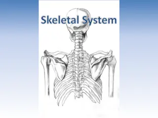
AXIAL SKELETON The axial skeleton is the part of the skeleton that consists of the bones of the head and trunk of a vertebrate. The word "Axial" is taken from the word "axis" and refers to the fact that the bones are located close to or along the central "axis"of the body. In the human skeleton, it consists of 80 bones and is composed of six parts; the skull (22 bones), the ossicles of the middle ear, the hyoid bone, the rib cage, sternum and the vertebralcolumn. The skeleton form the complete skeleton. Another definition of axial skeleton is the bones including the vertebrae, sacrum, coccyx, ribs, and sternum. axial skeleton together with the appendicular
Structure: Flat boneshouse the brain and other vital organs. This article mainly deals with the axial skeletons of humans; however, it is important to understand the evolutionary lineageof the axial skeleton. The human axial skeleton consists of 80 different bones. It is the medial core of the body and connects the pelvis to the body,wherethe appendix skeletonattaches. As the skeleton grows older the bones get weaker with the exception of the skull. The skull remains strong to protectthe brain from injury.
Human Skull: The human skull consists of the cranium and the facial bones. The cranium holds and protects the brain in a largespace calledthe cranial vault. The cranium is formed from eight plate-shaped bones which fit together at meeting points (joints) called sutures. In addition there are 14 facial bones which form the lower frontpart of the skull. Together the 22 bones that compose the skull form additional, smaller spaces besides the cranial vault, such as the cavities for the eyes, the internal ear, the nose, and the mouth.
COMPILEDAND CIRCULATED BY DR. POULAMI ADHIKARY MUKHERJEE, ASSISTANT PROFESSOR, DEPARTMENT OF ZOOLOGY, NARAJOLE RAJ COLLEGE The most important facial bones include the jaw or mandible, the upper jaw or maxilla, the zygomatic or cheek bone, and the nasal bone. Humans are born with separate plates which later fuse to allow flexibility as the skull passes through the pelvis and birth canal during birth. During development the eight separate plates of the immature bones fuse together into one single structure known as the Skull. The only bone that remains separate from the rest of the skull is the mandible. ZOOLOGY: SEM- IV, PAPER- C8T: COMPARATIVE ANATOMY OF VERTEBRATES, UNIT 2: SKELETAL SYSTEM
Ribcage: The rib cage is composed of 12 pairs of ribs plus the sternum for a totalof 25 separatebones. The rib cage functions as protection for the vital organs such as the heart and lungs. The ribs are shaped like crescents, with one end flattened and the other end rounded.
The rounded ends are attached at joints to the thoracic vertebrae at the back and the flattened ends come togetherat the sternum, in the front. The upper seven pairs of ribs attach to the sternum with costal cartilage and are known as true ribs. The 8th through 10th ribs have non-costal cartilage which connects them to the ribs above, and for this they are knownas "false ribs".
COMPILEDAND CIRCULATED BY DR. POULAMI ADHIKARY MUKHERJEE, ASSISTANT PROFESSOR, DEPARTMENT OF ZOOLOGY, NARAJOLE RAJ COLLEGE The last two ribs are called floating ribs because they do not attach to the sternum or to other ribs and simply hang free. The length of each rib increases from number one to seven and then decreases until rib pair number 12. The first rib is the shortest, broadest, flattest, and most curved. ZOOLOGY: SEM- IV, PAPER- C8T: COMPARATIVE ANATOMY OF VERTEBRATES, UNIT 2: SKELETAL SYSTEM
Vertebralcolumn: At birth the majority of humans have 33 separate vertebrae. However, during normal development several vertebrae fuse together, leaving a total of 24,in most cases. The confusion about whether or not there are 32-34 vertebrae stems from the fact that the two lowest vertebrae, the sacrum and the coccyx, are single bones made up of severalsmaller bones which have fused together. This is how the vertebrae are counted: 24 separate vertebrae and the sacrum, formed from 5 fused vertebrae and the coccyx, formed from 4 fused vertebrae.
If you count the coccyx and sacrum each as one vertebra, then there are 26 vertebrae. If the fused vertebrae are all counted separately, then the total numberof vertebraecomes to between32 and 34. The vertebral column consists of 5 parts. The most cranial (uppermost) part is made up by the cervical vertebrae (7), followed by thoracic (12), lumbar (5), sacral (5) and coccygeal vertebrae(4). Cervical vertebrae make up the junction between the vertebral column and the cranium. Sacral and coccygeal vertebras are fused and thus often called "sacral bone" or "coccygeal bone" as unit. The sacral bone makes up the junction between the vertebral column and the pelvic bones.


 - Left and right carpals (16) (wrist),")
 - Left and right carpals (16) (wrist),")