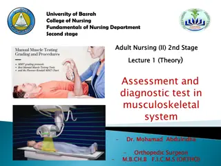
Understanding the Musculoskeletal System in the Human Body
The musculoskeletal system, comprising the muscular and skeletal systems, supports body movement, stability, and shape. It includes muscles, bones, tendons, ligaments, and joints. This system serves functions like providing support, protection to vital organs, mediating movement, and producing blood cells. The adult skeleton consists of 206 bones categorized into axial and appendicular divisions. Bones are vital for support and protection, and they vary in morphology such as long bones, short bones, flat bones, and irregular bones.

Download Presentation

Please find below an Image/Link to download the presentation.
The content on the website is provided AS IS for your information and personal use only. It may not be sold, licensed, or shared on other websites without obtaining consent from the author. Download presentation by click this link. If you encounter any issues during the download, it is possible that the publisher has removed the file from their server.
E N D
Presentation Transcript
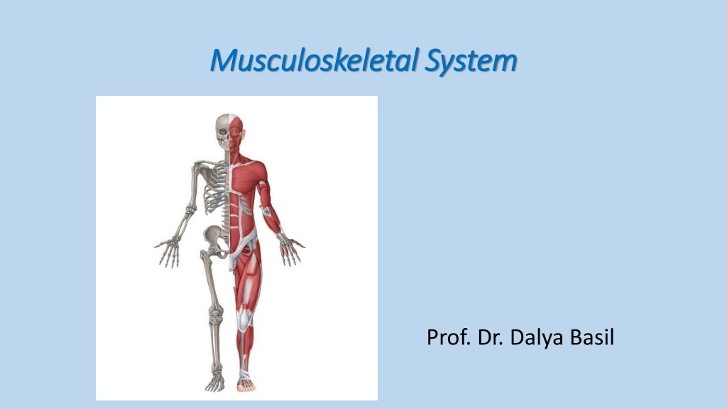
Musculoskeletal System Musculoskeletal System Prof. Dr. Dalya Basil
Musculoskeletal System Musculoskeletal System The musculoskeletal system (locomotor system) is a human body system that provides body movement, stability, shape, and support. It is subdivided into two broad systems: Muscular system, which includes all types of muscles in the body. Skeletal muscles, in particular, are the ones that act on the body joints to produce movements. Besides muscles, the muscular system contains the tendons which attach the muscles to the bones.
Musculoskeletal System Musculoskeletal System Skeletal system, whose main component is the bone. Bones articulate with each other and form the joints, providing the bodies with a hard- core, yet mobile, skeleton. The integrity and function of the bones and joints is supported by the accessory structures of the skeletal system; articular cartilage, ligaments, and bursae.
Function of the musculoskeletal system Function of the musculoskeletal system The musculoskeletal system serves the following functions: 1- Provides support to the body 2- Provides protection to the vital organs (brain, lungs, heart) 3- Mediates movement of the body 4- Blood cells production (by the bone marrow) 5- Other metabolic functions, Skeletal muscle is the primary site for glucose uptake and storage, and it is likewise a reservoir of amino acids that sustain protein synthesis in all other body sites.
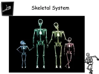
The skeletal system The skeletal system The adult human skeleton is composed of 206 bones and their associated cartilages. The bones are supported by ligaments, tendons, bursae, and muscles. The bones of the body are grouped within the two distinct divisions: Axial skeleton, that includes the bones along the long axis of the body. The axial skeleton consists of the vertebral column, bones of the head and bones of the thoracic cage. Appendicular skeleton, that involves the bones of the shoulder and pelvic girdle, as well as the bones of the upper and lower extremities.
The skeletal system The skeletal system
Bones Bones Bones are the hard parts of human body. They are rigid structures made of calcified dense connective tissue. Morphologically, the bones can be classified into: 1- Long bones: these are elongated structures, consisting of a shaft, & 2 enlarged ends. Example: the bones of the thigh & arm. 2- Short bones: shorter bones, example: phalanges of the hand & foot. 3- Flat bones: flattened structures, example: cranial bones & the sternum. 4- Irregular bones: the vertebrae & facial bones.
Bones Bones
Joints Joints Each bone of the musculoskeletal system is connected to one or more bones via a joint. The majority of joints allow movement. There are 3 types of joints: 1- Synovial joints: the main type in humans, it allows a good range of movement. Synovial joints are present generally in the upper & lower limbs. At the joint, the ends of the 2 bones forming the joint are covered by hyaline cartilage (to allow smooth movement & minimize friction). The bones are held together by strong connective tissue capsule, which is lined internally by a special membrane (the synovial membrane), which secretes a clear fluid (synovial fluid) that fills the joint cavity. Sometimes, synovial fluid sample is aspirated for diagnostic reasons. Also, medications can be injected into the synovial cavity in some joint diseases (eg: osteoarthritis).
Joints Joints
Joints Joints 2- Fibrous joints: here, the 2 bones are close to each other, with the thin space between them filled with fibrous tissue. This type of joints allow minimal or no movement, & is present for example- between skull bones. 3- Cartilaginous joints: here, the 2 bones are connected by a cartilage. Example: the intervertebral discs, which connect adjacent vertebral bodies. These joints allow a minimal range of movement.
Joints Joints Fibrous joints Cartilaginous joints
The Skeleton The Skeleton All together, the bones of the human body are called the "skeleton". The skeleton can be studied by dividing it into specific regions: the skull, vertebral column, thoracic cage, pelvis, bones of the upper limb, & bones of the lower limb. The skull (with the mandible) represents the bones of the head. It is made of a group of flat & irregular bones connected by fibrous joint with no movement (except for the joints between the mandible & the rest of the skull, the synovial temporomandibular joints). The skull part that makes the face bones is the (facial skeleton), while that contains the brain is the (cranium). The facial skeleton contains the 2 orbital cavities (containing the eyeballs), the nasal cavities (2 cavities separated by a midline septum) that connect the nasal openings (nostrils) to the pharynx, & the oral cavity between the mandible & the rest of the skull.
The skull The skull The skull Synovial temporomandibular joints
The vertebral column The vertebral column A strong bony structure made of 32 33 irregular bones (the vertebrae). The vertebral column gives the humans the erect posture, protects the spinal cord, carries the skull, the ribs, & the upper limbs, & is connected inferiorly to the pelvis, to which it transmits the upper body weight. The vertebral column is divided into the following regions: - Cervical region: in the neck, made of 7 vertebrae - Thoracic region: in the thorax, made of 12 vertebrae, connected to the 12 pairs of ribs - Lumbar region: in the lower back, made of 5 vertebrae - Sacral region (sacrum): 5 fused vertebrae that make the posterior part of the pelvis - Coccygeal region (coccyx): 3-4 small fused vertebrae at the lower tip of the column.
The Thoracic cage The Thoracic cage The skeleton of the chest, it is made of 12 pairs of ribs, connected posteriorly to the vertebral column & anteriorly to a flat bone called the "sternum". The anterior part of the rib (that articulates with the sternum) is made of hyaline cartilage, not bone, & called the "costal cartilage". The thoracic cage protects the heart & lungs, & gives a base on which the bones of the upper limb are attached.
The pelvis The pelvis A ring-like bony structure in the lower part of the trunk. It connects the vertebral column to the lower limbs. The pelvis is made of 3 bones: the right & left hip bones, & the sacrum & coccyx (of the vertebral column) situated between them posteriorly.
Bones of the upper limb Bones of the upper limb The upper limb bones are connected to the thoracic cage at the (sterno- clavicular joint), formed between the sternum & the clavicle. The arrangement of upper limb bones is as follows: - Clavicle: the bone in the anterior part of the shoulder region. It is connected to the scapula by the acromioclavicular joint. - Scapula: the flat triangular bone in the back of the shoulder, it is connected to the humerus by the shoulder joint. - The shoulder joint: a shallow synovial joint that allows a very wide movement range of the arm. - Humerus: the bone of the arm, connected to the bones of the forearm by the elbow joint.
Bones of the upper limb Bones of the upper limb - The elbow joint: the synovial joint between the humerus & forearm bones (radius & ulna). It allows only flexion-extension movement. - Forearm bones: the radius (laterally) & the ulna (medially). At their proximal & distal ends, these 2 bones are connected to each other by the proximal & distal radio-ulnar joints, respectively. - Wrist joint: the joint between the forearm bones & the carpal bones. - Carpal bones: 8 small bones at the wrist region.
Bones of the upper limb Bones of the upper limb - Metacarpal bones: the 5 bones of palm of the hand, connected to the carpal bones by the carpo-metacarpal joints, & to the phalanges by the metacarpo-phalangeal joints. - Phalanges: the short bones of the fingers. The thumb has 2 phalanges, & the other fingers have 3. The joints between the phalanges are the (interphalangeal joints)
Bones of the upper limb Bones of the upper limb
Bones of the lower limb Bones of the lower limb The lower limb bones are connected to the pelvis at the (hip joint), formed between the pelvis & the femur (the bone of the thigh). The arrangement of lower limb bones is as follows: - The hip joint: a deep synovial joint that allows a wide movement range of the thigh. - Femur: the bone of the thigh, connected to the bones of the leg by the knee joint. The femur is the longest bone in the body. - The knee joint: the synovial joint between the femur & leg bone (tibia), with a 3rd rounded bone in the front (the patella). It allows only flexion-extension movement, & carries most of the body weight.
Bones of the lower limb Bones of the lower limb - Leg bones: the tibia (larger, medially) & the fibula (smaller, laterally). At their proximal & distal ends, these 2 bones are connected to each other by the proximal & distal tibiofibular joints. - Ankle joint: the joint between the leg bones & the tarsal bones. - Tarsal bones: 7 small bones at the proximal part of the foot. - Metatarsal bones: the 5 bones of the distal part of the foot, connected to the tarsal bones by the tarsometatarsal joints, & to the phalanges by the metatarsophalangeal joints. - Phalanges: the short bones of the toes. The thumb has 2 phalanges, & the other toes have 3. The joints between the phalanges are the (interphalangeal joints)
Bones of the lower limb Bones of the lower limb
The Skeletal Muscles The Skeletal Muscles A skeletal muscle is a contractile living structure that is connected usually to 2 bones, & acts to move them towards each other when it contracts. The proximal attachment of a muscle to the bone is called the "origin", while the distal attachment is called the "insertion". Histologically, skeletal muscles belong to the "muscular tissues", which includes also smooth & cardiac muscles. Skeletal muscles are innervated by somatic motor nerves (cranial or spinal nerves). Skeletal muscle movement is under voluntary control. There are several skeletal muscles categorized according to their site:
Muscles of the head & neck Muscles of the head & neck - Muscles of facial expression: these are small muscles that originate from the facial bones & are inserted to the facial skin. They act to open & close the eyes, mouth, & to give the face its variable expressions. - Muscles of mastication: a group of muscles in the head that move the mandible & mediate mastication. - Tongue muscles: move the tongue during eating & talking. - Muscles of the pharynx: act to mediate swallowing. - Muscles of the larynx: act to create voice. - Other muscles of the head & neck: act to move the head & neck.
Muscles of the head & neck Muscles of the head & neck
Muscles of the upper limb Muscles of the upper limb - Muscle of the shoulder girdle: these are the muscles that move the shoulder. They originate from the axial skeleton & are inserted to the clavicle, scapula, or humerus. Examples: Pectoralis major muscle (flexes the arm towards the chest), deltoid muscle (abducts the arm away from the trunk). - Muscles of the arm: eg: biceps (anteriorly, flexes the elbow joint), triceps (posteriorly, extends the elbow joint). - Muscles of the forearm: anterior group (flexes the wrist joint), posterior group (extends the wrist joint) & the lateral group (flexes the elbow joint).
Muscles of the upper limb Muscles of the upper limb
Muscles of the Thorax Muscles of the Thorax Apart from the muscles moving the arm, the thorax has its own muscles that act to elevate or depress ribs during respiration. These include: - Intercostal muscles: short muscles between the ribs, mediate both inspiration & expiration. - Diaphragm: a big sheet of muscle between the thorax & abdomen, mediates inspiration only. - Accessary respiratory muscles: other muscles that help forceful respiration.
Muscles of the Thorax Muscles of the Thorax
Muscles of the abdominal wall Muscles of the abdominal wall These muscles move the trunk. They include: - Rectus abdominis: the longitudinal muscle in the front of the abdominal wall, it flexes the trunk anteriorly. - External oblique, internal oblique, & transversus abdominis: lying on the sides of the trunk, these muscles act to rotate the trunk on its axis or flex it laterally.
Muscles of the back Muscles of the back Paravertebral muscles (surrounding the vertebral column) are a group of strong muscles that extend the back & rotate the vertebral column.
Muscles of the lower limb Muscles of the lower limb - Muscles that move the hip joint from the pelvis & lower vertebral column & are inserted into the femur. They include the iliopsoas muscle anteriorly, & the gluteus muscles posteriorly. - Muscles of the thigh: anterior group (quadriceps muscle, extends the knee joint), medial group (adductor muscles, adduct the thigh), & posterior group (hamstring muscles, flex the knee) - Muscles of the leg: anterior group (extend the foot & toes), lateral group (evert the foot), & posterior group (flex the foot & toes).
Muscles of the lower limb Muscles of the lower limb