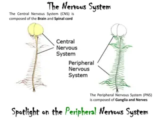
Understanding the Cardiovascular System
Explore the closed system of the cardiovascular system where blood is contained within the heart and vessels. Learn about the total blood volume and its significance for overall health and function.
Download Presentation
Please find below an Image/Link to download the presentation.
The content on the website is provided AS IS for your information and personal use only. It may not be sold, licensed, or shared on other websites without obtaining consent from the author. Download presentation by click this link. If you encounter any issues during the download, it is possible that the publisher has removed the file from their server.

Presentation Transcript
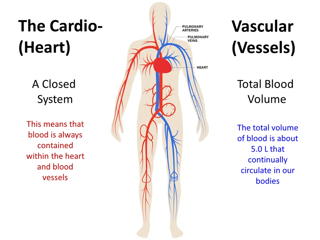
The Cardio- (Heart) Vascular (Vessels) A Closed System Total Blood Volume This means that blood is always contained within the heart and blood vessels The total volume of blood is about 5.0 L that continually circulate in our bodies
Functions of the Cardiovascular System Transport around the body of: O2from the lungs to body the tissues. CO2from body the tissues to the lungs. Delivery of Nutrients to cells (glucose, amino acids) Removal of Waste (+ toxins) from cells Hormone circulation (communication, growth, cycles) Defense healing and repair of body tissues Thermoregulation Blood is the warmest material in the body; delivery of it warms a region, absences cools it.
The Heart Pulmonary Systemic Circuits Chambers Pulmonary Trunk 3 1 Valves (one-way-flow) 4 2 Myocardiocytes
Volumes? Pressures? heart > lungs > heart (from RV to LA) heart > body > heart (from LV to RA) Artery = Vein =
The Heart has 4 Valves Purpose of Valves: To prevent retrograde Blood Flow Here are 4 valves within the Heart: The 2 atrioventricular valves (AV) are between the atria and ventricles. 1) Right AV (tricuspid) valve 2) Left AV (bicuspid/mitral) valve
The 2 semilunar valves of the heart 3) Aortic semilunar valve (located between the LV and the aorta) 4) Pulmonary semilunar valve (located between the RV and the pulmonary trunk)
Two Heart Sounds: Lub and Dup In a healthy heart, there two audible heart sounds: S1and S2. The 1st heart sound S1, the lub sound. The 2nd heart sound, S2, the dup sound.
Disorders of Heart Valves Normal Heart Valves Problem Heart Valves Problems Opening: Stenosis narrowing of valve, when a valve doesn't open completely. Turbulence = noise = murmur. Problems Closing: Prolapse overlapping or when valve doesn't close tightly. Also termed valvular insufficiency (regurgitation) Retrograde flow = noise = murmur.
There are 2 types of Cardiac Muscle Cells 1. Autorhythmic Myocardiocytes: 2. Contractile Myocardiocytes: Both of these myocardiocytes have Action Potentials (AP s) that differ from one another as will be seen in the graphs of the AP s that follow.
Myocardiocytes: 1) Autorhythmic 2) Contractile
Action Potentials for Autorhythmic Myocardiocytes Membrane Potential (mV) Time (msec) Additional things to Notice about this AP and Graph: Typically, no Hyperpolarization phase. *Parasympathetic innervation (via _________ nerve) can pull the membrane voltage below -60 mV. *Sympathetic innervation (via __________ nerve) truncates the repolarization phase to about -50 mV.
Action Potentials for Contractile Myocardiocytes Membrane Potential (mV) stimulus Time (msec) Things to Notice about this AP and Graph: Plateau Phase and what causes it. There is no real hyperpolarization phase in these cells. The absolute refractory period is almost all of the Plateau phase (about 250 msec, is very long!). What is the purpose pf the long absolute refractory in cardiac muscle? If tetanus were possible the heart could not fill, because that occurs during diastole, or relaxation.
Exercise Draw by hand (with all units, values, phases, which channels are open, closed): a) The action potential for an autorhythmic myocardiocyte; and b) The action potential for a contractile myocardiocyte. You need to know those graphs like the back of your hand! So, take a blank piece of scrap paper and draw them until you can do it all without looking at the graphs as a reference. Then go and test your understanding by answering Q s about them! (Next slide)
Some Questions on Myocardiocytes: 1. The Resting Membrane Potential in the autorhythmic myocardiocytes cells of the heart is a) -70 mV b) unstable, due to K+ efflux c) unstable, drifts from -60 mV d) -90 mV e) stable at -40 mV 2. The influx of which ion accounts for the plateau phase? a) sodium b) potassium c) chloride d) calcium 3. At the peak of the AP of contractile myocardiocytes, just before the plateau phase - a) the Na+ channels open b) the Ca2+ channels close c) the Na+ channels close, causing the membrane voltage to remain elevated d) the K+ channels close causing the membrane voltage to fall slightly e) opening of K+ channels, causes K+ efflux that drops the membrane voltage slightly 4. The absolute refractory period in contractile myocardiocytes: a) is to prevent contractions in cardiac muscle b) prevents temporal summation and therefore complete tetanus in cardiac muscle c) is a time when no other stimulus can cause an action potential, regardless of the strength d) b and c are correct e) a, b and c are correct 5. Autorhythmic myocardiocytes contract and generate force. T or F?
Calcium Induced Calcium Release in Myocardiocytes There are 2 Sources of Ca2+ in Myocardiocytes: 1) Ca2+ from the EFC ~10%. This must enter myocardiocytes first to release 2nd source. 2) Ca2+ release from the SR. This accounts for ~90% of the total Ca2+. It s called Ca2+- Induced Ca2+Release because Ca2+ needed from outside to release internal stores! The Ca2+ from ECF triggers release of Ca2+ from SR. Without ECF Ca2+, no Ca2+ released from SR. The Ca2+ enters cell from ECF by voltage gated Ca2+ channels (triggered by AP). Incoming Ca2+ binds ryanodine receptors on SR, opens Ca2+ channels on SR. Once Ca2+ released from SR it diffuses through cytosol to bind troponin! Ca2+ causes usual regulatory protein changes to allow cross-bridge cycling and contraction to occur. The 2 Sources of Ca2+ are put back from where they came! When signal for contraction stops, the Ca2+ goes back from to the 2 places it came from Back into the SR Back out into the ECF See figure on next slide.
How is Graded Contraction of Heart achieved? 3 ways to Generate More Force in myocardiocytes: 1. Proportional to amount of Calcium ions (Ca2+) [Ca2+]i => more crossbridges, more force & speed. 2. Modulated by Autonomic N.S. => Sym HR and Force => Para HR 3. Stretch-Length-Tension Relationship stretch, => Ca2+ entering => contraction force
2. ANS Sympathetic speeds heart rate by Ca2+ influx. Parasympathetic slows rate by K+ efflux, Ca2+ influx.
3. Sarcomere Stretch-Length-Tension Relationship When resting Cardiac Muscle is stretched, it contracts more forcefully! Like the length-tension relationship already seen in skeletal muscle, i.e., it s related to the degree of overlap between thick and thin filaments of sarcomere. Stretching myocardial cells also opens stretch-sensitive (mechanically gated) Ca2+ channels. This allows more Ca2+ into the cell = stronger contraction. The section on Cardiac Output relates back to this issue of *Frank-Starling s Law of the Heart *Starling s Law of the Heart: Stroke volume of the heart increases in response to an increase in volume of blood in the ventricles, before contraction, i.e., end diastolic volume (EDV), when all other factors remain constant. The Length-Tension relationship for both skeletal and cardiac muscle is shown in graph to the left. We do not need to know the specific values, just the pattern. That is, there is an optimal length (related to myofilament overlap) which give the greatest tension for both types of muscle.