Understanding Packed Cell Volume (PCV) in Blood Analysis

 undefined

Packed Cell Volume
(PCV)
Dr Versha Prasad
Packed Cell Volume
(PCV)
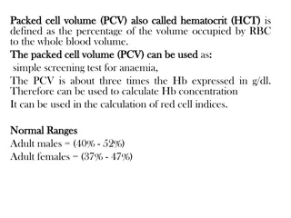
Packed cell volume (PCV) is the volume occupied by
the red cells when a sample of anticoagulated blood is
centrifuged.
It indicates relative proportion of red cells to plasma.
PCV is also called as hematocrit or erythrocyte volume
fraction.
It is expressed either as a percentage of original
volume of blood or as a decimal fraction.
USES OF PCV
Detection of presence or absence of anemia or
polycythemia.
Estimation of red cell indices (mean cell volume and
mean corpuscular hemoglobin concentration).
Checking accuracy of hemoglobin value (Hemoglobin
in grams/dl × 3 = PCV).
WINTROBE METHOD
Principle
Anticoagulated whole blood is centrifuged in a
Wintrobe tube to completely pack the red cells.
The volume of packed red cells is read directly from the
tube. An advantage with this method is that before
performing PCV, test for erythrocyte sedimentation rate
can be set up.
Equipment
1.
Wintrobe tube: This tube is about 110 mm in length
and has 100 markings, each at the interval of 1 mm.
Internal diameter is 3 mm. It can hold about 3 ml of
blood.
2.
Pasteur pipette with a rubber bulb and a sufficient
length of capillary to reach the bottom of the
Wintrobe tube.
3.
Centrifuge with a speed of 2300 g.
Specimen
Venous blood collected in EDTA (1.5 mg EDTA for 1 ml
of blood) or in double oxalate. Test should be
performed within 6 hours of collection.
Method
1.
Mix the anticoagulated blood sample thoroughly.
2.
Draw the blood sample in a Pasteur pipette and
introduce the pipette up to the bottom of the
Wintrobe tube. Fill the tube from the bottom exactly
up to the 100 mark. During filling, tip of the pipette is
raised, but should remain under the rising meniscus
to avoid foaming.
3.
Centrifuge the sample at 2300 g for 30 min (To
counterbalance a second Wintrobe tube filled with
blood from another patient or water should be placed
in the centrifuge).
4.
Take the reading of the length of the column of red
cells. Hematocrit can be expressed either as a
percentage or as a fraction of the total volume of
blood sample.
Anticoagulated blood-filled
Wintrobe hematocrit tubes
after centrifugation,
showing normal PCV, low
PCV (anemia), and thick
buffy coat layer.
Preparation of smear from buffy
coat.
Significance
In anemia, PCV is below the lower level of normal
range. PCV is raised in dehydration, shock, burns, and
polycythemia.
After centrifugation of anticoagulated whole blood,
three zones can be distinguished in the Wintrobe tube
from above downwards- plasma, buffy coat layer (a
small greyish layer of white cells and platelets, about 1
mm thick), and packed red cells.
Smears can be made from the buffy coat layer for
demonstration of lupus erythematosus (LE) cells,
malaria parasites, or immature cells.
REFERENCE RANGES
Adult males: 40-50%
Adult females (nonpregnant): 38-45%
Adult females (pregnant): 36-42%
Children 6 to 12 years: 37-46%
Children 6 months to 6 years: 36-42%
Infants 2 to 6 months: 32-42%
Newborns: 44-60%
CRITICAL VALUES
CRITICAL VALUES
Packed cell volume: < 20% or > 60%
Packed Cell Volume (PCV), also known as hematocrit, is a crucial parameter in blood analysis that indicates the relative proportion of red blood cells to plasma. It is used for detecting anemia, polycythemia, and assessing red cell indices. The Wintrobe method is commonly employed to measure PCV, involving centrifugation of anticoagulated whole blood in a specialized tube. Interpretation of PCV values helps in diagnosing various blood disorders and conditions affecting red cell volume.

Download Presentation

Please find below an Image/Link to download the presentation.
The content on the website is provided AS IS for your information and personal use only. It may not be sold, licensed, or shared on other websites without obtaining consent from the author. Download presentation by click this link. If you encounter any issues during the download, it is possible that the publisher has removed the file from their server.
E N D
Presentation Transcript
Packed Cell Volume (PCV) Dr Versha Prasad
Packed Cell Volume (PCV) Packed cell volume (PCV) is the volume occupied by the red cells when a sample of anticoagulated blood is centrifuged. It indicates relative proportion of red cells to plasma. PCV is also called as hematocrit or erythrocyte volume fraction. It is expressed either as a percentage of original volume of blood or as a decimal fraction.
USES OF PCV Detection of presence or absence of anemia or polycythemia. Estimation of red cell indices (mean cell volume and mean corpuscular hemoglobin concentration). Checking accuracy of hemoglobin value (Hemoglobin in grams/dl 3 = PCV).
WINTROBE METHOD Principle Anticoagulated whole blood is centrifuged in a Wintrobe tube to completely pack the red cells. The volume of packed red cells is read directly from the tube. An advantage with this method is that before performing PCV, test for erythrocyte sedimentation rate can be set up. Equipment 1. Wintrobe tube: This tube is about 110 mm in length and has 100 markings, each at the interval of 1 mm. Internal diameter is 3 mm. It can hold about 3 ml of blood. 2. Pasteur pipette with a rubber bulb and a sufficient length of capillary to reach the bottom of the Wintrobe tube. 3. Centrifuge with a speed of 2300 g.
Specimen Venous blood collected in EDTA (1.5 mg EDTA for 1 ml of blood) or in double oxalate. Test should be performed within 6 hours of collection. Method 1. Mix the anticoagulated blood sample thoroughly. 2. Draw the blood sample in a Pasteur pipette and introduce the pipette up to the bottom of the Wintrobe tube. Fill the tube from the bottom exactly up to the 100 mark. During filling, tip of the pipette is raised, but should remain under the rising meniscus to avoid foaming. 3. Centrifuge the sample at 2300 g for 30 min (To counterbalance a second Wintrobe tube filled with blood from another patient or water should be placed in the centrifuge). 4. Take the reading of the length of the column of red cells. Hematocrit can be expressed either as a percentage or as a fraction of the total volume of blood sample.
Anticoagulated blood-filled Wintrobe hematocrit tubes after centrifugation, showing normal PCV, low PCV (anemia), and thick buffy coat layer. Preparation of smear from buffy coat.
Significance In anemia, PCV is below the lower level of normal range. PCV is raised in dehydration, shock, burns, and polycythemia. After centrifugation of anticoagulated whole blood, three zones can be distinguished in the Wintrobe tube from above downwards- plasma, buffy coat layer (a small greyish layer of white cells and platelets, about 1 mm thick), and packed red cells. Smears can be made from the buffy coat layer for demonstration of lupus erythematosus (LE) cells, malaria parasites, or immature cells.
REFERENCE RANGES Adult males: 40-50% Adult females (nonpregnant): 38-45% Adult females (pregnant): 36-42% Children 6 to 12 years: 37-46% Children 6 months to 6 years: 36-42% Infants 2 to 6 months: 32-42% Newborns: 44-60% CRITICAL VALUES Packed cell volume: < 20% or > 60%