Techniques for Sample Spotting in Mass Spectrometry
Learn about different sample spotting techniques including Dried Droplet, Crushed Crystal, Thin Layer, and Sandwich methods used in mass spectrometry analysis. Each technique involves specific steps for preparing and applying samples on a sample plate before analysis. Ideal sample concentrations for peptides, proteins, oligonucleotides, and polymers are also highlighted.

Download Presentation

Please find below an Image/Link to download the presentation.
The content on the website is provided AS IS for your information and personal use only. It may not be sold, licensed, or shared on other websites without obtaining consent from the author. Download presentation by click this link. If you encounter any issues during the download, it is possible that the publisher has removed the file from their server.
E N D
Presentation Transcript
Sample spotting techniques Dried droplet Crushed crystal Thin layer Sandwich
Dried Droplet Most common method used with all matrices. Premix sample and matrix solution in small vial (500 L Eppendorf tube), spot 0.5 - 1 L onto sample plate and air dry. For small sample amounts, deposit 0.5 L on plate, then add 0.5 L matrix on top; mix and allow to dry.
Crushed Crystal Method Prepare a saturated solution of sinapinic acid (or CHCA) in 30% AcN. Deposit 1 L of this solution on the sample plate and allow to dry. Crush the crystals formed with a spatula or a foil- wrapped pencil eraser. Add 0.5 - 1 L of sample in saturated matrix on top of the crystal layer and allow to dry.
Thin Layer Use 10 mg/mL CHCA in acetone. Apply 0.5 L to plate and dry. Place 0.5 L sample in 0.1% TFA on top of the matrix layer and allow to dry. If sample is not acidified, 0.5 L 0.1% TFA can be added to the plate, then the sample.
Sandwich Method Deposit 0.5 L matrix(in acetone or methanol) on plate and let dry. Add 0.5 L sample in 0.1% TFA, then 0.5 L matrix (in 50% Acn with 0.1% TFA) and dry in air.
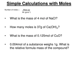
Ideal Sample Concentrations Peptides and proteins: 0.1 to 10 M Oligonucleotides: 10 to 100 M Polymers: 100 M