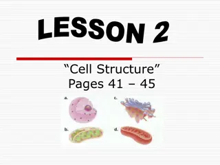
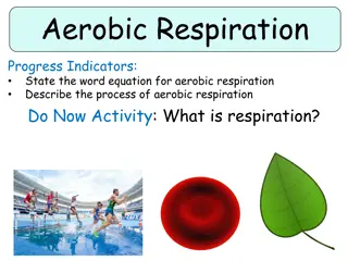
Understanding the Structure and Function of Mitochondria
Mitochondria, derived from Greek words meaning thread and granule, are essential organelles found in eukaryotic cells. They serve as energy powerhouses, metabolizing carbohydrates and fatty acids to produce adenosine triphosphate (ATP). The inner and outer membranes of mitochondria contain proteins and enzymes for various functions, including ATP synthesis. Mitochondria have their own DNA and genes, evolving from primitive bacteria. Cristae, folds in the inner membrane, play a crucial role in ATP production. Antibiotics can inhibit ATP synthesis in mitochondria.

Download Presentation

Please find below an Image/Link to download the presentation.
The content on the website is provided AS IS for your information and personal use only. It may not be sold, licensed, or shared on other websites without obtaining consent from the author. Download presentation by click this link. If you encounter any issues during the download, it is possible that the publisher has removed the file from their server.
E N D
Presentation Transcript
Mitochondria derives from two Greek words: mitos meaning thread and chondros meaning granule. Mitochondria are organelles typically ranging in size from 0.5 micrometer to 1 micrometer in length, found in the cytoplasm of eukaryotic cells. Mitochondria are specialized structures unique to the cells of animals, plants, and fungi. Prokaryotes have no mitochondria. Mito serve as batteries, powering various functions of the cell and the organism as a whole. Evidence shows that they evolved from primitive bacteria. Mitochondria have their own set of DNA and genes that encode proteins. The main function of mitochondria is to metabolize or break down carbohydrates and fatty acids to generate energy in the form of a chemical molecule called adenosine triphosphate. A secondary function of mitochondria is to synthesize proteins for their own use. The number of mitochondria in a cell depends on the cell's function. Cells with particularly heavy energy demands, such as muscle cells, have more mitochondria than other cells.
Structure of mitochondria Mitochondria contain the inner and outer membranes, separated by a space. Both the inner and outer membranes are constructed with tail-to-tail bilayers of phospholipids into which mainly hydrophobic proteins are embedded. The outer membrane contains proteins and lipids. The smooth outer membrane holds numerous transport proteins, which shuttle materials in and out of the mitochondrion. The outer membrane is 60 70 thick and permeable to small molecules including salts, adenine and nicotinamide nucleotides, sugars and coenzyme. The inner membrane contains all the enzymes and less lipid than the outer membrane. These membranes produce two separate compartments creating the intermembrane space (C-side) and the space enclosed by the inner membrane called matrix (M-side) The intermembrane space is usually 60 80 in width and contains some enzymes. The matrix however is very viscous and rich in protein, enzymes and fatty acids. A membrane component exhibits allotropy and changes its property when separated. https://www.youtube.com/watch?v=_XqBIGRlkg8
The inner membrane houses the respiratory chain and ATP synthesis, and is permeable to small neutral molecules such as water, oxygen, and carbon dioxide. Its permeability to charged molecules such as proton and ions is limited. The inner membrane has numerous folds called cristae, which have folded structure greatly increasing the surface area where ATP synthesis occurs. Transport proteins, molecules called electron transport chains, and enzymes that synthesize ATP are among the molecules embedded in the cristae. The cristae have the major coupling factors F1, (a hydrophilic protein) and Fo(a hydrophobic lipoprotein complex), which together comprise the ATPase complex activated by Mg+2. ATPase catalyses hydrolysis of ATP to adenosine diphosphate (ADP) and phosphate, while ATPsynthase produces ATP using the energy released by the redox reactions of the respiratory chain. Both reactions are inhibited by the antibiotics such as oligomycin.
Types of mitochondria Aerobic mitochondria- The mitochondria typical of mammalian cells respire O2during the process of pyruvate breakdown and ATP synthesis, generating water and carbon dioxide as end products. The Krebs cycle and the electron transport chain in the inner mitochondrial membrane enable the cell to generate about 36 moles (mol) of ATP per mole of glucose, with the help of O2 respiring mitochondria. Anaerobic mitochondria These do not use O2as the terminal acceptor during prolonged phases of the life cycle. These mitochondria allow the anaerobically growing cell to glean about 5 mol of ATP per mole of glucose with end products are carbon dioxide, acetate, propionate, and succinate. E.g. Fasciola hepatica Hydrogenosomes These lack an electron transport chain altogether. They synthesize ATP from pyruvate breakdown via simple fermentations that typically involve the production of molecular hydrogen as a major metabolic end product. They allow the cell to gain about 4 mol of ATP per mole of glucose. E.g. Trichomonas Mitosomes They are nconspicuous mitochondria that are not involved in ATP synthesis at all. These eukaryotes synthesize their ATP in the cytosol with the help of enzymes. They obtain 2-4 mol of ATP per mole of glucose. Their typical end products are carbon dioxide, acetate, and ethanol. E.g. Entamoeba histolytica
Why semiautonomous? 1.Mitochondria have their own DNA which can replicate independently. The mitochondrial DNA produces its own mRNA, tRNA and rRNA. 2.The organelles posses their own ribosomes, called mitoribosomes. 3.Mitochondria synthesize some of their own structural proteins. However, most of the mitochondrial proteins are synthesized under instructions from cell nucleus. 4.The organelles synthesize some of the enzymes required for their functioning. e.g. succinate dehydrogenase. 5.They show hypertrophy .i.e. internal growth.
DNA of mitochondria Mitochondria contain deoxyribonucleic acid (DNA) and ribosomes, protein-producing organelles in the cytoplasm. Within the mitochondria, the DNA directs the ribosomes to produce proteins as enzymes, or biological catalysts, in ATP production. This DNA is small and circular. It has only 16,500 or so base pairs in it. And it encodes different proteins that are specific for the mitochondrial. Mitochondrial DNA, unlike nuclear DNA, is inherited from the mother, while nuclear DNA is inherited from both parents. Mitochondrial division is important for the remodeling and rearrangement of mitochondrial networks, as well as for enabling mitochondrial segregation during cell division. By enabling genetic complementation, fusion of the mitochondria allows for two mitochondrial genomes with different defects within the same organelle to individually encode what the other lacks. In doing so, these mitochondrial genomes generate all of the necessary components for a functional mitochondrion. https://www.youtube.com/watch?v=HIR2weKk3gg
Origin of Mitochondria When the oceans were mostly anoxic from 1.8 billion years ago until about 580 million years ago because of the workings of marine, H2S-producing bacteria. Eukaryotes thus arose and diversified in an environment where anoxia was commonplace. Like eukaryotes themselves, mitochondria appear to have arisen only once in all of evolution. The best evidence for the single origin of mitochondria comes from a conserved set of clearly homologous and commonly inherited genes preserved in the mitochondrial DNA across all known eukaryotic groups.
Endosymbiont Theory Theory 1 The host that acquired the mitochondrion was an anaerobic nucleus-bearing cell, a full- fledged eukaryote that was able to engulf the mitochondrion actively via phagocytosis. The mitochondrial endosymbiont was an obligate aerobe, perhaps similar in physiology and lifestyle to modern Rickettsia species. The initial benefit of the symbiosis might have been the endosymbiont's ability to detoxify oxygen for the anaerobe host. Protection from O2is an unlikely symbiotic benefit.
Theory 2 The host that acquired the mitochondrion was a prokaryote, an archaebacterium outright. The ancestral mitochondrion was a metabolically versatile, facultative anaerobe (able to live with or without oxygen). The initial benefit of the symbiosis could have been the production of H2by the endosymbiont as a source of energy and electrons for the archaebacterial host, which is posited to have been H2dependent. The mechanism by which the endosymbiont came to reside within the host is unspecified. In this view, various aerobic and anaerobic forms of mitochondria are seen as independent, lineage-specific ecological specializations, all stemming from a facultatively anaerobic ancestral state. Because it posits that eukaryotes evolved from the mitochondrial endosymbiosis in a prokaryotic host, this theory directly accounts for the ubiquity of mitochondria among all eukaryotic lineages.
https://www.youtube.com/watch?v=c4JsEBI9u6I https://www.youtube.com/watch?v=Nxv6zD45at4