Exploring Ultrasound: Applications and Principles in Medical Imaging
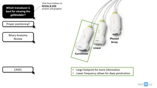
Utilizing ultrasound in medical imaging offers a non-invasive method to visualize soft tissues in the body. This technology involves sending ultrasound waves into the body, with reflections used to create images. The process involves calculating depths of layers based on wave return times. Understanding ultrasound frequencies across species is also key, with various applications discussed, including maternity scans and kidney stone detection. Additionally, the use of ultrasound in place of x-rays for certain scans is highlighted, showcasing its potential to capture images of soft tissues without damaging living cells.
- Ultrasound imaging
- Medical technology
- Reflection and transmission
- Non-invasive
- Soft tissue visualization
Download Presentation

Please find below an Image/Link to download the presentation.
The content on the website is provided AS IS for your information and personal use only. It may not be sold, licensed, or shared on other websites without obtaining consent from the author. Download presentation by click this link. If you encounter any issues during the download, it is possible that the publisher has removed the file from their server.
E N D
Presentation Transcript
3 Waves Part D3 Ultrasound Extension Only 1 Forces and Motion 2 Electricity 3 Waves 4 Energy 5 Solid Liquids Gases 6 Magnetism 7 Radioactivity 8 Astrophysics Animated Science 2018
Mr D Powell 2018 Animated Science 2018 Lesson 3 Exploring Ultrasound (2 hours) Triple Only Extra Reading Be able to recall simple examples of ultrasound such as a maternity scan or cleaning of items. 4 Be able to explain in detail how an A scan ultrasound scan works in terms of reflection and transmission and apply ideas to a context such as animals or humans 5/6 Complete detailed calculations to work out the distance from a transducer to an organ boundary 7/8 In the context of a kidney be able to explain and use an A-scan result in terms of intensity and reflection boundaries. Animated Science 2018
Ultrasound Ranges.... Task.. copy the simple diagram with some labels Baby Scans/ Kidney Stone detection 2 to 15 MHz. Human Range 20Hz to 20kHz Dolphin Hearing 1 130kHz Lowest frequency (Hz) 20 3000 300 100 30 50 1000 1000 Highest frequency (kHz) 20 120 45 3 50 46 100 Species Human Bat Rabbit Frog Cat Dog Mouse Dolphin Animated Science 130 2018
Animated Science 2018
Ultrasound Hospital Use Extension (Not Required) Ultrasound may be used instead of x-rays for certain scans, such as scan of unborn babies. Compared to x-ray photographs, ultrasound scans: Do not damage living cells Produce images of soft tissue 1. Ultrasound is sent into the patient's body. Some of the ultrasound is reflected at each boundary between different tissues or organs. 2. The depth of each layer is calculated using the time taken for each reflected wave to return. 3. The reflected waves (echoes) are usually processed to produce a picture of the inside of the body on a screen. Animated Science 2018
Ultrasound Hospital Use Extension (Not Required) When ultrasound waves reach a boundary between two media (substances) with different densities, they are partly reflected back. The remainder of the ultrasound waves continue to pass through. A detector placed near the source of the ultrasound waves is able to detect the reflected waves. It can measure the time between an ultrasound wave leaving the source and it reaching the detector. s = distance in metres, m v = speed in metres per second, m/s The distance travelled by an ultrasound wave can be calculated using this equation: t = time in seconds, s s = v t NB: but remember this is an echo so you must half it! An ultrasound machine can be used to detect cracks or flaws in materials such as metal. Animated Science 2018
Ultrasonic Physiotherapy Ultrasound is applied using a round-headed wand or probe that is put in direct contact with the patient's skin. Ultrasound gel is used on all surfaces of the head in order to reduce friction and assist in the transmission of the ultrasonic waves. Therapeutic ultrasound is in the frequency range of about 0.8-1.0 MHz. The sound waves that pass through the skin cause a vibration of the local tissues. This vibration can cause a deep heating locally though usually no sensation of heat will be felt by the patient. In situations where a heating effect is not desirable, such as a fresh injury with acute inflammation, the ultrasound can be pulsed rather than continuously transmitted. If the probe is held in one place for more than just a few seconds, a build up of the sound energy can result which can become uncomfortable. Interestingly, if there is even a very minor break in a bone in the area that is close to the surface, a sharp pain may be felt. This occurs as the sound waves get trapped between the two parts of the break and build up until becoming painful. In this way ultrasound can often be used as a fairly accurate tool for diagnosing minor fractures that may not be obvious on x-ray. Animated Science 2018
Ultrasonic Jewellery Cleaners An ultrasonic jewellery cleaner is an electronic device designed to remove dirt from rings, necklaces and other items of jewellery. These devices are readily found on home shopping channels or on the internet. To begin the cleaning process, the cleaning tank is filled with warm water or a cleaning solution. Cleaning solutions can consist of non-ionic surfactants, detergents or ammonia. Once on, the machine's motor produces ultrasonic energy, which is transmitted with vibrating energy waves. On average, ultrasonic jewellery cleaners emit at least 40,000 sound waves per second. 1. The vibrating motion of the ultrasonic waves creates microscopicbubbles in the water or cleaning solution in a process called cavitation 2. millions of tiny bubbles knock into one another and into the items resting in the cleaning tank. 3. The cavitation process gently knocks dirt off the jewellery. 4. The motion is very effective at penetrating the tiny crevices in jewellery that traditional cleaning cloths and topical cleaners cannot easily reach. Animated Science 2018
Ultrasonic Washing Machine LG recently put on sale a washing machine that doesn't require detergent to clean lightly soiled clothes. The machine is fitted with electrodes on the side of the tub that electrolyse the water, and an ultrasonic wave generator at the base of the machine. 1. The ultrasonic waves form tiny air bubbles, help loosen grime and grit on clothes in a purely mechanical action. 2. Electrolyzing the water produces active oxygen, or forms of oxygen such as hydrogen peroxide and ozone, and hypochlorousacid, a mild bleaching agent. Hypochlorous acid kills bacteria while active oxygen dissolves such dirt as the residue of body sweat. 3. LG claims users can half the cost of doing normal laundry. Reducing the amount of detergent sent into waste water streams is also environmentally friendly. Animated Science 2018
Breaking down kidney stones A high powered ultrasound wave is used to break down kidney stones and other stones in the body. The stones vibrate until they shake themselves apart and are then easily passed out of the body via the urethra. Animated Science 2018
Advantages / Disadvantages Animated Science 2018
Ultrasound Maths Bone = v*t = (2900ms-1 x 1000 x 1 x 10-6 s)/ 2 = 1.45m An ultrasound machine is used to investigate samples from a whale corpse found on the beach. An oscilloscope trace sees a return peak after the pulse is emitted. What is the distance from the surface to the part of the whale? d/t = s or s/t = v (vectors) Distance into the whale from transducer /m 1.45 0.404 0.314 Speed in medium ms-1 Time for return pulse in micro seconds / S Substance Bone 2900 1615 1000 Soft Tissue Blood Muscle Fat 400 250 200 1545 1450
Bone = v*t = (2900ms-1 x 1000 x 1 x 10-6 s)/ 2 = Soft = s/v = 0.4 x 2 /1615ms-1 = Blood = v=s/t = 2 x 0.314/ 400 x 1 x 10-6s = Muscle = v*t = (1545ms-1 x )/ 2 = Fat= = v*t = Maths Help d/t = m/s or ms-1 1 s = 1x10⁻⁶s = 0.000001 This ultrasound is designed to look the structure of the eye. The distance to the back of the eye scanned in this way was known to be 48mm. What would be the distance from the eye lens to the front of the model eye using this scale? Smaller pulses are due to.. A is the partial reflection as the waves enter the cornea, Extra distance in tissue results in more signal absorption Smaller fraction of signal reflected at second surface The pulse will be more spread over time The signal is diffracted B is a reflection of the pulse at the lens, 48mm = 3.9 sq 7.8 squares = 96mm or 1 sq = 12.3 1.1 square = 12.3mm *1.1 = 13.54mm 14mm = distance or simply 48mm * (1.1sq /3.9sq) = 14mm C is due to the reflection off the back of the eye. Further blips are due to the waves reflecting off bone in the skull.
Ultrasonic Speeds - Questions Bone = v*t = (2900ms-1 x 1000 x 1 x 10-6 s)/ 2 = 1.45m An ultrasound machine is used to investigate samples from a whale corpse found on the beach. An oscilloscope trace sees a return peak after the pulse is emitted. Soft = s/v = 0.4 x 2 /1615ms-1 = 500 x 10-6s Blood = v=s/t = 2 x 0.314/ 400 x 1 x 10-6s = 1570 ms-1 Muscle = v*t = (1545ms-1 x 250x 1 x 10-6 s)/ 2 = 0.193m What is the distance from the surface to the part of the whale? d/t = s or s/t = v (vectors) Fat= = v*t = (1450ms-1 x 200x 1 x 10-6 s)/ 2 = 0.145m Maths Help d/t = m/s or ms-1 1 s = 1x10⁻⁶s = 0.000001 Distance into the whale from transducer /m 1.45 0.404 0.314 0.193 Speed in medium ms-1 Time for return pulse in micro seconds / S Substance Bone 2900 1615 1570 1000 Soft Tissue Blood Muscle Fat 500 400 250 200 1545 1450 Animated Science 0.145 2018
Exam Question Example This ultrasound is designed to look the structure of the eye. A is the partial reflection as the waves enter the cornea, B is a reflection of the pulse at the lens, C is due to the reflection off the back of the eye. Further blips are due to the waves reflecting off bone in the skull. The distance to the back of the eye scanned in this way was known to be 48mm. What would be the distance from the eye lens to the front of the model eye using this scale? Smaller pulses are due to.. Extra distance in tissue results in more signal absorption Smaller fraction of signal reflected at second surface The pulse will be more spread over time The signal is diffracted 48mm = 3.9 sq 7.8 squares = 96mm or 1 sq = 12.3 1.1 square = 12.3mm *1.1 = 13.54mm 14mm = distance or simply 48mm * (1.1sq /3.9sq) = 14mm Animated Science 2018
Exam Questions The screen shows an ultrasound pulse which takes 32 millionths (32 S or 32 x 10-6S) to travel across each square of the grid. 1) How many internal boundaries are present in this sample... 2) How long does each pulse take to travel from the body surface to the nearest internal body 3) If the wavespeed is 1500ms-1. What is the distance from the body surface to the nearest tissue boundary? Animated Science 2018
Exam Questions The screen shows an ultrasound pulse which takes 32 millionths (32 S or 32 x 10-6S) to travel across each square of the grid. 1) How many internal boundaries are present in this sample... Two (excluding the initial pulse external boundary and far side boundary) 2) How long does each pulse take to travel from the body surface to the nearest internal body difference is 2.8 sq (there and back) t = * 2.8 * 32 millionths of a second = 44.8 S. 3) If the wavespeed is 1500ms-1. What is the distance from the body surface to the nearest tissue boundary? v = s/t so vt = s 1500ms-1 * 44.8 x 10-6 s = 0.067m = 6.7cm Animated Science 2018
The Eye real world ideas.... Extra Details Animated Science 2018
Extended Thinking... Animated Science 2018
Animated Science 2018
Summary Animated Science 2018
Extended ideas on Speed Animated Science 2018
B - Scans Extra Details Animated Science 2018
Piezoelectric Effect Extra Thinking Animated Science 2018
Ultrasound Answers.... (Self or Peer Assess) Questions.... 1) Describe the frequencies that ultrasound operates at (Basic) 1) 20,000Hz + as human hearing is 20 to 20,000Hz so beyond that. 2) The waves are partially reflected from a boundary i.e. Fat to water so are picked up by a detector. The pulse shown on an oscilloscope initially and the reflected pulse give the time there and back. So you half this to give the travel time for the wave and then the distance from the wave equation. s = x v x t = 0.5 x 1500ms-1 x 10 x 10-6S = 7.5 x 10⁻³m or 7.5mm 2) How can ultrasound be used to show the structure of a baby in a womb? 3) How can an oscilloscope be used to work out the distance across a babies head to determine if they may suffer from a genetic problem such as down syndrome. 4) If ultrasound travels at 1500ms-1 and a pulse difference is picked up as 10 S what is the distance to the probe (Harder) 3) 4) 4 My quick topic tip Animated Science 2018
Ultrasound Uses Answers.... (Self or Peer Assess) Questions.... 1) kidney stone, pre natal scan, blood flow 1. Give two examples of how ultrasound can be used in the human body. 2) See rev guide Lower energy as longer , non- ionising, not cancer causing, don t harm babies 2. Explain 1 example in detail 3) 3. Give two reasons why ultrasound is safer than x-rays? 4) Fuzzy compared to others but 3D Computed Tomography is a lot more radiation than X-ray. However, good for flesh as well as bone! 4. Comment on image quality of pictures compared to CT and X- rays (Harder) 4 My quick topic tip Animated Science 2018
Lesson 6 Exploring Ultrasound Extra Reading and Questions. Animated Science 2018
Lesson 3 Exploring Ultrasound Extra Reading and Questions. Animated Science 2018
Ultrasound Plenary / Review Task As a radiographer it is your task to make measurements of a foetus while it is in the womb. When you take a prenatal ultrasound scan, the echo of the pulse of ultrasound returns in 300 microseconds. v = 1540 m/s c = 3 x 10⁸ ms-1 1. If the sound travels at a speed of 1540 m/s in the fluids of the womb, you can work out the depth of the tissue that is returning the echo. 2. Ultrasound equipment produces ultrasound with a frequency between 2MHz and 18 MHz. What are the wavelengths of ultrasound at these two frequencies? 3. Why does the radiographer not recommend X-rays as a method of viewing the foetus? 4. Why does she use ultrasound instead? 5. When the ultrasound enters the bladder, the wave changes direction, producing an image of the organ on the monitor. What causes this change in the direction of the wave? http://en.wikipedia.org/wiki/Medica l_ultrasonography Animated Science 2018
Ultrasound Plenary / Review Task As a radiographer it is your task to make measurements of a foetus while it is in the womb. When you take a prenatal ultrasound scan, the echo of the pulse of ultrasound returns in 300 microseconds. v = 1540 m/s c = 3 x 10⁸ ms-1 1. If the sound travels at a speed of 1540 m/s in the fluids of the womb, you can work out the depth of the tissue that is returning the echo. 1) 1540ms-1 x 150 x 10-6s = 0.231m 2) c=f so c/f = 2. Ultrasound equipment produces ultrasound with a frequency between 2MHz and 18 MHz. What are the wavelengths of ultrasound at these two frequencies? =1540ms-1/2 x 106Hz = 7.7 x 10⁻⁴m or =1540ms-1/18 x 106Hz = 8.6 x 10⁻⁵m 3. Why does the radiographer not recommend X-rays as a method of viewing the foetus? 4. Why does she use ultrasound instead? 5. When the ultrasound enters the bladder, the wave changes direction, producing an image of the organ on the monitor. What causes this change in the direction of the wave? http://en.wikipedia.org/wiki/Medica l_ultrasonography Animated Science 2018


