Understanding Safety Protocols and Inflammation in Histotechnique Laboratory


Safety in the Histotechnique
Laboratory
 
PEL – Permissible Exposure Limit
TLV – Threshold Limit Value
OEL – Occupational Exposure Limit
 
 
*Complete penetration by alcohol will destroy all
infectious agents except prions.
Prions – are infectious agents that cause spongiform
encephalopathies such as Creutzfeld-Jakob disease
(CJD), scrapie and mad cow disease.
Normal steam sterilization does not inactivate these
particles, and common effective treatments like
sodium hypochlorite or phenol will create artifacts in
tissue.
 
 
Tissue from suspected patients with CJD can be
decontaminated by immersing the specimen in
formalin for 48 hours, followed by treatment in
concentrated formic acid for 1 hour and additional
formalin fixation for another 48 hours.
 
 
Small-dust particles generated from sectioning may
become airborne, particularly when performing
cryostat sections of fresh tissue.
Cryogenic sprays can magnify this risk, and therefore
should not be used to freeze potentially infectious
tissue.
 
Cytopathologic Changes in Disease
 
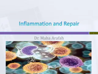
Inflammation – is the sum total of changes in the
living tissues, in response to an injurious agent,
including the local reaction and the repair of an injury.
- it is composed of a series of physiologic and
morphologic alterations in the blood vessels, blood
components and surrounding connective tissue, for
the purpose of protecting the body against injury.
 
Cardinal Signs of Inflammation


Rubor (Redness) – due to arteriolar and capillary
dilation with increased rate of blood flow towards the
site of injury, and concentration or packing of the red
cells in the capillaries causing increased viscosity and
consequent slowing of the blood flow (stagnation).
 
Tumor (Swelling) – due to increased capillary
permeability, allowing the extravasation of blood fluid,
associated with increased hydrostatic pressure within
the dilated arterioles and capillaries, causing localized
edema (tumor) accompanied by escape of blood cells
into the injured area, increased concentration of
plasma proteins, and accumulation of finely
particulate cellular debris caused by destruction of
tissue cells and bacteria
 
 
Calor (Heat) – due to transfer of internal heat to the
surface or site of injury, brought about by increased
blood content.
 
 
Dolor (Pain) – due to pressure upon the sensory nerve
by the exudate or tumor
 
 
Functio Laesa (Diminished Function) – may be due to
pain interference with nerve supply and to destruction
of the functioning units of the tissue.
 
Causes of Inflammation:
 
Living things (bacteria,parasites)
Viruses
Non-living agents (physical, mechanical, electrical, or
chemical stimuli)
 
Classification of Inflammation
 
A. duration of the inflammation
B. predominant type of exudate formed
C. location of the inflammation
 
A. Types of Inflammation according
to duration
 
1. Acute Inflammation
- usually, but not necessarily, of sudden onset and
characterized by the classic features of heat, redness,
swelling, pain and loss of function.
 
 
1. Acute Inflammation - It refers to an inflammatory
reaction in which the dominant anatomic changes are
vascular and exudative, hence, it is also called
Exudative Inflammation, with exudation of white
blood cells, predominantly the polymorphonuclear
leukocyte.
 
 
2. Chronic Inflammation – this involves the
persistence of the injurious agent for weeks or years,
characterized by proliferative (cell multiplication)
rather than exudative response, with a predominantly
mononuclear cell infiltration (macrophages,
lymphocytes and plasma cells).
 
 
2. Chronic inflammation – proliferation is chiefly
fibroblastic and vascular.
- polymorphonuclear leukocytes may also be present.
 
 
2. Chronic Inflammation – continued proliferation of
fibroblasts gives rise to considerable damage and
scarring with resultant deformities
 
Granulomatous Chronic
Inflammation
 
- this is characterized by formation of granuloma,
constituting a distinct histologic pattern of
inflammation in certain diseases like Tuberculosis,
Syphilis, Lymphogranuloma Inguinale, etc.
 
 
Granuloma – implies a tumor-like mass of granulation
tissue (actively growing fibroblasts and capillary
buds).
 
 
Subchronic inflammation – represents an intergrade
between acute and chronic inflammation
 
Types of Inflammation According to
Character
 
1. Serous Inflammation – serous exudation is
characterized by extensive outpouring of a watery,
low-protein fluid derived from either the blood serum
or secretions from the serosal mesothelial cells, i.e.
peritoneal, pleural or pericardial cavities.
- this is characteristic of certain forms of pericardial
tuberculosis with involvement of the pleura (pleurisy
with effusion).
 
2. Fibrinous Inflammation
 
- this is characterized by exudation of large amounts of
fibrinogen and the precipitation of masses of fibrin,
occuring in the more severe acute inflammations
associated with marked endothelial damage, also
found in diphteria, rheumatic pericarditis and in early
stages of pneumonia.
- polymorphonuclear leukocytes are usually trapped
within the fibrin meshwork
 
3. Catarrhal Inflammation
 
- this type affects mucous surfaces and is characterized
by hypersecretion of the mucosa with degenerative
changes in the epithelium, characteristic of
inflammatory involvement of the respiratory,
gastrointestinal and other mucus-secreting glands.
- this form of exudation is identified by large amounts
of faintly basophilic, amorphous, stringy mucoid
which usually contains white cells.
 
4. Hemorrhagic Inflammation
 
- this is characterized by the admixture of blood and
other elements of the exudate, the intensity of
hemorrhage varying from case to case, and may be
found in bacterial and other infections.
 
5. Suppurative or Purulent
Inflammation
 
- this is characterized by production of large amounts
of pus or purulent exudate.
- Pus – may be defined as a thick, creamy fluid
composed of large numbers of viable and necrotic
polymorphonuclear leukocytes and necrotic tissue
debris that is partially liqueified by proteolytic
digestion.
 
 
Suppuration – characterized by large numbers of
neutrophils and tissue debris and is most prominent in
the early stages of inflammation, its presence denoting
an acute reaction
 
Abnormalities in cell growth
 
Retroregressive changes: Organs or tissues smaller
than normal may be due to:
A. Developmental Defects:
1. Aplasia – is the incomplete or defective development
of a tissue or organ, represented only by a mass of fatty
or fibrous tissue, bearing no resemblance to the adult
structure
- commonly seen in one of paired structures such as
kidneys, gonads and adrenals
 
 
2. Agenesia – refers to the complete non-appearance of
an organ
3. Hypoplasia – refers to the failure of an organ to
reach or achieve its full mature or adult size due to
incomplete development.
4. Atresia – is the failure of an organ to form an
opening
 
 
Atrophy – refers to an acquired decrease in the size of a
normally developed or mature tissue or organ resulting
from reduction in cell size or decrease in total number
of cells or both.
 
Pathogenesis of Atrophy
 
Due to increased activity of the proteolytic enzymes
associated with deficient blood and lymphatic
circulation or increased metabolic activity leading to
accumulation of CO2 and organic acids in the cells,
ultimately causing loss of cell substance
 
Types of Atrophy
 
1. Physiologic atrophy – occurs as a natural
consequence of maturation, as in atrophy of the
thymus and lymphoid tissue during puberty. Sexual
organs and brain begin to undergo physiologic atrophy
at about 50 years of age
 
 
Senile Atrophy – occurs in old age characterized by
dry, lusterless, wrinkled skin due to atrophy of sweat
and sebaceous glands and loss of fat, gray hair, atrophy
of the ligaments, brittle bones which easily break, and
“arcus senilis” of the cornea.
 
 
2. Pathologic atrophy – refers to a decrease in size of
tissues or organs, outside the range of normal
variability, usually as a consequence of disease.
 
On the basis of apparent clinical causes,
pathologic atrophy may be classified into:
 
A. vascular atrophy (due to lack of nutrition) – occurs
if the blood supply to an organ or tissue becomes
reduced below critical levels, commonly encountered
in kidney and brain.
 - the most common cause is vascular narrowing due to
progressive arteriosclerosis, although, external
pressures may also play a role.
 
 
B. pressure atrophy – persistent pressure on the organ
or tissue may directly injure the cells or may
secondarily promote diminution of blood supply
leading to vascular atrophy.
 
 
C. starvation or hunger atrophy – produces a general
wasting of tissues due to excessive lack of nutritional
supply necessary for normal growth.
 
 
D. Atrophy of disuse – inactivity or diminished
function of a tissue or organ (as in long standing,
chronic illnesses, or inactivity of muscle fibers due to
plaster casts or neuropathy) may lead to narrowing of
the blood vessels with loss of nutrition and consequent
atrophic changes
 
 
E. Exhaustion atrophy – prolonged overwork,
especially of an endocrine organ (e.g. adrenals,
pituitary or thyroid) may produce initial enlargement
with ultimate slow progressive loss of parenchymal
elements due to excessive formation of acid
metabolites with subsequent increase of catabolic
enzymes
 
 
F. Endocrine Atrophy – diminished or absent
endocrine stimulation may produce functional atrophy
of organs which are mainly dependent on their
endocrine supply for maintenance of their normal
structures.
 
Pathogenic Changes seen in
Atrophy
 
Grossly, atrophic organs are smaller and firmer in
consistency due to increased amount of connective
tissue; vessels appear prominent and increased in
number due to proximity of vessels formerly separated
by larger masses of tissue
 
Pathogenic Changes seen in
Atrophy
 
Microscopically, atrophic cells are smaller than
normal; the tissue may appear to be relatively cellular
Atrophic parenchymal cells, particularly in the heart
and liver may contain yellow granular lipid-containing
pigments (lipochrome pigment or lipofuscin),
imparting on them a brownish discoloration on gross
inspection (“Brown Atrophy”).
 
Progressive Changes
Organs or Tissues larger than
normal
 
A. Hypertrophy – refers to an increase in size of tissues
or organs due to increase in the size of the individual
cells. Classified into various forms:
 
 
1. True hypertrophy – usually observed in the skeletal
muscles, heart, kidneys, endocrine organs (breast and
uterus) and smooth muscles of the hollow viscera (e.g.
intestinal tract), due to increased work load and
endocrine stimulation (e.g. during exercise and
pregnancy)
 
 
2. False hypertrophy – due to edema fluid and
connective tissue proliferation (e.g. in cirrhosis and
chronic hypertrophic salpingitis or appendicitis)
 
Salpingitis – inflammation of fallopian tubes
 
 
3. Compensatory hypertrophy – involves one of paired
organs when the other opposite organ has been
removed or suffered from functional insufficiency
 
B. Hyperplasia
 
Refers to an increased in size of an organ or tissue due
to increase in the number of cells resulting from
growth of new cells.
 
 
A. Physiological hyperplasia – occurring as a normal
phenomenon, e.g. in hyperplasia or hypertrophy of the
uterus during pregnancy.
 
 
B. Pathological hyperplasia – brought about by disease
as observed in hyperplasia of the lymphoid follicles
and peyer’s patches of the intestines in typhoid fever.
 
Degenerative Changes Due to
Aberrations of Cellular Growth
Patterns
 
A. Metaplasia – is a reversible change involving the
transformation in one type of adult cell to another.
 
1. Epithelial Metaplasia
 
Occurs in epithelium exposed to mechanical trauma
or chronic irritation of prolonged inflammation or
prolonged vitamin A deficiency; most commonly
leading to replacement of columnar cells by stratified
epithelium (seen in respiratory passages, linings of
gland ducts and mucosal lining of endocervix).
 
2. Mesenchymal Metaplasia
 
Occuring in connective tissue whereby fibroblasts are
transformed into more highly differentiated forms
such as osteoblasts, fat cells or tissue macrophages.
 
B. Dysplasia
 
Is the regressive alteration in adult cells manifested by
variation in size, shape and orientation, associated
with chronic inflammation and protracted irritation;
usually applied to epithelial cells.
It differs from metaplasia in that changes are not
towards development of another adult cell, but rather,
changes in structural component of the cell, e.g.
irregularity in size (some cells are larger, some are
smaller), loss or increase in size of nucleus, presence of
mitotic figures and disruption of normal architectural
pattern.
 
 
Dysplastic changes are reversible and do not inevitably
lead to tumor formation.
Removal of inflammation or irritations cause the
reversion of the epithelium to normal form.
 
C. Anaplasia (Dedifferentiation)
 
Is a marked regressive change in adult cells towards
more primitive or embryonic cell types, usually
utilized as a criterion toward malignancy.
This may develop in cells previously at the site of
dysplastic changes, but more commonly found in cells
which are apparently previously normal.
Cellular alterations resemble dysplasia, although more
marked, more disorganized and irreversible.
 
D. Neoplasia (Tumor)
 
Is the continuous abnormal proliferation of cells
without control, serving no useful purpose of function,
usually accompanied by increase in size and
pigmentation, mitosis, number, metaplastic and
anaplastic changes of the cell.
This condition therefore, represents a pathologic
overgrowth of the tissue.
 
General Characteristics of Tumor
Cells:
 
Tumor cells usually resemble the normal cells well
enough to make them identifiable as being derived
from it.
Some of them may also attain such a degree of
differentiation that they are capable of secreting
substances such as insulin, melanin, osteoid, or
parathyroid hormone.
 
 
There are cases where no resemblance at all can be
discerned.
These are the fully undifferentiated anaplastic,
malignant tumors.
 
 
One noticeable thing about tumor cells is their
autonomy, their capacity to imitate normal structures.
Tumor cells are parasitic, they will get nutrition and
multiply while the normal cells undergo necrosis and
death.
 
Tumor cells are usually divided into
two parts:
 
A. Parenchyma – refers to the active elements of the
tumor
B. Stroma – refers to the connective tissue framework
with lymphatic and vascular channels.
 
 
The supportive framework of a tumor may be derived
either from tumor or from the “host”.
Epithelial tumors have less tendency to produce
supportive tissue.
Tumors of connective tissue origin have an abundant
intercellular framework.
 
Differentiation of Tumors:
 
A. Depending upon capacity to produce death:
1. Benign Tumors – are those that do not produce
death.
A few exceptions will have to be mentioned to make
this statement relatively true.
Only in situations causing direct pressure or
encroachment on vital structures will benign tumors
to produce death – a glioma in the medulla oblongata
will be fatal (in the same situation, a hematoma or a
granuloma will just be as serious
 
 
2. Malignant Tumors – will produce death eventually,
however small they may be and wherever they may be
located.
 
B. Depending on Histologic
Characteristics
 
1. Medullary – where there are more cells than
supporting tissue; it is soft and very malignant.
2. Scirrhous Carcinoma – where there is more
connective tissue than cells, where the cells are
apparently trapped within fibrous tissue.
 
Grading of Tumors
 
Well-differentiated tumors as a rule are less malignant
than undifferentiated tumors.
1. differentiated cells – resembling normal cells
2. undifferentiated cells – younger forms (anaplastic)
 
There are four grades in Broder’s
Classification
 
Grade I – Differentiated cells – 100%-75%
             - Undifferentiated cells – 0%-25%
Grade II – Differentiated cells – 75%-50%
               - Undifferentiated cells – 25-50%
Grade III – Differentiated cells – 50%-25%
                - Undifferentiated cells – 50-75%
Grade IV – Differentiated cells – 25%-0%
                - Undifferentiated cells – 75%-100%
 
Value of Grading
 
A. as a guide for treatment – roughly, we can say that
lower grades are amenable to Surgery; the higher
grades are amenable to Radiation but each tumor must
be regarded individually.
B. As a prognostic side – the higher grades have
generally a poorer prognosis.
 
Limitations of Grading
 
A. While generally, tumors present a homogenous
appearance throughout, histologic grading may
sometimes be found to vary from section to section.
B. The higher grades of tumors are generally regarded
having more tendency to metastasize, but metastasis
may also be found in the more benign.
 
 
Usually the metastasis is of the same grade as the
primary tumor.
It has been found that sometimes the metastasis has a
lower grade; sometimes a higher grade than the
primary tumor.
 
 
Some tumors cannot be graded, as most of the
sarcomas.
 
Degeneration and Infiltration
 
Refers to the various alterations that reflect the
response of cells to factors causing injury and damage,
but not to cell death.
 
 
Degeneration – injury to the cell precedes and results
in an accumulation of metabolites
Infiltration – overloading of a healthy cell by
metabolites causes the injury
 
Alterations in lipid metabolism
induce a group of morphologic
changes called:
 
A. Fatty Degeneration
B. Fatty Infiltration
C. Fatty Metamorphosis
D. Fatty Phanerosis
 
Fatty Degeneration
 
Refers to the abnormal accumulation of fat within
previously injured parenchymal cells, most commonly
encountered in the heart, kidneys and liver, and may
be brought about by any of the following:
Anoxia, infections, intoxication, biologic agents,
endogenous toxins
 
 
The liver is the organ most commonly affected by fatty
degeneration, the cells in the center of the lobule
usually affected first since they are always the most
anoxic, being most remote from the portal and arterial
blood supply.
 
 
The phases or stages of cell degeneration, no matter
what the cause may be, are divided into four:
A. cloudy swelling
B. fatty degeneration
C. cell death or necrosis
D. calcification
 
Cloudy swelling
 
First and earliest manifestation of disease
A reversible process from the cells may return to
normal as soon as the causative factor is removed
If the injurious agent persists,  the condition will
progress to fatty degeneration and then to necrosis
 
Cloudy swelling
 
Grossly, the organ will appear increased in size,
although the normal shape is retained.
The tissue is whitish-cloudy in color, friable, with
bulging of the surface upon cutting.
Microscopically, the cells become swollen and the
usual fine granularity of the normal cytoplasm is thus
replaced by a coarse, irregular granulation.
Nuclei are usually normal
 
Fatty degeneration of the liver
 
Macroscopically, the condition of the liver is similar to
that in cloudy swelling with some additional features.
The organ is distinctly swollen and has more rounded
features than usual.
Slightly increased in weight, it is also abnormally soft,
so that it readily pits or tears.
 
Fatty degeneration of the liver
 
Microscopically, the lipoid constituents of the cells are
converted into visible, neutral fat or fatty acids, as a
consequence of cellular injury, hence the tissue is
characterized by the appearance of fat droplets within
the cells
 
Fatty Infiltration
 
Refers to the abnormal accumulation of fat within
healthy cells as the result of a systemic metabolic
derangement, affecting either the connective tissue
stroma of organs of the parenchymal cells.
 
Fatty metamorphosis
 
Is a descriptive term which refers merely to the
accumulation of fat within hepatic and parenchymal
cells, and therefore includes both changes seen in
Fatty Infiltration and Fatty Degeneration.
 
Hyaline Degeneration
 
Refers to a regressive change in cells in which the
cytoplasm takes on a homogenous, glassy appearance
The term “hyaline” is used in a purely descriptive sense
to characterize the physical appearance of the
alteration in the cell
It is a form of protein coagulation, usually an
indication of severe cell damage
 
 
Substances responsible for producing hyaline
degeneration may be classified, according to their
chemical reactions and gross and microscopic
appearance of tissues, into:
A. mucin
B. colloid
C. amyloid
D. glycogen
 
Necrosis
 
Means cell death which is due to disease or injury.
It is a rapid process that brings about death of a group
of cells or part of a tissue or organ, in relation with
living tissue.
It may be secondary to the earlier phases of cell
degeneration – i.e. cloudy swelling and fatty
degeneration, or may occur as a primary phenomenon.
 
Necrobiosis
 
Is a slower process, which refers to the physiologic
death of cells, indicating the death of a group of cells
with immediate regeneration of cells of the same type.
 
Somatic death
 
Refers to the death of the entire body or organism.
 
Microscopic changes in necrosis
 
A. nuclear changes
 - in general, the histologic changes that indicate death
of a cell are more prominent in the nucleus than in the
cytoplasm.
 - conversely, less severe injury, causing degenerations
and infiltrations which are usually compatible with
survival of the cell, tend to affect chiefly the cytoplasm
 
There are usually three major
changes that are observed in the
nucleus:
 
1. pyknosis – indicates reduction in size and
condensation of the nuclear material
2. karyorrhexis – indicates the segmentation and
fragmentation of the nucleus, whereby nuclear
contents are broken up and released into the
cytoplasm.
3. karyolysis – means the dissolution of the nucleus
where all basophilism is lost and the nucleus
disappears
 
Types of Necrosis according to
basic morphologic changes
 
1. coagulation necrosis
- this consists of more or less rapid coagulation of the
cytoplasm, probably brought about by intracellular
enzymes set free on the death of the cells.
- it is most commonly encountered when the arterial
supply is cut off, producing anemic or ischemic
infarction.
 
 
One particular example is Zenker’s degeneration of the
muscle, resulting from severe toxic action, occurring in
abdominal muscles, diaphragm and toxic muscles, in
typhoid fever and tetanus.
 
 
2. liquefaction necrosis (colliquative necrosis)
In contrast to coagulation necrosis, liquefaction
necrosis refers to a fairly total enzymatic dissolution of
the cells with complete destruction of the entire cell.
It is most commonly encountered in the brain; also in
all tissues in bacterial infections which lead to the
formation of pus; probably due to release of proteolytic
enzymes, causing hydrolysis of the damaged cells
 
 
3. fat necrosis
- this involves the peculiar destruction of adipose
tissue, particularly found in pancreatic degenerations.
-pancreatic lipase is released into the surrounding
tissues, splitting the adipose or neutral fats into fatty
acids and glycerol, without affecting the cell
membrane
 
 
4. caseous necrosis
 - in the special form of cell death produced by the
Tubercle Bacillus, the destroyed cells are converted
into a granular, friable mass made up of a mixture of
coagulated protein and fat, with total loss of cell detail.
It is called “caseous necrosis” because in the gross
state, the necrotic tissue has the appearance of soft,
friable cheese
 
 
Caseation necrosis is also observed in syphilis, and
other bacterial infections such as Tularemia and
Lymphogranuloma Inguinale
 
 
5. gangrenous necrosis (gangrene)
- refers to the massive death or necrosis of tissue,
caused by a combination of ischemia and
superimposed bacterial infection (necrosis plus
putrefaction)
Slide Note
Embed
Share

Safety in the histotechnique laboratory is crucial to prevent exposure to infectious agents like prions. Proper decontamination methods, such as formalin and formic acid treatment, are essential for handling tissues from suspected patients. Additionally, precautions should be taken to avoid airborne particles when sectioning tissues. Understanding inflammation and its cardinal signs, like rubor and tumor, is vital in recognizing tissue responses to injurious agents.


Uploaded on Jul 29, 2024 | 0 Views


Download Presentation

Please find below an Image/Link to download the presentation.

The content on the website is provided AS IS for your information and personal use only. It may not be sold, licensed, or shared on other websites without obtaining consent from the author. Download presentation by click this link. If you encounter any issues during the download, it is possible that the publisher has removed the file from their server.

E N D

Presentation Transcript


  1. Safety in the Histotechnique Laboratory PEL Permissible Exposure Limit TLV Threshold Limit Value OEL Occupational Exposure Limit

  2. *Complete penetration by alcohol will destroy all infectious agents except prions. Prions are infectious agents that cause spongiform encephalopathies such as Creutzfeld-Jakobdisease (CJD), scrapie and mad cow disease. Normal steam sterilization does not inactivate these particles, and common effective treatments like sodium hypochlorite or phenol will create artifacts in tissue.

  3. Tissue from suspected patients with CJD can be decontaminated by immersing the specimen in formalin for 48 hours, followed by treatment in concentrated formic acid for 1 hour and additional formalin fixation for another 48 hours.

  4. Small-dust particles generated from sectioning may become airborne, particularly when performing cryostat sections of fresh tissue. Cryogenic sprays can magnify this risk, and therefore should not be used to freeze potentially infectious tissue.

  5. Cytopathologic Changes in Disease Inflammation is the sum total of changes in the living tissues, in response to an injurious agent, including the local reaction and the repair of an injury. - it is composed of a series of physiologic and morphologic alterations in the blood vessels, blood components and surrounding connective tissue, for the purpose of protecting the body against injury.

  6. Cardinal Signs of Inflammation

  7. Rubor (Redness) due to arteriolar and capillary dilation with increased rate of blood flow towards the site of injury, and concentration or packing of the red cells in the capillaries causing increased viscosity and consequent slowing of the blood flow (stagnation).

  8. Tumor (Swelling) due to increased capillary permeability, allowing the extravasation of blood fluid, associated with increased hydrostatic pressure within the dilated arterioles and capillaries, causing localized edema (tumor) accompanied by escape of blood cells into the injured area, increased concentration of plasma proteins, and accumulation of finely particulate cellular debris caused by destruction of tissue cells and bacteria

  9. Calor (Heat) due to transfer of internal heat to the surface or site of injury, brought about by increased blood content.

  10. Dolor (Pain) due to pressure upon the sensory nerve by the exudate or tumor

  11. Functio Laesa (Diminished Function) may be due to pain interference with nerve supply and to destruction of the functioning units of the tissue.

  12. Causes of Inflammation: Living things (bacteria,parasites) Viruses Non-living agents (physical, mechanical, electrical, or chemical stimuli)

  13. Classification of Inflammation A. duration of the inflammation B. predominant type of exudate formed C. location of the inflammation

  14. A. Types of Inflammation according to duration 1. Acute Inflammation - usually, but not necessarily, of sudden onset and characterized by the classic features of heat, redness, swelling, pain and loss of function.

  15. 1. Acute Inflammation - It refers to an inflammatory reaction in which the dominant anatomic changes are vascular and exudative, hence, it is also called Exudative Inflammation, with exudation of white blood cells, predominantly the polymorphonuclear leukocyte.

  16. 2. Chronic Inflammation this involves the persistence of the injurious agent for weeks or years, characterized by proliferative (cell multiplication) rather than exudative response, with a predominantly mononuclear cell infiltration (macrophages, lymphocytes and plasma cells).

  17. 2. Chronic inflammation proliferation is chiefly fibroblastic and vascular. - polymorphonuclear leukocytes may also be present.

  18. 2. Chronic Inflammation continued proliferation of fibroblasts gives rise to considerable damage and scarring with resultant deformities

  19. Granulomatous Chronic Inflammation - this is characterized by formation of granuloma, constituting a distinct histologic pattern of inflammation in certain diseases like Tuberculosis, Syphilis, Lymphogranuloma Inguinale, etc.

  20. Granuloma implies a tumor-like mass of granulation tissue (actively growing fibroblasts and capillary buds).

  21. Subchronic inflammation represents an intergrade between acute and chronic inflammation

  22. Types of Inflammation According to Character 1. Serous Inflammation serous exudation is characterized by extensive outpouring of a watery, low-protein fluid derived from either the blood serum or secretions from the serosal mesothelial cells, i.e. peritoneal, pleural or pericardial cavities. - this is characteristic of certain forms of pericardial tuberculosis with involvement of the pleura (pleurisy with effusion).

  23. 2. Fibrinous Inflammation - this is characterized by exudation of large amounts of fibrinogen and the precipitation of masses of fibrin, occuring in the more severe acute inflammations associated with marked endothelial damage, also found in diphteria, rheumatic pericarditis and in early stages of pneumonia. - polymorphonuclear leukocytes are usually trapped within the fibrin meshwork

  24. 3. Catarrhal Inflammation - this type affects mucous surfaces and is characterized by hypersecretion of the mucosa with degenerative changes in the epithelium, characteristic of inflammatory involvement of the respiratory, gastrointestinal and other mucus-secreting glands. - this form of exudation is identified by large amounts of faintly basophilic, amorphous, stringy mucoid which usually contains white cells.

  25. 4. Hemorrhagic Inflammation - this is characterized by the admixture of blood and other elements of the exudate, the intensity of hemorrhage varying from case to case, and may be found in bacterial and other infections.

  26. 5. Suppurative or Purulent Inflammation - this is characterized by production of large amounts of pus or purulent exudate. - Pus may be defined as a thick, creamy fluid composed of large numbers of viable and necrotic polymorphonuclear leukocytes and necrotic tissue debris that is partially liqueified by proteolytic digestion.

  27. Suppuration characterized by large numbers of neutrophils and tissue debris and is most prominent in the early stages of inflammation, its presence denoting an acute reaction

  28. Abnormalities in cell growth Retroregressivechanges: Organs or tissues smaller than normal may be due to: A. Developmental Defects: 1. Aplasia is the incomplete or defective development of a tissue or organ, represented only by a mass of fatty or fibrous tissue, bearing no resemblance to the adult structure - commonly seen in one of paired structures such as kidneys, gonads and adrenals

  29. 2. Agenesia refers to the complete non-appearance of an organ 3. Hypoplasia refers to the failure of an organ to reach or achieve its full mature or adult size due to incomplete development. 4. Atresia is the failure of an organ to form an opening

  30. Atrophy refers to an acquired decrease in the size of a normally developed or mature tissue or organ resulting from reduction in cell size or decrease in total number of cells or both.

  31. Pathogenesis of Atrophy Due to increased activity of the proteolytic enzymes associated with deficient blood and lymphatic circulation or increased metabolic activity leading to accumulation of CO2 and organic acids in the cells, ultimately causing loss of cell substance

  32. Types of Atrophy 1. Physiologic atrophy occurs as a natural consequence of maturation, as in atrophy of the thymus and lymphoid tissue during puberty. Sexual organs and brain begin to undergo physiologic atrophy at about 50 years of age

  33. Senile Atrophy occurs in old age characterized by dry, lusterless, wrinkled skin due to atrophy of sweat and sebaceous glands and loss of fat, gray hair, atrophy of the ligaments, brittle bones which easily break, and arcus senilis of the cornea.

  34. 2. Pathologic atrophy refers to a decrease in size of tissues or organs, outside the range of normal variability, usually as a consequence of disease.

  35. On the basis of apparent clinical causes, pathologic atrophy may be classified into: A. vascular atrophy (due to lack of nutrition) occurs if the blood supply to an organ or tissue becomes reduced below critical levels, commonly encountered in kidney and brain. - the most common cause is vascular narrowing due to progressive arteriosclerosis, although, external pressures may also play a role.

  36. B. pressure atrophy persistent pressure on the organ or tissue may directly injure the cells or may secondarily promote diminution of blood supply leading to vascular atrophy.

  37. C. starvation or hunger atrophy produces a general wasting of tissues due to excessive lack of nutritional supply necessary for normal growth.

  38. D. Atrophy of disuse inactivity or diminished function of a tissue or organ (as in long standing, chronic illnesses, or inactivity of muscle fibers due to plaster casts or neuropathy) may lead to narrowing of the blood vessels with loss of nutrition and consequent atrophic changes

  39. E. Exhaustion atrophy prolonged overwork, especially of an endocrine organ (e.g. adrenals, pituitary or thyroid) may produce initial enlargement with ultimate slow progressive loss of parenchymal elements due to excessive formation of acid metabolites with subsequent increase of catabolic enzymes

  40. F. Endocrine Atrophy diminished or absent endocrine stimulation may produce functional atrophy of organs which are mainly dependent on their endocrine supply for maintenance of their normal structures.

  41. Pathogenic Changes seen in Atrophy Grossly, atrophic organs are smaller and firmer in consistency due to increased amount of connective tissue; vessels appear prominent and increased in number due to proximity of vessels formerly separated by larger masses of tissue

  42. Pathogenic Changes seen in Atrophy Microscopically, atrophic cells are smaller than normal; the tissue may appear to be relatively cellular Atrophic parenchymal cells, particularly in the heart and liver may contain yellow granular lipid-containing pigments (lipochrome pigment or lipofuscin), imparting on them a brownish discoloration on gross inspection ( Brown Atrophy ).

  43. Progressive Changes Organs or Tissues larger than normal A. Hypertrophy refers to an increase in size of tissues or organs due to increase in the size of the individual cells. Classified into various forms:

  44. 1. True hypertrophy usually observed in the skeletal muscles, heart, kidneys, endocrine organs (breast and uterus) and smooth muscles of the hollow viscera (e.g. intestinal tract), due to increased work load and endocrine stimulation (e.g. during exercise and pregnancy)

  45. 2. False hypertrophy due to edema fluid and connective tissue proliferation (e.g. in cirrhosis and chronic hypertrophic salpingitis or appendicitis) Salpingitis inflammation of fallopian tubes

  46. 3. Compensatory hypertrophy involves one of paired organs when the other opposite organ has been removed or suffered from functional insufficiency

  47. B. Hyperplasia Refers to an increased in size of an organ or tissue due to increase in the number of cells resulting from growth of new cells.

  48. A. Physiological hyperplasia occurring as a normal phenomenon, e.g. in hyperplasia or hypertrophy of the uterus during pregnancy.

  49. B. Pathological hyperplasia brought about by disease as observed in hyperplasia of the lymphoid follicles and peyer s patches of the intestines in typhoid fever.

Related


More Related Content

giItT1WQy@!-/#giItT1WQy@!-/#giItT1WQy@!-/#