Interactive Stereo Images and Molecular Biology Concepts
Explore the world of stereo images by learning how to view them in 3D using different techniques. Dive into molecular biology concepts such as DNA structure, nucleotide binding, and interactions with proteins through engaging visual aids and instructions. Enhance your understanding by identifying and labeling key components in these educational images.

Download Presentation

Please find below an Image/Link to download the presentation.
The content on the website is provided AS IS for your information and personal use only. It may not be sold, licensed, or shared on other websites without obtaining consent from the author. Download presentation by click this link. If you encounter any issues during the download, it is possible that the publisher has removed the file from their server.
E N D
Presentation Transcript
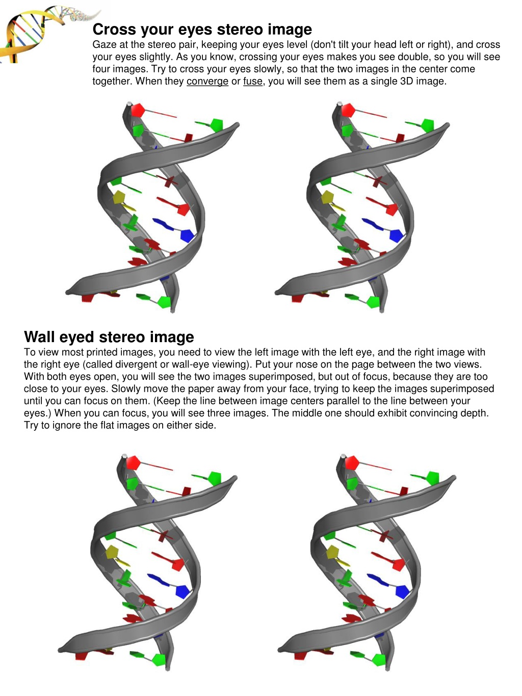
Cross your eyes stereo image Gaze at the stereo pair, keeping your eyes level (don't tilt your head left or right), and cross your eyes slightly. As you know, crossing your eyes makes you see double, so you will see four images. Try to cross your eyes slowly, so that the two images in the center come together. When they converge or fuse, you will see them as a single 3D image. Wall eyed stereo image To view most printed images, you need to view the left image with the left eye, and the right image with the right eye (called divergent or wall-eye viewing). Put your nose on the page between the two views. With both eyes open, you will see the two images superimposed, but out of focus, because they are too close to your eyes. Slowly move the paper away from your face, trying to keep the images superimposed until you can focus on them. (Keep the line between image centers parallel to the line between your eyes.) When you can focus, you will see three images. The middle one should exhibit convincing depth. Try to ignore the flat images on either side.
Identify and label on your handouts. Major Groove? Minor Groove? Phosphate Backbone? Stacked Bases?
This is Deoxyribose 1. Label the carbon atoms? 2. Where is the nucleotide base bound? 3. Where does the phosphate group of the next nucleotide bind? Fill in the blanks?
Draw in the Hydrogen-bonds 2 H-bonds 3 H-bonds Highlight the area's that interact specifically with a DNA binding proteins Q - what part of the DNA does the protein interact with?