Biochemical Quality Control Processes in Biotechnology
Quality control in biotechnology involves monitoring and regulating the levels of various components and impurities throughout the production process. Common methods of removal or purification include chromatography, filtration, and extraction. Scale-up considerations include changes in utility requirements, equipment setup, and process automation. Biochemical analysis techniques such as HPLC, IEF, ELISA, and SDS-PAGE are used for quality assessment during downstream processing of proteins. ELISAs are valuable for determining protein concentration and activity.

Download Presentation

Please find below an Image/Link to download the presentation.
The content on the website is provided AS IS for your information and personal use only. It may not be sold, licensed, or shared on other websites without obtaining consent from the author. Download presentation by click this link. If you encounter any issues during the download, it is possible that the publisher has removed the file from their server.
E N D
Presentation Transcript
Common Process Compounds and Methods of Removal or Purification Component Culture Harvest Level Final Product Level Conventional Method Therapeutic Antibody 0.1-1.5 g/l 1-10 g/l UF/Cromatography Isoforms Various Monomer Chromatography Serum and host proteins 0.1-3.0 g/l < 0.1-10 mg/l Chromatography Cell debris and colloids 106/ml None MF Bacterial pathogens Various <10-6/dose MF Virus pathogens Various <10-6/dose (12 LRV) virus filtration DNA 1 mg/l 10 ng/dose Chromatography Endotoxins Various <0.25 EU/ml Chromatography Lipids, surfactants 0-1 g/l <0.1-10 mg/l Chromatography Buffer Growth media Stability media UF Extractables/leachables Various <0.1-10 mg/l UF/ Chromatography Purification reagents Various <0.1-10mg/l UF
What Will Change During Scale-up? Process Development Considerations Utility requirements Water requirement Cleaning/Sanitizing solution requirements Buffer prep Number of steps in cell culture scale up Harvest techniques Column packing; distribution of introduced liquid at large columns Equipment bubble trap Automation of process Data collection Sample load
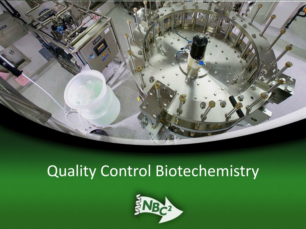
Quality Control Biochemistry HPLC (High Pressure Liquid Chromatography) IEF (Isoelectric Focusing) ELISA (Enzyme-Linked Immunosorbent Assay) SDS-PAGE (Sodium Dodecyl Sulfate-Polyacrylamide Gel Electrophoresis)
Quality Control Biochemistry: ELISAs and SDS-PAGE Each of these methods is important in the Downstream Processing of the Protein of Interest: IEF (Isoelectric Focusing): Use an SDS-PAGE gel box (or CE = capillary electrophoresis) to determine the pI or the pH at which the protein of interest is neutral. ELISAs: Use antibody reagents and a microtitre plate reader to determine the concentration and/or the activity of a protein of interest. SDS-PAGE: Use acrylamide gel electrophoresis to separate proteins according to molecular weight (a single band indicates purity if validated to do so).
ELISAs There are several types of ELISAs including direct (sandwich), indirect, competitive and activity ELISAs. ELISAs are read on a microtitre plate reader which is a mini-spectrophotometer that determines the absorption or transmission of a beam of light of a particular wave length passing through a solution of the protein of interest. Using standards to generate a standard curve, one can determine the concentration of the protein of interest in a sample.
HSA ELISA Results Spring 2009 Data HSA Standard Curve Concentration ng/ml OD 1.4 y = 0.0024x + 0.2992 R = 0.7191 1.2 0 0.071 1 6.25 0.169 25 0.426 0.8 OD OD 100 0.951 0.6 Linear (OD) 400 1.156 0.4 Sample 1 1.320 0.2 Sample 2 1.290 0 Sample 3 1.290 0 100 200 300 400 500 Concentration in ng/ml
ELISA Equipment Multi-Channel Pipettor Microtitre Plate Reader
ELISA Process To make an ELISA, one must utilize antibodies to the protein of interest. The first antibody recognizes the protein of interest. The second antibody recognizes another epitope on the protein of interest and carries an enzyme that will be used to quantify the protein of interest.
ELISA Process Colorimetric Reaction Colorless substrate Colored product TMB
ELISA Continued ELISA = Antibody Sandwich
Antibodies as Reagents ELISAS are Immunoassays which use an antibody (Ab) to detect and quantify substances Ab are extremely specific ADVANTAGE Ab can not be detected, need a marker: Radioactive labels (RIA) Enzymes (EIA) Horseradish Peroxidase; Alkaline Phosphatase Fluorescent Tag (FIA) Chemiluminescencent Tag
ELISA Animation The animation may be found at: usmlemd.wordpress.com/2007/06/12/elisa- test/
SDS-PAGE SDS-PAGE Gel Box SDS-PAGE Overview SDS Polyacrylamide gels (SDS-PAGE) are called denaturing gels because they contain sodium dodecyl sulfate (SDS), an ionic detergent that binds to the amino acid residues in the proteins. Due to its ionic properties, SDS confers a net negative charge on all the proteins, overcoming any intrinsic charge; in this way the proteins uniformly migrate toward the positive electrode. SDS also disrupts the secondary and tertiary structure of the proteins, essentially destroying their globular configuration and making them into linear molecules that then migrate in the electric field on the basis of their size. PAGE is a very powerful technique because even small differences in molecular weights produce distinguishable bands on a gel.
SDS-PAGE Continued Electrophoresis SDS-PAGE separate proteins based on molecular weight Isoelectric Focusing identify the pH at which a protein carries no net charge
SDS-PAGE Continued Sodium Dodecyl Sulfate - Polyacrylamide Gel Electrophoresis developed by Laemmli (1970)
SDS-PAGE Continued characterize (MW) quantify (densitometry) determine other proteins in a sample step in Western blot (used to identify)
SDS-PAGE Continued How to Detect Proteins? Coomassie Blue Stain (0.1 ug) Silver Stain (2 ng) How to Quantify Proteins? Densitometry
Molecular Weight Determination Run SDS PAGE with known standards (MW markers) Graph Measure distance unknown protein traveled Compare on standard curve
A280 Tryphophan Phenylalanine Tyrosine ALL ABSORB LIGHT AT 280 nm Crude, not necessarily quantitative Same amount of protein will show different A280 depending on amount of above amino acids
Bradford Assay SECOND MOST CITED PAPER IN SCIENCE JOURNALS Bradford, M. M. (1976) A Rapid and Sensitive Method for the Quantitation of Microgram Quantities of Protein Utilizing the Principle of Protein-Dye Binding. Anal. Biochem. 72 72:248-254. Coomassie Brilliant Blue G Dye
Protein Characterization Methods CHARACTERISTIC PURITY METHOD SDS-PAGE, 2D ELECTROPHORESIS, IEF, HPLC, MS, CAPILLARY ELECTROPHORESIS MOLECULAR WEIGHT SDS-PAGE, GEL FILTRATION CHROMATOGRAPHY, MS, ANALYTICAL ULTRACENTRIFUGATION MUCH VARIETY AA COMPOSITION, PEPTIDE MAPPING, N- TERMINAL SEQUENCING, COMPLETE AMINO ACID SEQUENCING X-RAY CRYSTALLOGRAPHY, NMR, ANALYTICAL ULTRACENTRIFUGATION, FLUORESCENCE SPECTROSCOPY DEPENDS ON TYPE OF MODIFICATION FUNCTION PRIMARY STRUCTURE SECONDARY, TERTIARY, QUARTENARY STRUCTURE POST-TRANSLATIONAL MODIFICATION
Quality Control Biochemistry: Some Additional Techniques Mass Sepctrometry Amino Acid Sequencing X-ray Crystallography Nuclear Magnetic Resonance
Mass Spectrometry Molecular Weight determination More accurate than SDS PAGE Less protein needed for analysis than SDS PAGE
Amino Acid Sequencing Edman Degradation
Amino Acid Composition Treat with HCl (hydrolysis) Separate individual aa by ion exchange chromatography Analyse with HPLC
3D Structure Determination X Ray Crystallography need to crystallize (Difficult) NMR small proteins 25kD
X-ray Crystallography X ray diffraction beam of x rays directed at protein (incident) beam is diffracted by electrons of atoms in protein (scattered) these beams hit a film detector computer analysis to create electron density map
Nuclear Magnetic Resonance uses radio frequency pulses energy is absorbed such that electrons move from ground to excited states atomic nuclei spin - create their own magnetic field emit radiation - differs based on atoms or chemical groups


")