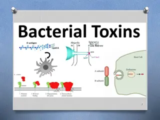
Microbiology Biochemical Tests for Bacterial Identification

Learn about important biochemical tests used in medical microbiology, including the Coagulase test for Staphylococcus aureus, the Catalase test for enzyme detection, and the Oxidase test for electron transport chain identification. Detailed methods and result interpretations are provided for each test.

Uploaded on Sep 17, 2024 | 0 Views
Download Presentation

Please find below an Image/Link to download the presentation.
The content on the website is provided AS IS for your information and personal use only. It may not be sold, licensed, or shared on other websites without obtaining consent from the author. Download presentation by click this link. If you encounter any issues during the download, it is possible that the publisher has removed the file from their server.
E N D
Presentation Transcript
Al-Mustaqbal University college 2 st Class, 1 st Semester Department of pharmacy Medical microbiology Lab 6: Biochemical test. Asst. Lec. Mariam A. Ali
1-Coagulase test Coagulase is an enzyme that clots blood plasma. This test is performed on Gram- positive, catalase positive species to identify the coagulase positiveStaphylococcus aureus. Coagulase is a virulence factor of S. aureus. The formation of clot around an infection caused by this bacteria likely protects it from phagocytosis. This test differentiates Staphylococcus aureus from other coagulase negative Staphylococcus species. Method Suspend one colony from the suspected pure culture in 0.5 ml of plasma from man. Incubate at 37 C. Read the test after 4 h. If the result is negative Positive reaction if the plasma coagulates and the coagulate is stable. Negative reaction if the plasma does not coagulate.
2-Catalase test This test is used to identify organisms that produce the enzyme, catalase. This enzyme detoxifies hydrogen peroxide by breaking it down into water and oxygen gas. The bubbles resulting from production of oxygen gas clearly indicate a catalase positive result. The sample on the right below is catalase positive. TheStaphylococcus spp. and the Micrococcus spp. are catalase positive. The Streptococcus and Enterococcus spp. are catalase negative.
Method Spread the bacteria on an agar plate and incubate the plate over night (18-24 h) under appropriate conditions. Collect bacteria from one colony with a sterile inoculating loop (of plastic or platinum) and apply the bacteria on a microscope slide. If the bacteria are collected from a blood agar plate, one has to avoid contamination of agar, because hemoglobin also contains heme groups which can cause a false positive reaction. Add one drop of 3% H2O2to the bacteria and observe the suspension. Positive test resultant: Gas formation (O2) in the form bubbles shows that the bacterium has a catalase. Negative test resultant: No gas formation.
3-Oxidase Test This test is used to identify microorganisms containing the enzyme cytochrome oxidase (important in the electron transport chain). It is commonly used to distinguish between oxidase negative Enterobacteriaceae and oxidase positive Pseudomadaceae. Cytochrome oxidase transfers electrons from the electron transport chain to oxygen (the final electron acceptor) and reduces it to water. In the oxidase test, artificial electron donors and acceptors are provided. When the electron donor is oxidized by cytochrome oxidase it turns a dark purple. This is considered a positive result. In the picture below the organism on the right (Pseudomonas aeruginosa) is oxidase positive.
Method Apply two drops of the oxidase reagent onto a piece of filter paper. Transfer bacteria from one colony with a plastic or platinum loop onto the spot with the oxidase reagent. The colonies should have been incubated at the appropriate temperature for 18-24 h . Positive test resultat: Dark blue-purple colour change within 10-30 sec. Negative test resultat: No colour change or colour change after more than 30 sec.
4- Simmons Citrate Agar This is a defined medium used to determine if an organism can use citrate as its sole carbon source. It is often used to differentiate between members ofEnterobacteriaceae. In organisms capable of utilizing citrate as a carbon source, the enzyme citrase hydrolyzes citrate into oxaoloacetic acid and acetic acid. The oxaloacetic acid is then hydrolyzed into pyruvic acid and CO2. If CO2is produced, it reacts with components of the medium to produce an alkaline compound (e.g. Na2CO3). The alkaline pH turns the pH indicator (bromthymol blue) from green to blue. This is a positive result (the tube on the right is citrate positive). Klebsiella pneumoniae and Proteus mirabilis are examples of citrate positive organisms. Escherichia coli and Shigella dysenteriae are citrate negative.
Method Inoculate a tube containing citrate medium with a small amount of bacteria. It is also possible to streak or perform a deep inoculation into "Simmons citrate tube". Incubate at 30-37 C during 24-48 h. Positive test result: growth in citrate medium or growth with color change to blue in Simmon's citrate tube. Negative test result: no growth in citrate medium and no color change (still green color) in Simmon's citrate tube.
5- Triple Sugar Iron Agar (TSI) and Kliger's Iron Agar (KIA) General determine if bacteria can ferment glucose and/or lactose and if they can produce hydrogen sulfide or other gases. In addition, TSI detects the ability to ferment sucrose. These characteristics various Enterobacteriacae, including Salmonella and Shigella, which are intestinal pathogens. Composition of Triple Sugar Iron Agar (TSI) Lactose, Sucrose and Glucose in the concentration of 10:10:1 (i.e. 10 part Lactose (1%), 10 part Sucrose (1%) and 1 part Glucose (0.1%)). TSI is similar to Kligler s iron agar (KIA),except that Kligler s iron agar contains only two carbohydrates: glucose (0.1%) and lactose (1%). 0.1% Glucose: If only glucose is fermented, only enough acid is produced to turn the butt yellow. The slant will remain red help distinguish
1.0 % lactose/1.0% sucrose: a large amount of acid turns both butt and slant yellow, thus indicating the ability of the culture to ferment either lactose or sucrose. Iron: Ferrous sulfate: Indicator of H2S formation Phenol red: Indicator of acidification (It is yellow in acidic condition and red under alkaline conditions). It also contains Peptone which acts as source of nitrogen. (Remember that whenever peptone is utilized under aerobic condition ammonia is produced) Procedure for Triple Sugar Iron Agar (TSI) Test With a sterilized straight inoculation needle touch the top of a well-isolated colony Inoculate TSI Agar by first stabbing through the center of the medium to the bottom of the tube and then streaking on the surface of the agar slant. Leave the cap on loosely and incubate the tube at 35 C in ambient air for 18 to 24 hours.
So the expected results of TSI Agar test are: Triple Sugar Iron Agar Test Results: Alkaline slant/Alkaline butt (K/K) i.e. Red/Red = glucose, lactose and sucrose non- fermented Alkaline slant/acidic butt (K/A); Red/Yellow = glucose fermentation only, gas (+ or -), H2s (+ or -) Acidic slant/acidic butt (A/A); Yellow/Yellow = glucose, lactose and/or sucrose fermented gas (+ or -), H2s (+ or -). UseThe test can be used for differentiation of, among Enterrococcus bacteria and other bacteria such as Campylobacter spp.
6-Indole test General Bacteria, which express the enzyme tryptophanase can hydrolyze the amino acid tryptophan to indole, pyruvic acid and ammonia. Presence of indole can be shown by means of Kov c's reagent in the so-called indole test. Method Suspend one colony from a pure culture of the bacterium to be investigated, in a suitable medium (for instance LTLSB or tryptophan medium). Incubate the medium at 37 C during 20-28 h. Add a few drops of Kov c's reagent. Positive test result: The indole reagent change colour to ring red. Negative test result: The indole reagent remains pale yellow. Use Confirmation of Brachyspira spp. in combination with other tests of suspected coli-strains. Typing (species determination) E.
7-Mixed acid fermentation (Methyl red) General Some bacteria can ferment glucose to a mixture of the following organic acids: formic acid, acetic acid and lactic acid. This is called mixed acid fermentation and it causes highly decreased pH in the medium. Mixed acid fermentation can, therefore, be detected by addition of the pH indicator methyl red (MR). The test method is sometimes called the MR test. Method Suspend a bacterial colony from a pure culture in the MR/VP medium. Incubate at 30 C during 3 days or at 37 C during 48 h. Add 2-3 drops of a solution of methyl red. Positive test result: red colour change Negative test result: no colour change. Use Members of the family Enterobacteriaceae sometimes have mixed acid fermentation.
8-Urease test This test is used to identify bacteria capable of hydrolyzing urea using the enzyme urease. It is commonly used to distinguish the genus Proteus from other enteric bacteria. The hydrolysis of urea forms the weak base, ammonia, as one of its products. This weak base raises the pH of the media above 8.4 and the pH indicator, phenol red, turns from yellow to pink. Proteus mirabilis is a rapid hydrolyzer of urea (center tube pictured here). The tube on the far right was inoculated with a urease negative organism and the tube on the far left was uninoculated.
Urea Broth method: Sterilize the loop in the blue flame of the Bunsen burner till red hot until it is red-hot and then allowed to cool. Take out a loopful organism from the broth culture tube with the cooled loop aseptically. Again flame the neck of the tube and replace the tube in the test tube rack. Take a sterile urea broth tube, remove the cap and flame the neck of the tube. Inoculate the urea broth with the inoculation loop containing the organism from broth culture. Again flame the neck of the urea tube and place it in the test tube rack. Incubate for 24-48 hours at 37 C. Obtain the broths from the incubator and observe the colour.
9-Voges-Proskauer (VP) test General The VP test shows if the bacterium has butanediol fermentation and can split glucose to acetoin via pyruvat and further to 2,3-butanediol according to: 2 pyruvate + NADH --> 2CO2+ 2,3-butanediol. If KOH (potassium hydroxide) is added, acetoin will be converted to diacetyl (= 2,3-butanedione), which reacts with alpha-naphtol and forms a pink complex. Method Suspend one colony from the pure culture, which is to be investigated, in VP/MR medium. Incubate at 30-37 C during 24-48 h. Add 0.2 ml of 40% KOH and then 0.6 ml of alpha-naphtol solution.
Positive test result: color change to pink. Negative test result: no color change. Voges-Proskauer test, where the tube to the right shows a positive test result. Use Klebsiella spp. and Enterobacter spp. has the capacity to perform butanediole fermentation in contrast to Escherichia coli, Salmonella spp. and Shigella spp.


 and Kliger's Iron Agar (KIA)")


")


 test")