Understanding Gestational Trophoblastic Disease: Hydatidiform Mole & Choriocarcinoma
Gestational trophoblastic disease encompasses hydatidiform mole, invasive mole, and choriocarcinoma. This condition involves abnormal fertilization of the oocyte, resulting in various presentations including abnormal growth of the uterus, severe nausea, vaginal bleeding, and symptoms resembling hyperthyroidism or preeclampsia. Early detection and management are crucial to prevent potential complications and ensure optimal outcomes.
- Gestational trophoblastic disease
- Hydatidiform mole
- Choriocarcinoma
- Molar pregnancy
- Pregnancy complications

Download Presentation

Please find below an Image/Link to download the presentation.
The content on the website is provided AS IS for your information and personal use only. It may not be sold, licensed, or shared on other websites without obtaining consent from the author. Download presentation by click this link. If you encounter any issues during the download, it is possible that the publisher has removed the file from their server.
E N D
Presentation Transcript
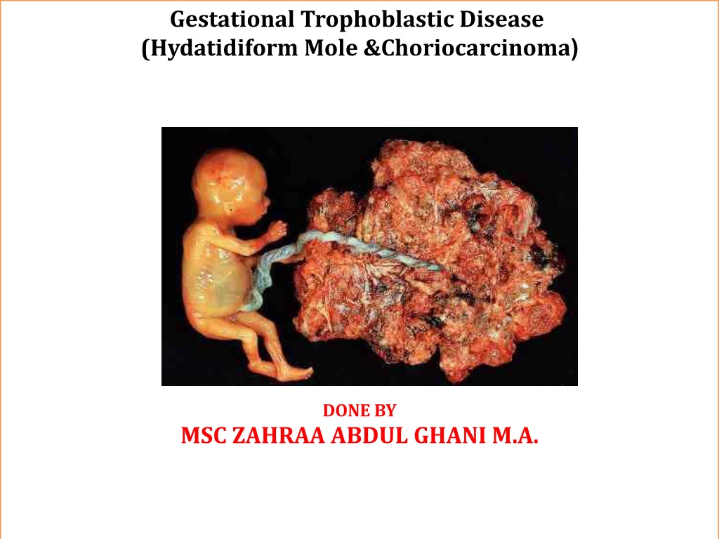
Gestational Trophoblastic Disease (Hydatidiform Mole &Choriocarcinoma ( DONE BY MSC ZAHRAA ABDUL GHANI M.A.
Case study A 36-year-old woman presents with vaginal bleeding at 8 weeks 3 days gestation. She has never been pregnant before. Bright red spotting commenced 7 days ago, which she thought was normal in early pregnancy. However since then the bleeding is now almost as heavy as a period. There are no clots. She has no abdominal pain. Systemically she has felt nausea for 3 weeks and has vomited occasionally. Examination The heart rate is 68/min and blood pressure is 108/70 mmHg. The abdomen is soft and non-tender. Speculum reveals a normal closed cervix with a small amount of fresh blood coming from the cervical canal. Bimanually the uterus feels bulky and soft, approximately 10 weeks in size. There is no cervical excitation or adnexal tenderness. Urinary pregnancy test: positive Figure 46.1 shows the transvaginal ultrasound findings.
Gestational trophoblastic disease is a spectrum of disorders that includes hydatidiform mole, invasive mole, and choriocarcinoma. Causes HM, or molar pregnancy, results from abnormal fertilization of the oocyte (egg). It results in an abnormal fetus. The placenta grows normally with little or no growth of the fetal tissue. The placental tissue forms a mass in the uterus. On ultrasound this mass often has a grape-like appearance, as it contains many small cysts. Chance of mole formation is higher in older women. A history of mole in earlier years is also a risk factor. Molar pregnancy can be of 2 types: -Partial molar pregnancy. There is an abnormal placenta and some fetal development. - Complete molar pregnancy. There is an abnormal placenta and no fetus. Partial moles tend to follow a benign course, while complete moles have a greater tendency to become choriocarcinomas.
Symptoms Symptoms of a molar pregnancy may include: 1- Abnormal growth of the uterus, either bigger or smaller than usual 2- Severe nausea and vomiting 3- Vaginal bleeding during the first 3 months of pregnancy 4-Symptoms of hyperthyroidism, including heat intolerance, loose stools, rapid heart rate, restlessness or nervousness, warm and moist skin, trembling hands, or unexplained weight loss 5-Bilaterally enlarged cystic ovaries are sometimes palapable ,as a result of ovarien hyperstimulation due to excess of hCG. 6-Symptoms similar to preeclampsia that occur in the first trimester or early second trimester, including high blood pressure and swelling in the feet, ankles, and legs (this is almost always a sign of a hydatidiform mole, because preeclampsia is extremely rare this early in a normal pregnancy)
Choriocarcinoma may be manifested by continued or recurrent uterine bleeding after evacuation of a mole or following delivery, abortion, or ectopic pregnancy. The presence of an ulcerative vaginal tumor, pelvic mass, or evidence of distant metastatic tumor may be the presenting observation. The diagnosis is established by pathologic examination of curettings or by biopsy.
Tests done may include: 1-hCG (quantitative levels) blood test 2-Abdominal or vaginal ultrasound of the pelvis 3-Chest x-ray 4-CT or MRI of the abdomen (imaging tests) 5-Complete blood count (CBC) 6-Blood clotting tests 7-Kidney and liver function tests Laboratory findings A serum hCG( above 40,000 mU/mL ) or a urinary hCG value in excess of 100,000 units/24 h increases the likelihood of hydatidiform mole.
Imaging Ultrasound has virtually replaced all other means of preoperative diagnosis of hydatidiformmole. A pregnancy ultrasound will show a snowstorm appearance with an abnormal placenta, with or without some development of a baby.A preoperative chest film is indicated to rule out pulmonary metastases of trophoblast. Treatment The uterus should be emptied as soon as the diagnosis of hydatidiformmole, removal of the abnormal tissue with a dilation and curettage (D & C) will most likely be suggested. D & C may also be done using suction. This is called suction aspiration (The method uses a suction cup to remove contents from the uterus). A hysterectomy (surgery to remove the uterus) may be an option for older women who DO NOT wish to become pregnant in the future. Sometimes a partial molar pregnancy can continue. A woman may choose to continue her pregnancy in the hope of having a successful birth and delivery. However, these are very high-risk pregnancies. Risks may include bleeding, problems with blood pressure, and premature delivery (having the baby before it is fully developed). In rare cases, the fetus is genetically normal. Women need to completely discuss the risks with their provider before continuing the pregnancy.
Ovarian cysts should not be resected nor ovaries removed; spontaneous regression of theca lutein cysts will occur with elimination of the mole. If malignant tissue is discovered at surgery or during the follow-up examination, chemotherapy is indicated. Thyrotoxicosis indistinguishable clinically from that of thyroid origin may occur. While hCG usually has minimal TSH-like activity, the very high hCG levels associated with moles result in the release of T3 and T4 and cause hyperthyroidism. Patientsthyrotoxicon this basis should be stabilized with (B- blockers prior to induction of anesthesia for their surgical evacuation. Surgical removal of the mole promptly corrects the thyroid overactivity. Follow up: After treatment, the hCG level should be followed. It is important to avoid another pregnancy and to use a reliable contraceptive for 6 to 12 months after treatment for a molar pregnancy. This time allows for accurate testing to be sure that the abnormal tissue does not grow back. Women who get pregnant too soon after a molar pregnancy are at high risk of having another molar pregnancy.