Ribosomes: Structure and Function


RIBOSOMES
INTRODUCTION
The ribosomes are small, dense, rounded and granular particles of the rib
nucleoprotein.
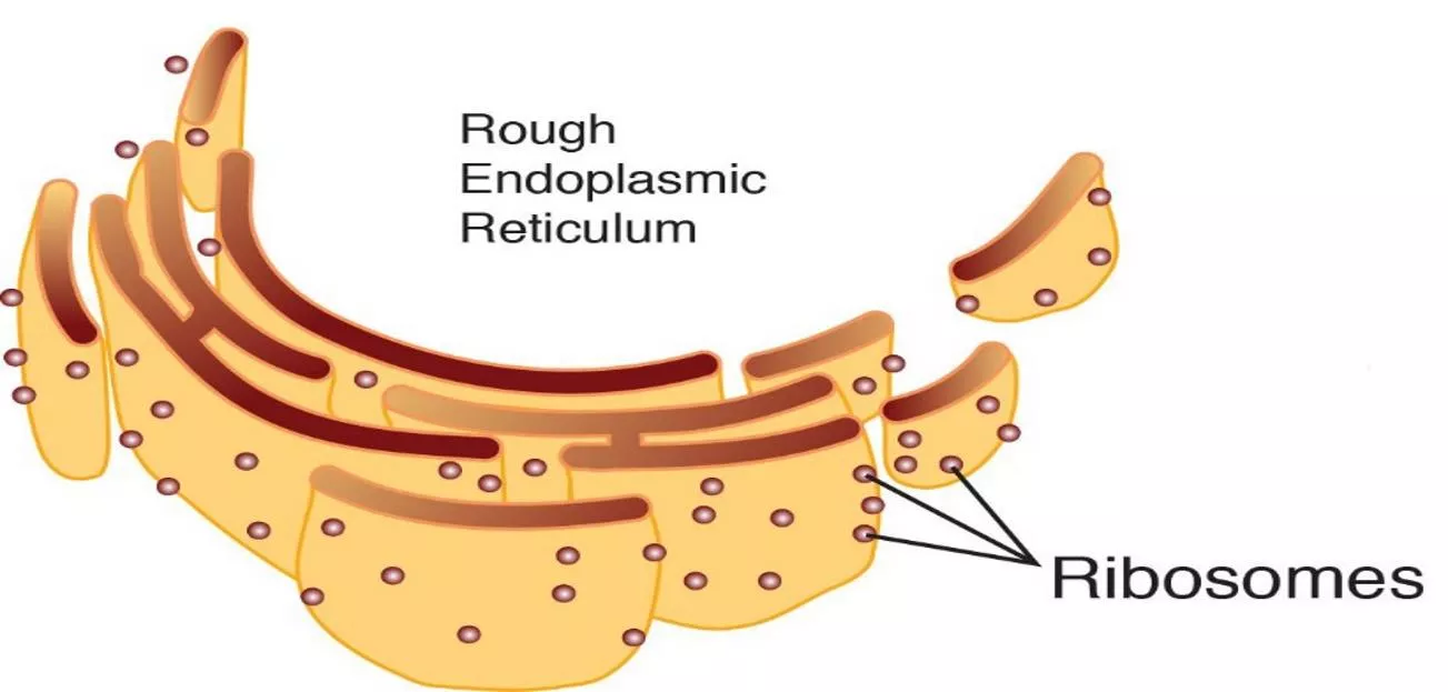
They occur either freely in the matrix of mitochondria, chloroplast and cytoplasm
or remain attached with the membranes of the endoplasmic reticulum and nucleus.
Ribosomes are remarkable organelles of cell.
They were studied before they were discovered.
In later case the membranes are called 'rough surfaced membranes' (Palade and
porter, 1954), or ergastoplasm or alpha cytomembranes (Sjostrand, 1956, 1962).
The membranes without ribosomes are called 'smooth profiles'.
Mature mammalian erythrocytes have no ribosomes.
.

Ribosomes are of two basic types, 70s and 80s ribosomes.
The 'S' refers to Svedberg units.
MORPHOLOGY
The ribosomes are oblate spheroid structures of 150 to 250 A in diameter.
Each ribosome is porous, hydrated and composed of two subunit is large in size and
has a dome-like shape, while the other ribosomal subunit is smaller in size and occurring
above the large subunit and forming a cap-like structure
.
The 70s ribosome consists of two subunit, viz, 50s and 30s.
The 50s ribosomes subunit is large in size and has the size of 160 A to 180 A.
The 30s ribosomal subunit is smaller in size and occurs above the 50s subunit like cap
.
The 80s ribosome also consists of two subunits, viz, 60s and 40s.
The 60s ribosomal subunit is dome-shaped a large in size.
In the ribosomes which remain attached with the membranes of endoplasmic reticulum
and nucleus, etc., the 60s subunit forming a cap-like structure.
Both the subunit remain separated by a narrow cleft.
The two ribosomal subunits remain united with each other due to high
concentration of the Mg++ions.
When the concentration of Mg++ions reduces in the matrix, both ribosomal
subunits get separated.
Actually in bacterial cells the two subunits are found to occur freely in the
cytoplasm and they unite only during the process of
protein synthesis.
At high concentration of Mg++ions in the matrix, the two ribosomes (called
monosomes
) become associated with each other and known as the
dimer.
Further, during protein synthesis many ribosomes are aggregated due to common
messenger RNA and from the
polyribosomes
or
polysomes.
ULTRA STRUCTURE
Molecular organization and function of ribosomes have been studied more intensively
in prokaryotes than in eukaryotes.
Fine or ultra-structure of 70s ribosomes is very complex.
In it, the RNA and proteins are intertwined and arranged in a complex manner in the
two subunits.
Since the positive protein charges are not sufficient to balance the many negative
charges in the phosphate of the RNA, so ribosomes are strongly negative and bind cations
and basic stains.
Consequently,
negative staining
of ribosomes has led to better understanding of the
fine structure of these or, ganelles.
Recently following two models have been suggested to explain the three-dimensional
structure of prokaryotic or 70s ribosomes:
Stoffler and Wittmann's Model (Quasi-Symmetrical Model,1977)
According to this model the 30s ribosomal subunit has an elongated, slightly bent
prolate shape.
It is a bipartite structure.
A transverse hollow or cleft divides the 30s subunit into two parts, a smaller
head
and larger
body
, giving in the appearance of a telephone receiver or embryo.
In electron microscopy 50s ribosomal subunit showed various shapes depending on
structure seen in different views such as formed-maple leaf, lateral-kidney shaped or rear
view-rounded.
In a frontal view, the 50s subunit appears bilaterally symmetrical and shows three
protuberances arising from a rounded base (maple leaf structure).
of the large subunit.
The 50s subunit is often compared with an armchair, with the
rounded base forming a
vaulted seat
, the central protuberance forming the
back
and the lateral protuberances the
arms
of chair.
A
tunnel
is formed between the hollow of the small subunit and
vaulted seat.
The large subunit consists of a
ridge
, a
central protuberance
and a
stalk
.
The ridge and the central protuberance are separated with the help of a valley.
This completely asymmetrical model of ribosome has been suggested by
James
A. Lake
(1981).
The smaller subunit has a
head
, a
base
and a
platform
.
The platform separates the head form the base by the help of a
cleft
.
This cleft is an important functional region; it is suggested to be the site of codon-
anticode interaction and as a part of binding site for initiation factors of
protein
synthesis
.
Lake's Model (Asymmetrical Model, 1981)
Three Dimensional Model of 80s Ribosome
In spite of the difference in overall sizes (manifested in the greater
molecular weights, sedimentation constants, sizes and numbers of RNAs and
proteins), the cytoplasmic ribosomes
of eukaryotes (80s) are remarkably similar in
morphology to those units of prokaryotes.
As in 30s subunits of prokaryote ribosomes, the 40s ribosomal subunit of
eukaryotes is divided into head and base segments by a transverse groove.
The 60s ribosomal subunit is generally rounder in shape than the small
subunit, although its one side is flattened; this is the side that becomes confluent
with the small subunit during the formation of the monomer or monosome.
Dissociation and reconstitution (self-assembly) of the ribosomes.
To understand the three-dimensional organization of ribosomal proteins in
the ribosomes and also for the investigation of interactions between the molecules of
rRNA and proteins, following classical experiment of dissociation and reconstitution of
Nomura
and
Traub
(1968) can be considered.
This experiment involves to take purified 30s ribosomal subunits, dissociate
them by chemical means into their component RNAs and proteins and then allow them
to reassociate under appropriate ionic conditions.
Dissociation of 30s subunit may be achieved by treatment with four molar
urea and two molar LiCl, Which separate the proteins.
BIOCHEMISTRY
The ribosomes are chemically composed of RNA and proteins as their major
constituents; both occurring approximately in equal pro-portions in smaller as well as
larger subunit.
However, the 70s ribosomes contain more RNA (60 to 40%) than the
proteins (36 to 37%), e.g., the ribosomes of E. coli contain 63% rRNA and 37% protein.
While the 80s ribosomes have 40 to 44% RNA and 60 to 56% proteins; ribosomes of
pea seeding contain 40% RNA and 60% proteins.
There is no lipid content in ribosomes.
Ribosomal RNAs
The 70s ribosomes contain three types of rRNA, viz.,
23S rRNA, 16S rRNA,
5S rRNA.
The 23S and 5S rRNA occur in the large 50S ribosomal subunit, while the
16S rRNA occurs in the smaller 30S ribosomal subunit.
Thus, the 23S rRNA consists of 3300 nucleotides, 16S rRNA contains 1650
nucleotides and 5S rRNA includes 120 nucleotides in it (
Brownlee, 1968; Fellner,
1972).
The 80S ribosomes contain four types of rRNA, viz.,
28S rRNA
(or
25-26 r
RNA
in plants, fungi and protozoa),
18S rRNA, 5S rRNA
and
5.8S rRNA
.
The 28S, 5S and 5.8S rRNAs occur in the larger 60S ribosomal subunit, while
the 18S rRNA occurs in the smaller 40S ribosomal subunit.
About 60 per cent of the rRNA is helical (i.e., double stranded) and contains
paired bases.
Ribosomal Proteins
According to
Nomura
(1968, 1973) and
Garett
and
Wittmann
(1973) each 70S
Ribosome of E. coli is composed of about 55
ribosomal Proteins
.
Out of these 55 proteins, about 21 different molecules have been isolated from the
30S ribosomal subunit, and some 32 to 34 proteins from the 50S ribosomal subunit.
Most of the recent knowledge about the structure of ribosomal proteins has been
achieved by dissociation of ribosomal subunits into their component rRNA and protein
molecules.
When both 50S and 30S ribosomal subunit are dissociated by centrifuge both of
them in a gradient of 5M cesium chloride then there are two inactive core particles (40S and
23, respectively) Which contain the RNA and some proteins called
core proteins (CP)
at the
same time several other proteins-the so-called
spilt proteins (SP)
are released from each
particle.
Metallic Ions
The most important low molecular weight components of ribosomes are the
divalent metallic ions such as Mg++, Ca++ and Mn++.
Different rRNA molecules evidently play a central role in the
catalytic activities of ribosomes in the process of
protein synthesis
.
Various ribosomal proteins have been found to mainly enhance the
catalytic function of the rRNA in the ribosomes.
FUNCTIONS
Ribosomes take part in protein synthesis.
Two or more ribosomes simultaneously engaged in protein synthesis
on the same mRNA strand form polyribosomes.
The ribosome functions as a template, bringing together different
components involved in the synthesis of proteins.
Interaction of the tRNA-amino acid complex with mRNA, which brings
about translation of the genetic code, is coordinated by the ribosomes.
Ribosomes also have a protective function.
The mRNA strand which passes between the two subunits of the ribosomes is
protected from the action of the enzymes of the nucleus (nucleases).
Similarly the nascent polypeptide chains passing through the tunnel or channel
between the subunits are protected against the action of protein-digesting
enzymes
.
Ribosomes, essential organelles in cells, exist in two types - 70s and 80s. They play a crucial role in protein synthesis. Structurally, ribosomes are composed of two subunits held together by Mg++ ions. The ultrastructure of ribosomes, particularly in prokaryotes, is intricate, with RNA and proteins intricately arranged in a complex manner. Ribosomes are negatively charged and bind cations and basic stains, aiding in their visualization and study.

Download Presentation

Please find below an Image/Link to download the presentation.
The content on the website is provided AS IS for your information and personal use only. It may not be sold, licensed, or shared on other websites without obtaining consent from the author.If you encounter any issues during the download, it is possible that the publisher has removed the file from their server.
You are allowed to download the files provided on this website for personal or commercial use, subject to the condition that they are used lawfully. All files are the property of their respective owners.
The content on the website is provided AS IS for your information and personal use only. It may not be sold, licensed, or shared on other websites without obtaining consent from the author.
E N D
Presentation Transcript
RIBOSOMES INTRODUCTION The ribosomes are small, dense, rounded and granular particles of the rib nucleoprotein. They occur either freely in the matrix of mitochondria, chloroplast and cytoplasm or remain attached with the membranes of the endoplasmic reticulum and nucleus. Ribosomes are remarkable organelles of cell. They were studied before they were discovered. In later case the membranes are called 'rough surfaced membranes' (Palade and porter, 1954), or ergastoplasm or alpha cytomembranes (Sjostrand, 1956, 1962). The membranes without ribosomes are called 'smooth profiles'. Mature mammalian erythrocytes have no ribosomes. .
Ribosomes are of two basic types, 70s and 80s ribosomes. The 'S' refers to Svedberg units.
MORPHOLOGY The ribosomes are oblate spheroid structures of 150 to 250 A in diameter. Each ribosome is porous, hydrated and composed of two subunit is large in size and has a dome-like shape, while the other ribosomal subunit is smaller in size and occurring above the large subunit and forming a cap-like structure. The 70s ribosome consists of two subunit, viz, 50s and 30s. The 50s ribosomes subunit is large in size and has the size of 160 A to 180 A. The 30s ribosomal subunit is smaller in size and occurs above the 50s subunit like cap. The 80s ribosome also consists of two subunits, viz, 60s and 40s. The 60s ribosomal subunit is dome-shaped a large in size. In the ribosomes which remain attached with the membranes of endoplasmic reticulum and nucleus, etc., the 60s subunit forming a cap-like structure. Both the subunit remain separated by a narrow cleft.
The two ribosomal subunits remain united with each other due to high concentration of the Mg++ions. When the concentration of Mg++ions reduces in the matrix, both ribosomal subunits get separated. Actually in bacterial cells the two subunits are found to occur freely in the cytoplasm and they unite only during the process of protein synthesis. At high concentration of Mg++ions in the matrix, the two ribosomes (called monosomes) become associated with each other and known as the dimer. Further, during protein synthesis many ribosomes are aggregated due to common messenger RNA and from the polyribosomes or polysomes.
ULTRA STRUCTURE Molecular organization and function of ribosomes have been studied more intensively in prokaryotes than in eukaryotes. Fine or ultra-structure of 70s ribosomes is very complex. In it, the RNA and proteins are intertwined and arranged in a complex manner in the two subunits. Since the positive protein charges are not sufficient to balance the many negative charges in the phosphate of the RNA, so ribosomes are strongly negative and bind cations and basic stains. Consequently, negative staining of ribosomes has led to better understanding of the fine structure of these or, ganelles. Recently following two models have been suggested to explain the three-dimensional structure of prokaryotic or 70s ribosomes:
Stoffler and Wittmann's Model (Quasi-Symmetrical Model,1977) According to this model the 30s ribosomal subunit has an elongated, slightly bent prolate shape. It is a bipartite structure. A transverse hollow or cleft divides the 30s subunit into two parts, a smaller head and larger body, giving in the appearance of a telephone receiver or embryo. In electron microscopy 50s ribosomal subunit showed various shapes depending on structure seen in different views such as formed-maple leaf, lateral-kidney shaped or rear view-rounded. In a frontal view, the 50s subunit appears bilaterally symmetrical and shows three protuberances arising from a rounded base (maple leaf structure). of the large subunit.
The 50s subunit is often compared with an armchair, with the rounded base forming a vaulted seat, the central protuberance forming the back and the lateral protuberances the arms of chair. A tunnel is formed between the hollow of the small subunit and vaulted seat.
Lake's Model (Asymmetrical Model, 1981) This completely asymmetrical model of ribosome has been suggested by James A. Lake (1981). The smaller subunit has a head, a base and a platform. The platform separates the head form the base by the help of a cleft. This cleft is an important functional region; it is suggested to be the site of codon- anticode interaction and as a part of binding site for initiation factors of protein synthesis. The large subunit consists of a ridge, a central protuberance and a stalk. The ridge and the central protuberance are separated with the help of a valley.
Three Dimensional Model of 80s Ribosome In spite of the difference in overall sizes (manifested in the greater molecular weights, sedimentation constants, sizes and numbers of RNAs and proteins), the cytoplasmic ribosomes of eukaryotes (80s) are remarkably similar in morphology to those units of prokaryotes. As in 30s subunits of prokaryote ribosomes, the 40s ribosomal subunit of eukaryotes is divided into head and base segments by a transverse groove. The 60s ribosomal subunit is generally rounder in shape than the small subunit, although its one side is flattened; this is the side that becomes confluent with the small subunit during the formation of the monomer or monosome.
Dissociation and reconstitution (self-assembly) of the ribosomes. To understand the three-dimensional organization of ribosomal proteins in the ribosomes and also for the investigation of interactions between the molecules of rRNA and proteins, following classical experiment of dissociation and reconstitution of Nomura and Traub (1968) can be considered. This experiment involves to take purified 30s ribosomal subunits, dissociate them by chemical means into their component RNAs and proteins and then allow them to reassociate under appropriate ionic conditions. Dissociation of 30s subunit may be achieved by treatment with four molar urea and two molar LiCl, Which separate the proteins.
BIOCHEMISTRY The ribosomes are chemically composed of RNA and proteins as their major constituents; both occurring approximately in equal pro-portions in smaller as well as larger subunit. However, the 70s ribosomes contain more RNA (60 to 40%) than the proteins (36 to 37%), e.g., the ribosomes of E. coli contain 63% rRNA and 37% protein. While the 80s ribosomes have 40 to 44% RNA and 60 to 56% proteins; ribosomes of pea seeding contain 40% RNA and 60% proteins. There is no lipid content in ribosomes.
Ribosomal RNAs The 70s ribosomes contain three types of rRNA, viz., 23S rRNA, 16S rRNA, 5S rRNA. The 23S and 5S rRNA occur in the large 50S ribosomal subunit, while the 16S rRNA occurs in the smaller 30S ribosomal subunit. Thus, the 23S rRNA consists of 3300 nucleotides, 16S rRNA contains 1650 nucleotides and 5S rRNA includes 120 nucleotides in it (Brownlee, 1968; Fellner, 1972). The 80S ribosomes contain four types of rRNA, viz., 28S rRNA (or 25-26 r RNA in plants, fungi and protozoa), 18S rRNA, 5S rRNA and 5.8S rRNA. The 28S, 5S and 5.8S rRNAs occur in the larger 60S ribosomal subunit, while the 18S rRNA occurs in the smaller 40S ribosomal subunit. About 60 per cent of the rRNA is helical (i.e., double stranded) and contains paired bases.
Ribosomal Proteins According to Nomura (1968, 1973) and Garett and Wittmann (1973) each 70S Ribosome of E. coli is composed of about 55 ribosomal Proteins. Out of these 55 proteins, about 21 different molecules have been isolated from the 30S ribosomal subunit, and some 32 to 34 proteins from the 50S ribosomal subunit. Most of the recent knowledge about the structure of ribosomal proteins has been achieved by dissociation of ribosomal subunits into their component rRNA and protein molecules. When both 50S and 30S ribosomal subunit are dissociated by centrifuge both of them in a gradient of 5M cesium chloride then there are two inactive core particles (40S and 23, respectively) Which contain the RNA and some proteins called core proteins (CP) at the same time several other proteins-the so-called spilt proteins (SP) are released from each particle.
Different rRNA molecules evidently play a central role in the catalytic activities of ribosomes in the process of protein synthesis. Various ribosomal proteins have been found to mainly enhance the catalytic function of the rRNA in the ribosomes. Metallic Ions The most important low molecular weight components of ribosomes are the divalent metallic ions such as Mg++, Ca++ and Mn++.
FUNCTIONS Ribosomes take part in protein synthesis. Two or more ribosomes simultaneously engaged in protein synthesis on the same mRNA strand form polyribosomes. The ribosome functions as a template, bringing together different components involved in the synthesis of proteins. Interaction of the tRNA-amino acid complex with mRNA, which brings about translation of the genetic code, is coordinated by the ribosomes. Ribosomes also have a protective function.
The mRNA strand which passes between the two subunits of the ribosomes is protected from the action of the enzymes of the nucleus (nucleases). Similarly the nascent polypeptide chains passing through the tunnel or channel between the subunits are protected against the action of protein-digesting enzymes.


")

")

 of the")