The Nervous System: Structure and Function
The Nervous System
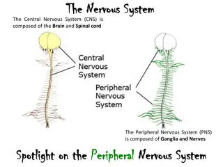
I. General organization of nervous system
A.
CNS
1. brain
2. spinal cord
B. PNS
1. sensory
2. motor
a. Somatic
b. ANS
-sympathetic
-parasympathetic
II. Nervous Supporting Cells - neuroglia
A.
Astrocytes
1.
Connect to
capillaries
2.
Mopping up
chemical
environment of
brain as far as
potassium ions and
neurotransmitters
B. Microglia
•
1. spider-like
phagocytes
•
2. debris, dead
brain cells, bacteria
C. Ependymal cells
•
1. lines cavities in CNS
•
2. beating of cilia moves
cerebrospinal fluid
•
3. fluid nourishes and cushions
CNS
D. Oligodendrocytes
•
1. wrap axons of nerve cells
with fatty layer
•
2. produces myelin sheath
•
3. speeds conduction
E. Glia cells in general
•
1. resemble neurons
•
2. not excitable
•
3. supportive cells
•
4. capable of repeated mitosis
•
5. gliomas-glial tumors
III. Neurons
A. Structure
•
1. cell body
•
2. nissl bodies-rer
•
3. dendrites
•
4. axon
•
5. axon hillock
•
6. axon collateral
•
7. axon terminals
•
8. neurotransmitters
•
9. synaptic cleft
B. Myelin sheath
•
1. functions
•
2. PNS-Schwann cell
•
3. Node of Ranvier
•
4. Can form a pathway for
regrowth of damaged
axon
•
5. multiple sclerosis
C. Neurons classified by function
•
1. afferent
•
2. interneuron
•
3. efferent
•
4. ganglia
•
5. nuclei
D. Neurons classified by structure
•
1. multipolar
•
2. bipolar
•
3. unipolar
IV. Neuron physiology
•
A. Resting membrane
potential
•
B. Action potential-nerve
impulse
C. Propagation of action potential
•
1. diagram on board
•
2. a lot like dominoes
•
3.
http://www.youtube.com/watch?v=7tBWl4GE8rk&NR=1
D. Anatomy of a synapse
•
1. presynaptic membrane
•
2. synaptic cleft
•
3. postsynaptic membrane
•
4. synaptic vesicles
•
5. receptor sites for transmitter
substance
E. Physiology of synapse
•
1. action potential arrives
•
2. Calcium ion channels
open
•
3. synaptic vesicles fuse
with membrane
•
4. transmitter substance
released
•
5. diffusion of transmitter
substance
•
6. binding to receptors
•
7. creates a graded
potential
•
8. may bring postsynaptic
membrane to threshold
•
9. nerve gas-blocks
cholinesterase
F. You tube of synaptic events
•
http://www.youtube.com/watch?v=z3F5dfmQ
3hk
V. Functional Anatomy of the Brain
A. Introduction
•
1. difficult to talk about
•
2. two fistfuls of pinkish/gray
•
3. wrinkled
•
4. consistency of cold
oatmeal
•
5. three pounds
•
6. hugely complex
•
7. four basic regions
–
a. Cerebral hemispheres
–
b. Diencephalon
–
c. Brain stem
–
d. cerebellum
B. Cerebral hemispheres
•
1. most important part
•
2. overshadows
diencephalon and brain
stem
•
3. mushroom cap covers
top of stalk
•
4. gyri
•
5. sulci
•
6. fissures-ie longitudinal
cerebral fissure
7. Lobes of cerebrum
•
a. Frontal lobe
controls mainly motor
function
•
b. Primary motor area
is on the precentral
gyrus -governs
conscious motor
control which can be
mapped
Motor homunculus
c. Motor homunculus
•
-specific regions of
the precentral
gyrus control
specific body parts
•
-finer the
movements, the
more brain area
needed to control
those movements
d. Premotor area
•
-learned
repetitive
tasks
•
Typing,
playing piano
•
Athletes
learn tasks
by visualizing
motions
•
Ingrained in
this area
e. Broca’s area
•
speech center
•
Usually located left cerebral hemisphere
•
Damage here causes inability to speak
8. Other important areas of cerebral hemispheres
•
a. Primary somatic sensory area
•
b. Visual area in occipital lobe
•
c. Complex memory in the temporal lobe
•
d. Note close proximity to olfactory area
•
e. Anterior association area-higher intellectual reasoning and
socially acceptable behavior
9. Sensory homunculus
C. Diencephalon
1.
Thalamus
a. Encloses third vent.
b. Screens incoming
sensory messages
2.
Hypothalamus
a. ANS center for body
temperature and water
balance
b. Regulates pituitary
3.
Epithalamus
a. Pineal gland
b. Choroid plexus
D. Brain stem
•
1. size of thumb
•
2. midbrain
•
3. pons
•
4. medulla
•
5. interchange
for sensory and
motor paths
•
6. nuclei for
respiratory,
blood pressure,
heart rate, RAS
E. Cerebellum
1.
Cauliflower
shape
2.
Controls
balance and
equilibrium
3.
Produces
smooth and
coordinated
muscular
contractions
VI. Protection of the
brain
•
A. Meninges
•
1. dura mater
•
2. arachnoid
•
3. pia mater
•
B. CSF
•
1. produced
choroid plexi
•
2. flow
•
3. functions
•
4. hydrocephalus
The nervous system is a complex network divided into the central nervous system (CNS) and peripheral nervous system (PNS). Neuroglia, or supporting cells, play vital roles in maintaining the health and function of neurons. Neurons, the fundamental units of the nervous system, vary in structure and function. Explore the intricate world of neurons, neuroglia, and the physiological aspects of neuron function.

Uploaded on Sep 18, 2024 | 1 Views
Download Presentation

Please find below an Image/Link to download the presentation.
The content on the website is provided AS IS for your information and personal use only. It may not be sold, licensed, or shared on other websites without obtaining consent from the author.If you encounter any issues during the download, it is possible that the publisher has removed the file from their server.
You are allowed to download the files provided on this website for personal or commercial use, subject to the condition that they are used lawfully. All files are the property of their respective owners.
The content on the website is provided AS IS for your information and personal use only. It may not be sold, licensed, or shared on other websites without obtaining consent from the author.
E N D
Presentation Transcript
I. General organization of nervous system A. CNS 1. brain 2. spinal cord B. PNS 1. sensory 2. motor a. Somatic b. ANS -sympathetic -parasympathetic
II. Nervous Supporting Cells - neuroglia A. Astrocytes 1. Connect to capillaries 2. Mopping up chemical environment of brain as far as potassium ions and neurotransmitters
B. Microglia 1. spider-like phagocytes 2. debris, dead brain cells, bacteria
C. Ependymal cells 1. lines cavities in CNS 2. beating of cilia moves cerebrospinal fluid 3. fluid nourishes and cushions CNS
D. Oligodendrocytes 1. wrap axons of nerve cells with fatty layer 2. produces myelin sheath 3. speeds conduction
E. Glia cells in general 1. resemble neurons 2. not excitable 3. supportive cells 4. capable of repeated mitosis 5. gliomas-glial tumors
III. Neurons A. Structure 1. cell body 2. nissl bodies-rer 3. dendrites 4. axon 5. axon hillock 6. axon collateral 7. axon terminals 8. neurotransmitters 9. synaptic cleft
B. Myelin sheath 1. functions 2. PNS-Schwann cell 3. Node of Ranvier 4. Can form a pathway for regrowth of damaged axon 5. multiple sclerosis
C. Neurons classified by function 1. afferent 2. interneuron 3. efferent 4. ganglia 5. nuclei
D. Neurons classified by structure 1. multipolar 2. bipolar 3. unipolar
IV. Neuron physiology A. Resting membrane potential B. Action potential-nerve impulse
C. Propagation of action potential 1. diagram on board 2. a lot like dominoes 3. http://www.youtube.com/watch?v=7tBWl4GE8rk&NR=1
D. Anatomy of a synapse 1. presynaptic membrane 2. synaptic cleft 3. postsynaptic membrane 4. synaptic vesicles 5. receptor sites for transmitter substance
E. Physiology of synapse 1. action potential arrives 2. Calcium ion channels open 3. synaptic vesicles fuse with membrane 4. transmitter substance released 5. diffusion of transmitter substance 6. binding to receptors 7. creates a graded potential 8. may bring postsynaptic membrane to threshold 9. nerve gas-blocks cholinesterase
F. You tube of synaptic events http://www.youtube.com/watch?v=z3F5dfmQ 3hk
V. Functional Anatomy of the Brain A. Introduction 1. difficult to talk about 2. two fistfuls of pinkish/gray 3. wrinkled 4. consistency of cold oatmeal 5. three pounds 6. hugely complex 7. four basic regions a. Cerebral hemispheres b. Diencephalon c. Brain stem d. cerebellum
B. Cerebral hemispheres 1. most important part 2. overshadows diencephalon and brain stem 3. mushroom cap covers top of stalk 4. gyri 5. sulci 6. fissures-ie longitudinal cerebral fissure
7. Lobes of cerebrum a. Frontal lobe controls mainly motor function b. Primary motor area is on the precentral gyrus -governs conscious motor control which can be mapped
c. Motor homunculus -specific regions of the precentral gyrus control specific body parts -finer the movements, the more brain area needed to control those movements
d. Premotor area -learned repetitive tasks Typing, playing piano Athletes learn tasks by visualizing motions Ingrained in this area
e. Brocas area speech center Usually located left cerebral hemisphere Damage here causes inability to speak
8. Other important areas of cerebral hemispheres a. Primary somatic sensory area b. Visual area in occipital lobe c. Complex memory in the temporal lobe d. Note close proximity to olfactory area e. Anterior association area-higher intellectual reasoning and socially acceptable behavior
C. Diencephalon 1. Thalamus a. Encloses third vent. b. Screens incoming sensory messages 2. Hypothalamus a. ANS center for body temperature and water balance b. Regulates pituitary 3. Epithalamus a. Pineal gland b. Choroid plexus
D. Brain stem 1. size of thumb 2. midbrain 3. pons 4. medulla 5. interchange for sensory and motor paths 6. nuclei for respiratory, blood pressure, heart rate, RAS
E. Cerebellum 1. Cauliflower shape 2. Controls balance and equilibrium 3. Produces smooth and coordinated muscular contractions
VI. Protection of the brain A. Meninges 1. dura mater 2. arachnoid 3. pia mater B. CSF 1. produced choroid plexi 2. flow 3. functions 4. hydrocephalus