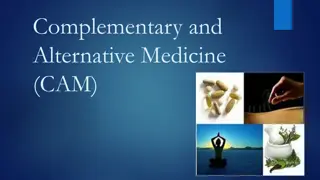
Innovative Uses of 3D Printing in Medicine
Explore the groundbreaking applications of 3D printing in the medical field, from creating custom implants and prosthetics to precise surgical planning and educational models. Dr. Thomas Boland's pioneering work in organ printing and the current process of 3D printing using CT or MRI scans are highlighted. Discover how this technology revolutionizes medical training, patient care, and surgical interventions.

Download Presentation

Please find below an Image/Link to download the presentation.
The content on the website is provided AS IS for your information and personal use only. It may not be sold, licensed, or shared on other websites without obtaining consent from the author.If you encounter any issues during the download, it is possible that the publisher has removed the file from their server.
You are allowed to download the files provided on this website for personal or commercial use, subject to the condition that they are used lawfully. All files are the property of their respective owners.
The content on the website is provided AS IS for your information and personal use only. It may not be sold, licensed, or shared on other websites without obtaining consent from the author.
E N D
Presentation Transcript
January 2016 Narayanan R
Additive manufacturing Biotexture modelling
Thomas Boland Chief Science Officer and co- founder of TeVido BioDevices in association with Clemson University, Dr. Boland s work has been cited more than 800 times. He has received numerous awards. He was the first to retrofit a standard inkjet printer and use cells as ink. By printing layer by layer he could achieve 3D printing of organs by 2003
How is 3D printing done now? Series of successive slices of CT scan or MRI are made These are converted into layers of plastic, ceramic, glass or even tissue They are constructed into graspable 3D digital models
Potential uses of 3D Models in Medicine Exact simulation and better understanding of structural pathology & anatomy Appreciation of tissue planes for dissection Demonstration & Teaching
Potential uses Models of fetus in utero to assess anomalies Depiction of fascial planes for dissection & repair (eg. Perineal repair) Depiction of injuries (eg. Ureteric injury) Precise models of lesions, tumors etc Pre operative surgical planning Education of patients ) hematomas & tumours
Custom made for perfect fit Dentures Vaginal dilators Hearing aids Cartilage & Bones Implants & stents Prosthetic limbs Organs for transplantation Complex regeneration of damaged tissues & organs Surgical instruments
PINNA SKIN SKIN HEARING AIDS PROSTHESIS PROSTHESIS
HEART VALVES HEART VALVES Kidney KIDNEY
Paramedics on Facebook shared Medical Addicts(Medicine, Surgery, Obstetrics and Gynaecology)'s photoLooks completely realistic, doesn't it? This is actually a SYNTHETIC human cadaver created by SynDaver Labs. It's designed to help medical students and professionals when there's a shortage of donated human bodies. Life-like replicas of complete cadavers made through 3D printing help in anatomy dissection
Blind mother sees her son thanks to 3D printing
3D Printing Combined With Sonogram Revolutionary Breakthrough In Obstetrics: 3D Printing Combined With Sonogram: Depiction Of Congenital defects
Case study Broad ligament hematoma due to extension of uterine angle following difficult emergency caesarean section performed after full dilatation of cervix -Yoong W et al, Royal college of Ob Gyn 2015
B B A MRI data set first transferred to a dedicated workstation. A. Broad ligament haematoma (10x10 cm) B. Uterus
MRI data set of a broad ligament haematoma
a (a)Using rapid prototyping, (a ) 3D model is printed showing haematoma (A) and its relation to the uterus (B) A B (b) Lateral view of 3D model showing broad ligament haematoma (A) and its relation with the uterus (B). b
3D Model of vasculature in Conjoined Twins
The Mata Twins at birth Knatalye Hope and Adeline Faith
Many internal organs were shared (chest wall, lungs, pericardial sac, diaphragm, liver, intestines, colon and pelvis). Using 3D printing the twin organs were printed showing all the parts in detail which helped in separation of twins. combination of volumetric CT, 3-D modeling, and 3-D printing will become a standard part of preparation for surgical separation of conjoined twins in future -Dr Darrel Cass, Texas Childrens Hospital
Cancer Cervix Customised 3D implant for intracavitary brachytherapy: Radioactive source can be precisely guided through the channels
Even if you happen to lose your face See what 3D printing can do!
Tokyo researchers have successfully taken goat fetuses and put them in synthetic wombs for continued gestation! The fetuses have stayed in the environment for up to three weeks, but Yoshinori Kuwabara, chairman of the Department of Obstetrics and Gynecology at Juntendo University in Tokyo, has said that this can be extended or applied to human beings! Ectogenesis was a term created by British scientist J. B. S. Haldane in 1924. #science #biology Ectogenesis Japanese researchers have grown a goat embryo inside a 3D printed uterus