Digestive System
Digestive System
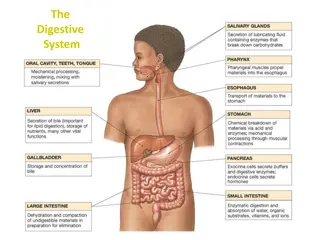
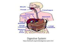
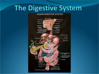
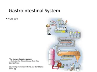
Organs of the alimentary Canal
•
Mouth
•
Pharynx
•
Esophagus
•
Stomach
•
Small intestine
•
Large intestine
* Salivary glands, liver, pancreas and gall bladder are accessory organs
•
Main functions:
1.
digestion
•
Food is broken down:
•
Mechanically (chewing, bowl
movements)
•
Chemically (enzymes, acids)
2. Absorption
•
Food is absorbed from small
intestine
blood
body
3. Excretion
Mouth
•
Food enters DT through the oral cavity (lined
with mucous membranes)
•
Lips (labia)
protect its opening
•
Cheeks
form the lateral walls
•
Hard palate
forms anterior roof
•
Soft palate
forms posterior roof
•
Uvula:
fingerlike projection of soft palate
•
Tongue
occupies the floor of the mouth
•
Lingual frenulum (fold of mucous membrane)
secures tongue to floor of moth and limits its
posterior movements
Mouth cont.
•
Both types of digestion
mechanical and chemical
•
Salivary glands secrete
enzymes that break
carbohydrates
•
Teeth help break down food
by chewing
Mouth (salivary glands)
•
Three pairs of salivary glands secrete saliva
into the mouth
•
Parotid glands:
•
Located close to our ears
•
Submandibular glands
•
below the mandible
•
Sublingual glands
•
Under the tongue
•
Saliva is a mixture of mucus and serous fluids
•
Mucus moistens +helps bind food together
into a mass called bolus which eases
swallowing
•
The serous portion contains an enzyme,
amylase which begin starch digestion
•
Saliva also contains Abs which inhibit bacteria
Pharynx and Esophagus
•
Food passes from oral cavity
to the pharynx
esophagus
stomach
•
Esophagus is lined with
mucous membranes to allow
food to pass easily
•
Epiglottis flips over the
trachea to keep food from
entering the trachea
Pharynx and Esophagus cont.
Swallowing:
•
A bolus of food is pressed backward into the pharynx by the tongue.
This is the only step that is voluntary - the remaining steps occur by
reflex.
•
Once the bolus reaches the pharynx several actions are initiated, the
epiglottis swings backward to cover the larynx.
•
The tongue presses backward and a peristaltic contraction in the
pharynx propels the bolus into the esophagus, where the actual act of
swallowing takes place.
Esophagus Histology
•
Mucosa:
•
Innermost, moist layer that lines the cavity (lumen) of the organ
•
Consists of surface epithelium (stratified squamous)+ small amount of CT (lamina
propria) +SM layer
•
*beyond the esophagus all epithelium is simple columnar
•
Sub mucosa:
•
covers the mucosa
•
Soft C.T layer contains BV, nerve endings, lymphatics
•
Muscularis externa:
•
2 Layer of SM cells (longitudinal muscle layer + circular muscle layer)
•
Serosa:
•
Outer most layer
•
Single layer of serous fluid producing cells- visceral peritoneum- covers the external
surface of the esophagus
•
Mesentery is a membrane extension of the visceral peritoneum
The digestive system consists of the alimentary canal and accessory organs like the salivary glands, liver, pancreas, and gall bladder. It functions to break down food mechanically and chemically, absorb nutrients in the small intestine, and excrete waste. The process starts in the mouth, where food enters through the oral cavity, aided by various organs like the tongue, salivary glands, pharynx, and esophagus, progressing through the entire digestive tract. Understanding these organs and their functions is vital for optimal health.

Download Presentation

Please find below an Image/Link to download the presentation.
The content on the website is provided AS IS for your information and personal use only. It may not be sold, licensed, or shared on other websites without obtaining consent from the author.If you encounter any issues during the download, it is possible that the publisher has removed the file from their server.
You are allowed to download the files provided on this website for personal or commercial use, subject to the condition that they are used lawfully. All files are the property of their respective owners.
The content on the website is provided AS IS for your information and personal use only. It may not be sold, licensed, or shared on other websites without obtaining consent from the author.
E N D
Presentation Transcript
Organs of the alimentary Canal Mouth Pharynx Esophagus Stomach Small intestine Large intestine * Salivary glands, liver, pancreas and gall bladder are accessory organs
Main functions: 1. digestion Food is broken down: Mechanically (chewing, bowl movements) Chemically (enzymes, acids) 2. Absorption Food is absorbed from small intestine blood body 3. Excretion
Mouth Food enters DT through the oral cavity (lined with mucous membranes) Lips (labia) protect its opening Cheeks form the lateral walls Hard palate forms anterior roof Soft palate forms posterior roof Uvula: fingerlike projection of soft palate Tongue occupies the floor of the mouth Lingual frenulum (fold of mucous membrane) secures tongue to floor of moth and limits its posterior movements
Mouth cont. Both types of digestion mechanical and chemical Salivary glands secrete enzymes that break carbohydrates Teeth help break down food by chewing
Mouth (salivary glands) Three pairs of salivary glands secrete saliva into the mouth Parotid glands: Located close to our ears Submandibular glands below the mandible Sublingual glands Under the tongue Saliva is a mixture of mucus and serous fluids Mucus moistens +helps bind food together into a mass called bolus which eases swallowing The serous portion contains an enzyme, amylase which begin starch digestion Saliva also contains Abs which inhibit bacteria
Pharynx and Esophagus Food passes from oral cavity to the pharynx esophagus stomach Esophagus is lined with mucous membranes to allow food to pass easily Epiglottis flips over the trachea to keep food from entering the trachea
Pharynx and Esophagus cont. Swallowing: A bolus of food is pressed backward into the pharynx by the tongue. This is the only step that is voluntary - the remaining steps occur by reflex. Once the bolus reaches the pharynx several actions are initiated, the epiglottis swings backward to cover the larynx. The tongue presses backward and a peristaltic contraction in the pharynx propels the bolus into the esophagus, where the actual act of swallowing takes place.
Esophagus Histology Mucosa: Innermost, moist layer that lines the cavity (lumen) of the organ Consists of surface epithelium (stratified squamous)+ small amount of CT (lamina propria) +SM layer *beyond the esophagus all epithelium is simple columnar Sub mucosa: covers the mucosa Soft C.T layer contains BV, nerve endings, lymphatics Muscularis externa: 2 Layer of SM cells (longitudinal muscle layer + circular muscle layer) Serosa: Outer most layer Single layer of serous fluid producing cells- visceral peritoneum- covers the external surface of the esophagus Mesentery is a membrane extension of the visceral peritoneum


")