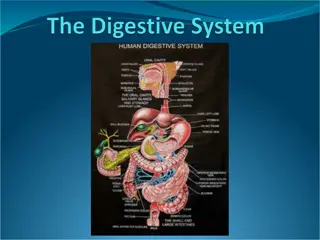
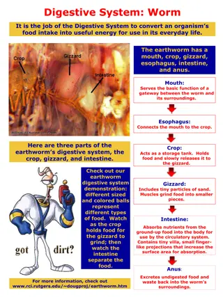
Histological Structure of Digestive System in Domestic Animals
The histology of the digestive system in domestic animals, specifically focusing on the teeth and their structures, including short (brachydont) and long (hypsodont) types. Details include the composition of enamel, dentine, cementum, and dental pulp, highlighting the differences in tooth morphology between species like humans, carnivores, ruminants, and pigs. The layers and characteristics of tooth anatomy are explained, from the crown to the root, along with the histological features like dentine tubules, odontoblasts, and periodontal fibers. The esophagus is also mentioned briefly.

Download Presentation

Please find below an Image/Link to download the presentation.
The content on the website is provided AS IS for your information and personal use only. It may not be sold, licensed, or shared on other websites without obtaining consent from the author.If you encounter any issues during the download, it is possible that the publisher has removed the file from their server.
You are allowed to download the files provided on this website for personal or commercial use, subject to the condition that they are used lawfully. All files are the property of their respective owners.
The content on the website is provided AS IS for your information and personal use only. It may not be sold, licensed, or shared on other websites without obtaining consent from the author.
E N D
Presentation Transcript
Histology Digestive system By: Dr. Ammar Ismail
Teeth There are two types of teeth in the domestic animal, short and long teeth . Short teeth (Brachydont ): which are stopped to growth when erupted to the mouth cavity and its usually consist of crown which covered by enamel until the neck , neck and one or more root which covered by cementum and under the enamel and cementum the dentine extended which surrounded the pulp cavity .This type of teeth is found in the human , carnivores , incisive of ruminants and incisive of pig except the canine of wild pig.
Teeth Long teeth (Hypsodont ): Which very long teeth and its growth continuous during the life of animal and its characterized by have no crown and neck but it have long body . and the external surface of tooth under the gum or upper the gum is covered by cementum and under the cementum found the enamel along the body of tooth and under the last there is dentine layer . This teeth found in the trusks or canine of wild pig and teeth of horse and cheek teeth of ruminants
Histological Structure of tooth The tooth consist of four histological layers which include enamel , dentine , cementum , dental pulp. Dentine : Its form the bulk of crown and root . Composed of calcified organic matrix similar to that of bone, the inorganic constitutes a large proportion of the matrix of dentine than that of bone, therefore teeth are harder than bone. From the pulp cavity minute parallel tubules, called dentine tubules radiate to the periphery of the dentine in longitudinal section of tooth. Periodontal : fibrogenic layer Odentoblast : columnar cell located beneath the dentin it has ability to produce the organic matrix of dentin
Enamel : The crown of the tooth is covered by enamel .Its hardest substance in the body tissues. composed of parallel enamel rods, highly calcified material. Cementum : The root is invested by a thin layer of cementum which is generally thicker toward the apex of root .The cementum is an amorphous calcified tissue into which the fibers of periodontal membrane are anchored. Dental pulp : Composed of connective tissue cells and fibers , matrix , blood and lymphatic vessels and nerves . Translucent substance
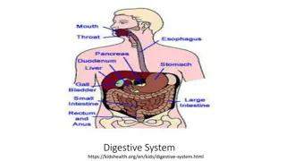
Esophagus : Contain all the layers of typical tubular organ of digestive system(typical tunics) : The mucosa consist of stratified squamous epithelium, lamina propria and muscularis mucosa . The degree of keratinazation of stratified squamous epithelium varies with the species , its non keratinized in carnivores, slightly keratinized in pig , keratinized in horse , highly keratinized in ruminants. The epithelium is supported by lamina propria of collagenous and few elastic fibers which contain diffuse lymph tissue . The muscularis mucosa well developed composed of smooth muscle fibers .The sub mucosa consist of loose connective tissue . Both the mucosa and sub mucosa may be present mucous or seromucous secreting glands . Tunica muscularis : consist of skeletal muscles in ruminants and dogs , while in the horse the first two third is skeletal but the last third is smooth but in the pig the first third is skeletal , the second third is mixed striated and smooth and the last third is smooth . Tunica Adventitia or serosa : The cervical region is surrounded by tunica adventitia of loose connective tissue containing blood ,lymphatic vessels and nerves .The thoracic region is surrounded by mediastinal pleura (serous membrane ) .The abdominal region covered by peritoneal serosa
Stomach : It s the most dilated region of the alimentary canal , its sac like responsible for digestion (enzymatic hydrolytic breakdown food ) .The stomach is lined extensively by mucosa in where as animals have in addition to glandular region glandular region lined with stratified epithelium structure chemical and of glandular carnivores herbivorous , , anon squamous
Non Glandular Region: Its greatest developed in Ruminants and is subdivided into rumen omasum. While it's absent in the carnivores and small region in Pig . In the Horse the non glandular region extends distance and end at the margoplicatus . The lining epithelium of non glandular region is stratified squamous and may be keratinized or non , reticulum and a considerable
Glandular Region : The mucosa of the glandular region has gastric longitudinal folds of mucosa and sub mucosa which disappear in the distended stomach .The epithelial lining of stomach invaginated into the mucosa , to forming gastric pits .the epithelium of stomach is secretary simple columnar and the lamina propria contain large number of gastric glands . According to the type and nature of gastric gland secretion the gastric mucosa divided into three region : 1 Cardiac region . 2 Fundic region . 3 Pyloric region
Cell types in the gastric gland The gland are diffuse with few cells in the cardia but are abundant and cellular in the fundus .There are five cell types in the gastric gland : 1 Stem cell: located at the neck of the gland , it divides to replaced the surface epithelium . 2 Parital (Oxyntic ) cells are large polyhedral with central nucleus cytoplasm , they secrete the hydrochloric acid . 3 Mucous neck cell : at the neck of gland , secrete mucous . 4 Chief (zymogenic , peptic ) small basophilic cells secrete the enzyme pepsinogen which converted into pepsin by the gastric acid 5 Entero endocrine population that are identified with specialized silver stain and are also known as argantaffin cells . The chemical messengers ( serotonin , gastrin , somatostain and entero glycogen and eosinophilic cell: Are diffuse
Stomach : The lamina propria is loose cellular connective tissue with lymphatic cells present as a local population and part of gut associated lymphoid tissue. The muscularis mucosa is composed of several layers of smooth muscle fibers. The sub mucosa is a glandular loose connective tissue .The muscularis externa consist of three layers of smooth muscle, oblique, circular and longitudinal .the serosa is covered by mesothelial cells continuous with the visceral periton.