The Digestive System Processes
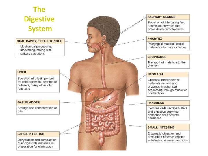
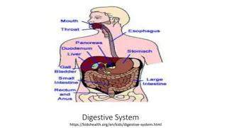
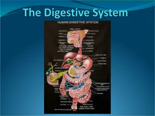
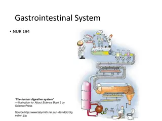
The digestive system is responsible for the breakdown, absorption, and elimination of food. It involves processes such as ingestion, propulsion, mechanical and chemical digestion, absorption, and defecation. Starting from the mouth with salivary glands and oral cavity, it progresses through the esophagus and stomach, where churning and enzymatic digestion take place. This systematic journey through different organs ensures the body gets the necessary nutrients for energy and maintenance.

Download Presentation

Please find below an Image/Link to download the presentation.
The content on the website is provided AS IS for your information and personal use only. It may not be sold, licensed, or shared on other websites without obtaining consent from the author.If you encounter any issues during the download, it is possible that the publisher has removed the file from their server.
You are allowed to download the files provided on this website for personal or commercial use, subject to the condition that they are used lawfully. All files are the property of their respective owners.
The content on the website is provided AS IS for your information and personal use only. It may not be sold, licensed, or shared on other websites without obtaining consent from the author.
E N D
Presentation Transcript
The Digestive System
Digestive Processes Ingestion taking food/drink into mouth. Propulsion movement through alimentary canal (swallowing, peristalsis). Mechanical Digestion Physical breakdown of food (chewing, churning).
Chemical Digestion Enzymatic breakdown of food (from complex to simple building blocks). Absorption transport of digested products from lumen of G.I. tract to blood and lymph (inside body). Defecation elimination of indigestable and waste products from body (feces).
Salivary Glands Intrinsic (inside oral cavity) e.g., lips & cheeks. Extrinsic (outside oral cavity): Mumps 1) Parotid (largest) - a serous gland. 2) Submandibular - a serous gland. 3) Sublingual (smallest) - a mucus gland Lingual amylase (breaks down starch). Lysozyme antibacterial agent in saliva.
The Oral Cavity Typical Adult Teeth (in one quadrant) Incisors Canines Premolars Molars Times 4 quadrants: Total: 4 x 8 = 32 2 1 2 3 = 8
The Esophagus - is a Muscular tube. ~ 10 inches long. Transitions from skeletal to smooth muscle. (hence voluntary to involuntary) Mucous glands in tela submucosa (layer) to lubricate bolus. Outermost layer is Adventitia or Serosa. outside peritoneal cavity inside peritoneal cavity
Stomach acidic (pH 2) storage of chyme. Mechanical Digestion continued (churning). Has 3 muscle layers, for churning. Enzymatic Digestion of proteins occurs here (Pepsin breaks down proteins). Only Absorption of alcohol and aspirin. Rugae allows for expansion when volume of contents increase.
Can you identify the 4 Tunics? Unique in the GI tract, the stomach has 3 muscle layers:
Production and secretion of gastric juices controlled by CNS. e.g., Vagus nerve Parietal cells: make Hydrocholic acid (HCl) in gastric glands. Chief cells: make Pepsinogen, which is cleaved to pepsin ( HCl), to digest proteins. Gastric Gland
Small Intestine: Chemical Digestion/Absorption Duodenum Jejunum Ileum Increase Surface Area for Absorption 1) Plicae Circulares 2) Villi (Intestinal) 3) Microvilli Lacteals absorption lipids Intestinal glands Goblet cells Stem cells
Villi of Small Intestine Vascular Arcade (from superior mesenteric a.) Mesentery Proper - is a double layered serous membrane attached to the small intestine. Roles: - Supports branches of blood vessels. - Supports lymphatics of the jejunum and ileum. - Supports nerves of the jejunum and ileum.
The Mesentery Proper of the Small Intestine The Greater Omentum
Distinguishing features of each region of the Small Intestine
The Large Intestine Begins as pouch inferior to end of ileum Ends at anus. Functions of Large Intestine: 1) Reabsorb water and compact feces. 2) Absorb vitamins (helps make Vit K) electrolytes. 3) Stores fecal matter. The Cecum: Contains the Ileocecal valve and connected to appendix. The Colon: Ascending, Transverse, Descending, Sigmoid.
The Colon Lack of villi Abundance of goblet cells Abundance of mucous-secreting intestinal glands
Anatomy between the Liver (makes Bile), the Gallbladder (stores and concentrates bile), and the Pancreas (makes/releases pancreatic juices) into the Duodenum.
Histology of the Pancreas Exocrine Gland: Pancreatic Juices Amylases Proteases Lipases Endocrine Gland: Makes Hormones Insulin Glucagon Somatostatin Gastrin
A Mucous Membrane Lines entire digestive tract Moistened by glandular secretions Simple or stratified depending on area of tract Pleated for expansion ( Surface Area)
These are the 4 Layers! 1. Tunica Mucosa is a Mucous Membrane (wet, absorbs, protects) 1) Epithelium 2) Lamina propria 3) Muscularis mucosae 2. Tela Submucosa Areolar Connective Tissue Contains: Submucosal plexus (nervous innervation) Blood and Lymphatic Vessels
3. Tunica Muscularis Externa Smooth muscle layers 1) Inner Circular Layer 2) Outer Longitudinal Layer What are some exceptions? Myenteric Plexus 4. Tunica Serosa (or Adventitia*) Serous membrane aka visceral peritoneum * Name depends on location: a)Inside peritoneal cavity = serosa b)Outside peritoneal cavity = adventitia
Layers: How are they different in each diagram above? 1. _______________ 2. ______________ 3. _______________ 4. _______________
The Peritoneum: Two layers Visceral peritoneum (a.k.a. serosa) Parietal peritoneum - Lines inner surfaces of body wall Mesenteries: Fused double sheets of peritoneal membrane to suspend portions of digestive tract: e.g. Greater omentum Lesser omentum Mesentery proper Mesocolon
Retroperitoneal Structures these are attached to posterior abdominal wall, and are not in the peritoneal cavity. e.g. - Most of the Duodenum - Ascending colon - Descending colon - Pancreas
Horizontal section through the upper abdomen showing the position of the liver relative to other visceral organs.


 storage of chyme.")


 Surgery")


, the Gallbladder")