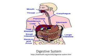
The Human Digestive System

 Gastrointestinal System
NUR 194
 
'
T
h
e
 
h
u
m
a
n
 
d
i
g
e
s
t
i
v
e
 
s
y
s
t
e
m
'
i
l
l
u
s
t
r
a
t
i
o
n
 
f
o
r
 
A
b
o
u
t
 
S
c
i
e
n
c
e
 
B
o
o
k
 
3
 
b
y
S
c
i
e
n
c
e
 
P
r
e
s
s
Source:http://www.labyrinth.net.au/~daviddic/dig
estion.jpg
Quick Review: A&P
GI tract – 25 ft pathway from mouth to anus
Function of GI tract:
Break down food particles into molecules for digestion
Absorb into the bloodstream the small molecules
produced by digestion
Eliminate undigested and unabsorbed foodstuffs and
other waste products from the body
Mouth
Food entry
Process of digestion begins with
chewing (mastication) to break down
foods into small particles that can be
swallowed and mixed with digestive
enzymes
Saliva:
First secretion that comes in contact with
food; contains water and mucus; lubricant
for swallowing
Contains salivary amylase (enzyme:
ptyalin) digests starches
Produced in 3 pairs of glands: parotid,
submaxillary, and sublingual.
Approx. 1L of saliva is produced daily
Pharynx
The pharynx or throat extends from the soft palate to the esophagus.
It is lined with mucous membrane and contains 3 pairs of organs: the
adenoids, the lingual tonsils, and the tonsils.
Esophagus →
Responsible for food passage from
mouth to stomach
Distends
Smooth muscle contracts in
rhythmic sequence (peristalsis)
from upper esophagus to stomach
to propel food along
Upper Esophageal Sphincter 
(UES)
(entry). Closed at rest to prevent
air entering the esophagus during
respiration.
passes through diaphragm at
opening called diaphragmatic
hiatus
Lower esophageal sphincter
(LES)
relaxes to allow food to enter
stomach then 
closes tightly to
prevent reflux of stomach
contents back into esophagus.
about 24cm or 10 inches long
Stomach→
Lined with smooth muscle that promotes gastric
motility.
Expandable pouch, stores and mixes food with
secretions; holds approx. 1500ml
Parietal cells line wall and secrete hydrochloric acid
(HCl)
Breaks down food
Kills most ingested bacteria
Gastric secretions contain pepsin for protein
digestion
Gastric mucosa secretes intrinsic factor to combine
with Vitamin B
12
 in food to be absorbed in ileum
Parietal cells also produce 
intrinsic factor, which
works to facilitate the absorption of Vit B12.
Absence of the intrinsic factor causes pernicious
anemia.
Chyme: food particles mixed with gastric secretions
Stomach
Inlet: esophagogastric junction
surrounded by smooth muscle
(lower esophageal sphincter or
cardiac sphincter) which
contracts and closes off
stomach from esophagus
4 anatomic regions:
Cardia (entrance)
Fundus
Body or corpus
Pylorus or antrum: outlet
Outlet: Pyloric sphincter – circular
smooth muscle that controls
the opening of the stomach to
the intestine
3 phases of gastric secretion
Cephalic- begins with sight, smell, and taste of food
and is regulated by the vagus nerve.
Gastric- begins with the presence of food in the
stomach.
Intestinal- begins as the chyme passes from the
stomach into the duodenum, causing distention.
Secretin :  a hormone that inhibits further acid
production and decreases gastric motility.
Pancreas
 
Gland that lies behind the stomach and extends to the spleen
2 cellular bodies within Pancreas:
Exocrine: 80% of the organ and cells secrete enzymes needed for digestion of
carbohydrates, fats, and proteins (trypsin, chymotrypsin, amylase and lipase
Endocrine- produces glucagon & insulin.
Liver
Largest organ of body –other than skin
Located in RUQ of abdomen.
Primary functions:
Forms & secretes bile which is necessary for fat breakdown.
Bile empties into the duodenum at sphincter of Oddi.  If sphincter is
closed, the bile is stored in the gallbladder.
Small Intestine→
Longest segment – about 2/3 the total
length of GI tract
Approx 7000 cm of surface area for
secretion and 
absorption
 (the process
by which nutrients enter bloodstream
through intestinal wall --primary fx of
small intestine).
3 anatomic parts:
Duodenum (upper)
Sphincter of Oddi
Jejunum (middle)
Ileum (lower)
Cecum: junction between small and
large intestine; ileocecal valve controls
passage of contents into large intestine
& prevents reflux of bacteria into small
intestine.
Appendix located at junction of small
and large intestine
Small intestine
The common bile duct and pancreatic duct join to form the ampulla
of Vater, emptying into the duodenum at the duodenal papilla. This
papillary opening is surrounded by muscle known as the sphincter of
Oddi.
Sphincter of Oddi leads to the jejunum.
Small Intestine 
Duodenum: contains intestinal glands & receives secretions from pancreas, liver, and
gallbladder that aid in digestion (contain enzymes and bile)
Intestinal glands
:
secrete mucus, electrolytes, and enzymes: Coat cells and protect mucosa from
HCl
Hormones, neuroregulators, and local regulators control rate of secretion
production; influence GI motility
Pancreas:
Secretions have alkaline pH due to high bicarbonate concentration; neutralizes
acid entering from stomach
Digestive enzymes: Trypsin (protein digestion); Amylase (starch); lipase (fats)
Liver:
secretes bile (emulsify fats making them easier to digest and absorb) know all
functions of the liver –Chapter 55
Gallbladder: Bile storage
Two types of contractions- stimulated by presence of chyme
Segmentation: mixing waves; churns
Intestinal peristalsis: propels contents of small intestine to colon
Vitamins and minerals: absorbed essentially unchanged
Absorption begins in jejunum 
– diffusion and active transport
Large Intestine
Functions: movement, continuation of absorption and
beginning of elimination
Ileocecal valve allows passage of residual waste material
from terminal ileum into proximal colon—restricts reflux
of contents back into ileum,
Bacteria is major component of large intestine
2 colonic secretions: an electrolyte solution (bicarb to
neutralize) and mucus (protects mucosa and provides
adherence of fecal mass)
 
Waste materials reach and distend rectum in about 12
hours
Large Intestine
Components
:
Begins at cecum.
Ascending segment (R-side of
abdomen)
Transverse
 (
right to left of
upper abdomen)
Descending segment (L side of
abdomen)
Terminal portion: sigmoid and
anus
Anus -- striated muscles form
internal and external anal
sphincters
Waste Products
Feces – 75% fluid, 25% solid materials: foodstuffs, inorganic
materials, water and bacteria
Brown color comes from breakdown of bile
Chemicals formed by bacteria are largely responsible for odor
Approx 150 ml of gases are contained in GI tract: methane; hydrogen
sulfide; and ammonia—either absorbed into portal circulation and
detoxified by liver, or expelled through rectum as flatus
Process of elimination: Stimulation of rectum causes rectal distention
which reflexively initiates contraction of rectal muscles causing the
internal anal sphincter to relax (autonomic nervous system). The
external anal sphincter is under conscious control of the cerebral
cortex—once allowed to relax will allow contents to be expelled.
Contraction of the abdominal muscles (bearing down or straining)
facilitates the emptying of the colon.
Average frequency of defecation: once a day, but varies among
individuals
GI Changes with Aging
Stomach: Decreased gastric HCL
 
-Atrophic gastritis due to bacterial overgrowth
   
Encourage bland foods high in vitamins & iron
   
Assess for epigastric pain
Large Intestine: Decreased peristalsis
Constipation & Impaction
High fiber diet
Increased fluid intake (1500ml) if not contraindicated
Encourage activity as tolerated
Pancreas: Pancreatic duct change
Steatorrhea: excess fat in stool.
Small frequent feedings
Assess for diarrhea
Liver: Decreased enzyme activity & cholesterol synthesis
Depresses drug metabolism leading to accumulation of drug and possible toxicity
Assess for adverse effects of drugs
Know Chart 55-1
Assessment
Take a complete history; focus on symptoms common to GI dysfunction Read Gordon’s functional health
patterns-Chart 55-2
Family & Genetic HX * Ex: Familial adenomatous Polyposos(FAP)-rpedisposition to colon cancer
Previous hx of GI disease
Past and current medication use. Including OTC & herbal products
Previous or current treatments
Previous surgeries
Nutritional/Dietary hx:
Food Allergies
Tobacco and alcohol use
Changes in appetite or eating patterns
Anorexia (loss of appetite for food); dysphagia (difficulty swallowing); N/V; dyspepsia
(indigestion/heartburn)
Unexplained weight gain or loss
Stool characteristics
Socioeconomic, psychosocial, spiritual, or cultural factors
Cultural: food restrictions, use of ingredients. Fasting
80-90% African Americans are lactose intolerant. Lack of enzyme lactase which converts dairy to
glucose and galactose. (s/s: bloating, cramping, diarrhea)
PE: Abdominal girth; tenderness/guarding; rectal exam; pelvic exam
Diagnostics: CBC, urinalysis, abdominal x-rays, EKG, pregnancy test for the female
PQRST
P: precipitating or palliative
Q: quality or quantity
R: region or radiation
S: severity scale
T: timing
Symptoms Common to GI
Dysfunction
Pain: can be major symptom; assess character,
frequency, location, pattern, time of pain—be
aware of common sites of referred pain
Indigestion: Upper abdominal discomfort or
distress; most common symptom; Peristaltic activity
may be contributing; BMs may or may not relieve
pain. Fatty  foods often perpetrator—remain in
stomach longer. Coarse vegetables and highly
seasoned foods may cause distress.
Symptoms
Intestinal Gas: Accumulation of gas may leading to belching
(expulsion from stomach thorough mouth) or flatus (from
rectum)
Complaint of bloating, distension, or “full of gas”
Excessive flatulence may be a symptom of gallbladder disease or food
intolerance.
Nausea and Vomiting:
Nausea : usually triggered by odors, activity, or food intake
Emesis may vary in color and content; may contain undigested food
particles or blood (hematemesis).
Blood: If bright red signal of hemorrhage; if dark or has coffee-ground
appearance it has been subjected to gastric enzymes
Symptoms
Change in Bowel Habits and Stool Characteristics
May signal colon disease
Diarrhea: abnormal increase in frequency or liquidity of stool
Constipation: decrease in frequency of stools; stools that are hard, dry, or of
smaller volume than normal
Symptoms
Stool Characteristics – color and appearance
Color: can be affected by foods or meds
Blood: can present in several ways
Tarry-black: blood shed in upper GI tract
Bright red: blood enters in lower portion of GI
Dark red: passing rapidly through GI tract
Streaking of blood on surface of stool/toilet tissue: may be lower
rectal or anal bleeding
Common abnormalities of stool:
Bulky, greasy, foamy; foul in odor; gray with silvery sheen
Light gray or clay-colored caused by absence of urobilin
Mucus threads or pus visible on gross inspection
Small, dry, rock-hard masses called scybala—may be streaked with
blood from rectal trauma during passage
Loose, watery stool that may or may not be streaked with blood
Physical Assessment
Examination includes:
Mouth: tongue, buccal mucosa, teeth, gums –note any
ulcerations, nodules, swelling, inflammation; color changes
Abdomen
Rectum - –anal and perineal areas: rash or excoriation;
fissures or fistulas; hemorrhoids. Digital exam: masses or
tender areas
Performance of the exam must be followed in this order so
intestinal activity & bowel sounds are not increased prior to
exam.
Inspection
Auscultation
Percussion
Palpation
Inspection
Skin changes
May result from GI Tract disorders such as liver and biliary obstructions
Scars – trauma, previous surgeries
Contour and symmetry
Bulging
Distension
Peristaltic waves
Cullen’s sign
Auscultation
Bowel Sounds – all 4 quads – using the diaphragm
of the stethoscope (best for high-pitch and
gurgling):
Increased bowel sounds-borborygmus
Character
Location
Frequency
Normal – sound is heard every 5 to 20 seconds
Hypoactive – one or two sounds heard in 2 minute period
Hyperactive: 5 to 6 sounds heard in less than 30 seconds
Absent: No sounds heard in 3 to 5 minutes
BRUITS
Percussion and Palpation
Percussion:
Tympany (high pitched) or dullness
Palpation: 2 types:
Light: detects large masses & areas of tenderness.
Rebound tenderness (Blumberg’s sign-used to further evaluate areas
of pain).
: exert pressure over an area and release quickly—is there pain on
withdrawal of pressure? If so, report to health care provider.
Note any areas of tenderness or guarding for further evaluation
during deep palpation. If rigidity noted, indicative of peritoneal
inflammation.
Deep: identify masses in 4 quads. Current practice is that only MD or
APN’s perform deep palpation. Used to determine size & shape of organs
& masses.
Diagnostics
Blood Tests: CBC, PT, Carcinoembryonic antigen (CEA and the CA19-9);
total calcium, Potassium, Albumin levels
AST, ALT, amylase and lipase bilirubin, ammonia.
 Serum Cholesterol and Triglycerides
Urine Tests: amylase; urobilinogon
Stool Tests ( FOBT)-Fecal Occult Blood Test , FIT, Stool samples for ova/
parasites; stool test for clostridium difficile
Know:  Labs: Chart 55-3 for normal ranges
Diagnostics cont’d
Abdominal Ultrasound
Plain films of the abdomen
Upper GI Study
Lower GI study
PTC
CT
Endoscopy
Endoscopy: direct visualization of GI tract with a flexible fiberoptic
endoscope.
Used to evaluate bleeding, ulceration, inflammation, tumors, and cancer
of esophagus, stomach, biliary system, or bowel. May obtain specimans
for biopsy or studies (H.pylori).
 Requires Informed consent by patient prior to procedure.
EGD- examines esophagus, stomach & duodenum. May be used to dilate an
esophageal stricture (during procedure)
ERCP- includes exam of liver, gallbladder, bile ducts, and pancreas to identify cause &
location of obstruction(s). Uses contrast so x-rays may be obtained. May obtain
biopsies, or remove gallstones.Lasts 30 minutes to 2 hours. Reqiures Iv access for
moderate sedation drugs and contrast.
 Colonoscopy
Colonoscopy: endoscopic exam of entire large bowel..  Recommended every 10 years for all persons age 50+.
More frequently if family hx of colon cancer. Evaluates for causes of chronic diarrhea, source of GI bleeding.
Prep:
 
Clear liquid diet 12-24 hours  prior  to procedure.
  NPO 6-8 hours prior—may have water.
Avoid aspirin, NSAIDS, and anticoagulants (with MD order) for several days
Diabetic patients should check With MD re: diabetic meds as pt is NPO
Night before procedure, Pt drinks Sodium Phosphate (Phospho-Soda) to clean bowel. May be repeated the
morning of procedure. May prescribe Go-Lytely as a bowel cleanser the day before as well. Chill drinks  to
improve taste. Have pt drink quickly to avoid N/V. Watery diarrhea begins in about 1 hour. Additional
laxatives or cleansing enemas may be required.
Procedure:
IV access for moderate sedation. Versed. Air is instilled to prmoote better viewing.
Endoscopy passed through rectum to cecum.
Atropine Sulfate : available in case of bradycardia from vasovagal response.
After-Care:
NPO until sedation wear off and pt is fully alert. VS every 15 minutes until pt is stable. Siderails up until Pt
fully alert. Monitor for rectal bleeding or pain. May expect abdominal cramping for several hours. Monitor
for bowel perforation, hypovolemic shock.
Chart 55-4.
Tests cont.
Gastric Analysis, Gastric Acid Stimulation Test(Histolog), pH
monitoring
Ultrasonography,
Endoscopic ultrasonography
Liver-spleen scan
The nurse’s role
Is to prepare the client, explain the procedure, and provide the
necessary post procedure care.
Read and know the procedures so you can explain them to the client.
Oral and Esophageal Disorders
Chapter 56
Examining the Lips, Mouth, and Gums:
Examples of Lip Conditions :
 
Cold Sore
  
Chancre
   
Contact 
  
       
Dermititis
Stomatitis/Candidiasis
Stomatitis is a painful ulceration of the oral mucosa
that appear as inflammation and denudation of the
oral mucosa, known as canker sores
Candidiasis is a fungal/yeast infection resulting from
an overgrowth of normal flora. Type of secondary
stomatitis. Overgrowth may be caused by long term
antibiotic use, chemotherapy, radiation, and HIV.
White plaque on tongue. Red and painful underneath.
Mouth
 
Hairy Leukoplakia
Candidiasis
Lichen planus
 
Mouth and Gums
Periodontitis
Aphthous
Stomatitis
(Canker Sore)
Salivary Glands
Acute Sialadenitis:
Inflammation of salivary glands
Causes: dehydration; radiation therapy, malnutrition,
improper oral hygiene. Invasion of 
staph a.
;
 streptococcus
viridans
;
  
or 
pneumococcus
S/Sx: Pain, swelling, purulent discharge
Tx: Antibiotics; massage, hydration, corticosteroids;
surgery to drain; analgesics, warm compresses, use of
lemon slices & fruit/citrus flavor candy to stimulate
    saliva
Best prevented by good routine oral hygiene.
Xerostomia-very dry mouth . Usually caused by
exposure of glands to radiation.
Esophagus
Chapter 57
Dysphagia: difficulty swallowing
Most common symptom of esophageal disease
Odynophagia: acute pain on swallowing
Motility disorders:
Achalasia: rare disorder. absent or ineffective peristalsis;
accompanied by the  esophageal sphincter  that fails to
relax during swallowing resulting in sensation of food
sticking in lower esophagus.
Diffuse spasm: Motor disorder with unknown cause;
stress produces contractions of the esophagus
Esophagus
 Gastroesophageal Reflux Disease (GERD)
 
Caused by an incompetent lower esophageal sphincter
,
pyloric stenosis, or a motility disorder  that allows
backflow of gastric or duodenal contents
S/Sx: Pyrosis (burning sensation in the esophagus); dyspepsia
(indigestion); regurgitation; dysphagia; odynophagia- Chart 57-1
& Table 57-1
Tx: Low-fat diet; avoid caffeine, tobacco, beer, milk;
peppermint/spearmint; carbonated beverages; avoid eating 3
hours before bedtime; maintain a healthy weight; avoid tight-
fitting clothes; 
elevated head and/or HOB
; surgical intervention
for severe cases not responding to medical intervention. Chart
57-2
GERD
Tx: Meds:
Antacids;
Histamine receptor blockers (prevent or block the production of
gastric acid): Cimetidine (Tagamet); Famotidine (Pepcid);
Nizatidine (Axid); Ranitidine (Zantac)
Proton Pump Inhibitors (decrease gastric acid production): Prevacid
(lansoprazole), Aciphex (rabeprazole); Prilosec (Omeprazole); Nexium
(esomeprazole magnesium)
Prokinetic Agents (accelerate gastric emptying): Urecholine
(bethanechol); Motililium (domperidone) and Reglan
(metroclopramide)
Untreated GERD can lead to Barrett’s Esophagus – a
precancerous condition
Esophagus
Hiatal Hernia: opening in the diaphragm (through which the
esophagus passes) becomes enlarged and the upper part of the
stomach moves up into the lower part of the thorax.
2 major types:
Sliding HiatalHernia
Heartburn, regugitation, chest pain, dyshapgia, belching
Paraesophageal Hernia
Feels full after eating, breathless after eating, feeling of suffocation, chest
pain mimics angina, s/s worse in recumbent position.
Diverticulum: (Of esophagus). Herniation of mucosa into surrounding tissue. S/S:
dysphagia, reflux, bad breath, nocturnal cough.DX by EGD. Small meals of
semisoft foods, elevate HOB. May require surgical removal:
Gastric Disorders-Chapter 58
Gastritis
: inflammation of the gastric (stomach) mucosa;
membranes become edematous and hyperemic
(congested with fluid and blood) and begin to erode.
Leads to ulceration and a potential for hemorrhage
Acute: lasts hours to a few days; 
infection with Helicobactor
pylori
. usually caused by eating contaminated foods; highly
seasoned foods; 
overuse of aspirin or NSAIDs
; excessive
alcohol intake; bile reflux; radiation therapy; or the ingestion
of a strong acid or alkali.
S/Sx: Acute: abdominal pain; headache; N&V; anorexia; hiccoughing
Tx: Usually self-limiting; mucosa able to repair itself; refrain from
food and alcohol until resolved; non-irritating diet during recovery; IV
fluids. Acids or alkalis: dilution and neutralization; acids (common
antacids), alkali (diluted vinegar or lemon juice)
Gastric Disorders
Gastritis:
Chronic: repeated exposure to irritating agents; recurring
episodes of acute gastritis
Causes: Prolonged inflammation-- benign or malignant ulcers;
bacteria 
Helicobacter pylori; 
autoimmune diseases (pernicious
anemia) B12 deficiency; caffeine; medications; alcohol; smoking;
reflux of intestinal contents.
S/Sx: anorexia; heartburn; belching; N&V; sour taste in the mouth
Tx: Modify diet; promote rest; reduce stress; pharmacotherapy—
antibiotics; proton pump inhibitors; bismuth salts (Pepto-Bismol).
Gastric and Duodenal Ulcers
Peptic Ulcer: musocsal lesion of the stomach or duodenum.
Peptic Ulcer Disease (PUD): occurs when mucosal defenses are
impaired and no longer protect the epithelium from the effects
of acid and pepsin.
Types of Peptic Ulcers:
Gastric: occurs when break in mucosal barrier and HCL acid
injuries lining. Delays gastric emptying resulting in regurgitation
of duodenal contents. Deep & penetrating ulcers.
Duodenal: chronic break in duodenal mucosa and leaves a scar
after healing. High gastric acid secretion.. Most common ulcer.
Stress Ulcers: acute lesions that occur after an acute medical
crisis or trauma. Gastritis may lead to stress ulcers.
Review Table 58-2
 
Gastric and Duodenal Ulcers
Stress and anxiety formerly thought to be the cause
Caused by infection with gram-negative bacteria
Helicobacter pylori
Familial tendency may predispose; Blood Type O at
greater risk; also associated with chronic pulmonary
disease or chronic renal disease
Associated with chronic use of NSAIDs, alcohol
ingestion, and excessive smoking
Gastric and Duodenal Ulcers
S/Sx:
May come and go; days to weeks to months and then
disappear then reappear often without identifiable cause
Dull, gnawing pain; usually relieved with eating
Burning sensation 
in midepigastrium or back
Pyrosis (heartburn); belching
N&V (vomiting usually is indicative of complication and
often follows a bout of pain and bloating; usually consists
of undigested food )
Constipation or diarrhea (usually as a result of medication
or diet regimen)
Gastric and Duodenal Ulcers
Assessment
: Pain and epigastric tenderness;
abdominal distention
Diagnostics
: Barium study of Upper GI; Endoscopy;
Stool testing for occult blood
; biopsy and histology
with culture, breath test, or serological testing for
H. pylori
Tx
:
 
Medications:
 
Antibiotics for 
h. pylori
; histamine
2 (H
2
) receptor antagonists; proton-pump inhibitors
for ulcers not associated with 
h. pylori
. Also,
lifestyle changes and surgical intervention
Surgical Intervention read p.1278
Nursing Considerations
Relieving pain: meds; avoid aspirin, caffeine; regular meals;
relaxation techniques to manage stress and pain; smoking
cessation
Reduce anxiety: Answer questions, explain diagnostic tests;
administer meds on schedule; encourage family support.
Maintain optimal nutrition: Assess for malnutrition and wt
loss; teach about importance of complying with med and
dietary regimen.
Promote self-care—teach clients to manage disease and
factors that help or aggravate; medication regimen; dietary
considerations; smoking cessation
Nursing Considerations
Monitor and manage potential complications:
Hemorrhage (goal: to avoid hemorrhagic shock)—
hematemesis (may include NG tube insertion and care);
correct blood loss; assess for faintness, dizziness, or nausea;
Frequent VS; assess for tachycardia, hypotension, tachypnea;
monitor Hgb and Hct; urinary output; monitor O
2
 sats and
administer O
2
Perforation (erosion of ulcer through gastric serosa into the
peritoneal cavity) and Penetration (erosion of ulcer through
gastric serosa into adjacent structures)– 
requires immediate
surgery as peritonitis sets in quickly; sudden severe upper
abdominal pain (may be referred), Vomiting and collapse;
extremely tender and rigid abdomen
; hypotension and
tachycardia; Tx: NG insertion and care; monitor fluid &
electrolyte balance.
Nursing Considerations
Pyloric Obstruction (gastric outlet obstruction) – area distal to pyloric
sphincter scarred and stenosed
S/Sx:
N &V
Constipation
Epigastric fullness
Anorexia
Weight loss (later)
Irritable Bowel Syndrome (IBS)
One of the most common GI problems
More common in women than men
No anatomic or biochemical abnormalities identified to explain
symptoms; no evidence of inflammation or tissue changes
Factors associated with IBS:
Heredity
Psychological stress
Depression and anxiety
High-fat diet
Stimulating or irritating foods
Alcohol consumption
Smoking
IBS
Results from functional disorder of intestinal motility
(peristaltic waves  affected at specific segments of the
intestines and in the intensity at which they propel) that may
be related to:
Neurologic regulatory system
Infection or irritation
Vascular or metabolic disturbance
S/Sx: Wide variability; Range in intensity and duration from
mild and infrequent to severe and continuous
Primary symptom: alteration in bowel patterns: constipation,
diarrhea or a combination of both
Pain – often precipitated by eating and relieved by defecation
Bloating
Abdominal distention
IBS: Assessment and Diagnostics
Definite Dx: Tests that prove the absence of structural or other
disorders
Stool studies, X-ray, proctoscopy: to r/o colon diseases
Barium enema and colonoscopy: reveal spasm, distention, mucus
acculmulation
Manometry; Electromyography—study intraluminal pressure changes
generated by spasm
Medical and Nursing Management
Medical:
Relieve abdominal pain
Control constipation or diarrhea
Elimination and gradual reintroduction of foods suspected of being irritants (e.g.
caffeine; beans; fried foods; spicy foods; alcohol)
High-fiber diet
Exercise -reduces anxiety and increases intestinal motility
Stress reduction or behavior-modification programs
Anti-diarrheals (loperamide – Imodium) and hydrophilic colloids (i.e., bulk) to
control diarrhea and fecal urgency
Antidepressants: anxiety and depression
Anticholinergics and calcium channel blockers to 
↓ smooth muscle spasm
(decrease cramping and constipation
Nursing:
Reinforce good dietary habits:
eat at regular times; chew thoroughly
Avoid fluids with meals (causes distention)
Discourage alcohol use and smoking
Intestinal Obstruction
Occurs when intestinal contents cannot pass through
the GI tract –partial or complete blockage of the lumen
Medical emergency; Requires prompt treatment
If untreated, complete obstruction can cause death
within hours from shock and vascular collapse
Pathophysiology
Mechanical or non-mechanical blockage of the lumen
Fluid, air, gas collects near site
Peristalsis increases temporarily to break through the
blockage
Intestinal mucosa is injured and distention at and above
obstruction site occurs
Venous blood flow is impaired and normal absorptive
processes cease
Water, sodium, potassium secreted by bowel into fluid
pooled in the lumen
Intestinal Obstruction
Causes: Mechanical and non-mechanical.
Mechanical-occlusion of lumen. Most common in the ileum.
E.g. adhesions, strangulated hernias; carcinomas, volvulus (twisting
of bowel itself); intussusception (portion of bowel telescopes or
invaginates into an adjacent bowel portion)
Non-mechanical caused from neurologic or vascular
disorder. E.g. Paralytic ileus; electrolyte imbalances
Pseudo-obstruction: Looks like an mechanical
obstruction but shows no obstruction on x-rays.
Vascular: very rare and caused by emboli.
Etiology and Pathophysiology
Review daily bowel activity:
When obstruction occurs in pylorus metabolic alkalosis will
occur from loss of HCl. Also from use of NG tube suction.
Small bowel obstruction will cause dehydration and
electrolyte imbalance (if occurs proximal colon - most of the
fluids have been absorbed).
Solid fecal material accumulates until discomfort occurs.
Adhesions, hernias can cause obstructions as well.
Strangulation and gangrene can occur after two different
spots are obstructed.
Intestinal Obstruction
Clinical Manifestations:
Vary depending on obstructed area.
N & V, abdominal pain, distention, inability to
pass flatus, and obstipation
Assessment and Diagnostics
H&P, Abdominal x-rays, Barium enemas,
Upper GI series, Colonscopy, CBC,
Electrolytes, amylase, BUN; Stool tests to
check for occult blood.
Treatment
Decompression of Intestines with removal of gas and
fluid.
Maintenance of fluid;  and correction of electrolyte
imbalance.
Removal of obstruction.
NG tubes inserted, colonscopy to reduce voluvulus. IVF
and/or TPN.
Most mechanical obstructions are corrected surgically.
Nonsurgical approach is to use colonscopy to remove
polyps, dilate strictures, remove tumors or destroy
tumors via laser.
Nursing Management
Nursing Care:
NPO
NG tube to low-pressure, intermittent suction
Semi-Fowler’s position
Mouth and nose care
IV and medication therapy
Monitor VS; electrolytes
Watch for s/sx of shock
Monitor BS and signs of returning peristalsis
Monitor wound site (post-op)
Patient Teaching: TC & DB; ostomy care
Hemorrhoids
Dilated portions of veins in the anal canal
Very common
By age 50, 50% of people have hemorrhoids to some extent
May be internal (occurring above the internal sphincter) or external
(occurring outside the external sphincter).
Symptoms: bleeding, pruritus, prolapse, and pain.
Etiology & Pathophysiology
Thought to develop as a result of shearing forces
during defecation.
When straining, supporting tissues in the anal canal
weaken and the venules become dilated & blood
flow is impaired.  If the blood clots, a thrombosed
external hemorrhoid results
Most common cause of bleeding with defecation
Precipitating factors: pregnancy, prolonged
constipation, straining when having a BM, heavy
lifting, prolonged standing & sitting, portal HTN (as
found in cirrhosis).
Clinical Manifestations
Internal
:
May be asymptomatic but will have pain if they become
constricted.
Pain usually described as a chronic, dull aching discomfort,
particularly when the hemorrhoids have prolapsed.
Blood on toilet tissue when wiping
External:
Reddish-blue & seldom bleed or cause pain unless a vein
ruptures.
If thrombosed: become inflamed & painful.
S/Sxs are intermittent pain, pain on palpation, itching,
burning, bleeding noted on toilet paper when wiping.
Assessment and Diagnostics
Internal
: digital exam, anoscopy, or sigmoidoscopy.
External
: diagnosed by visual inspection & digital exam.
Therapy should be directed toward the causes & the client’s
symptoms.
Treatment
High fiber diet
Increased fluid intake
Creams
Suppositories
Impregnated pads containing anti-inflammatory
agents, astringents & anesthetics (e.g. witch hazel) to
shrink the mucous membranes and provide comfort
Stool softeners
Sitz baths
Ice packs followed by warm packs may provide
comfort for thrombosed external hemorrhoids
Treatment
Topical: shrinking the tissues with 5% phenol in oil or a
combination of quinine & urea.
Internal hemorrhoids: band ligation: through an anoscope
the hemorrhoid is identified & then ligated with a rubber
band.
Infrared coagulation where infrared or electrical current
produces local inflammation.
Cryotherapy where the hemorrhoid is frozen
Laser treatment
Hemorrhoidectomy when there is prolapse, excessive pain
or bleeding or large hemorrhoids.  The surgeon cauterizes,
clamps, or excises the hemorrhoids.
Nursing Management
Patient Teaching:
Constipation prevention techniques
Avoid prolonged standing or sitting
Proper use of OTC drugs
Seek medical care if excessive pain & bleeding occurs
Sitz baths for 15-20 mins 2-3 x per day for 7-10 days is helpful;
started 1-2 days postop
Pain can be severe: narcotics can be given initially
Topical preparations
Packing in the rectum: usually removed the first or second day
post-op
Provide privacy
Stool softeners.
Malabsorption
(
Impaired absorption of fats, carbohydrates, proteins, minerals, and vitamins
(especially B
12
).
Diseases of the small intestine are the most common cause of
malabsorption
Pathophysiology:
Mucosal (transport) disorders: celiac sprue; reginal enteritis
Infectious Diseases: small bowel bacterial overgrowth; tropical sprue; Whipple’s
disease (bacterial invasion of intestinal mucosa)
Luminal problems: bile acid deficiency; Zollinger-Ellison syndrome
Post-operative: following gastric or intestinal resection
Disorders of specific nutrients: dissacharidase deficiency leading to lactose
intolerance
S/SX: Diarrhea of frequent, loose bulky, foul-smelling, grayish stools with
increased fat content (steatorrhea); weakness; abdominal pain; 
↑flatus;
weight loss; decreased sense of well-being; malnutrition with s/sx of vitamin
and mineral deficiency (e.g. easy bruising; anemia; osteoporosis)
Assessment and Diagnostics
Stool studies: fat content
Lactose tolerance tests
Schilling test
Bile acid breath test: assess absorption of bile salts
Hydrogen breath tests-evaluate carbohydrate absorption
Pancreatic secretion/fuction test to test for insufficiency
Endoscopy of small intestine to biopsy (enzyme activity;
infection; destruction of mucosa)
Barium enema
Labs: CBC (anemia)
Medical and Nursing Management
Medical:
Avoid dietary substances that aggravate malabsorption
Nutrient supplementation
Dietary therapy (e.g.celiac sprue: reducing gluten intake)
Antibiotics
Antidiarrheals
Parenteral fluids to treat dehydration
Nursing:
Patient and Family teaching: diet and use of nutrient supplements;
Nutrient deficiencies– osteoporosis (calcium)
Monitor for fluid and electrolyte imbalances
Malabsorption Syndrome: Sprue
Types-nontropical and tropical kinds. Also called celiac
sprue, celiac disease, gluten-induced enteropathy.
Atrophy and flattening of the villi which reduces absorption
in small intestines.
Cause is hypersensivity response from gluten which leads to
inflammation of the mucosa.
Usually peaks in childhood. But can occur at anytime.
Signs includes-steatorrhea, diarrhea, wt loss, abdominal
distention, excessive flatulence. You might see glossitis  in
multi-vitamin deficiency
Sprue: Diagnosis and Care
Stool analysis and biopsy. Barium enema.
Care-depends on cause:
Nontropical – Gluten-free diet. Corticosteriods may be given when diet does
not work.
Tropical sprue– broad spectrum antibiotic and folic acid therapy. On
remission will remain on folic acid.
Malabsorption Syndrome: Lactose
Deficiency
Lactase enzyme is deficient or absent from the body.
S/SX:
Lactose intolerance, bloating, flatulence, crampy abdominal
pain, and diarrhea.
Usually occurs 30 minutes to several hours after ingestion.
Care
Lactose tolerence test
Lactose free diet
Lactaid OTC prior to intake of lactose containing foods.
Malabsorption Syndrome: Short
Bowel Syndrome
Results from resection of small intestines.
Rapid intestinal transit
Impaired digestion and absorption
Fluid and electrolyte loss
Varying symptoms: depends on the amount of colon resected
Short Bowel Syndrome:
Clinical Manifestations
and Management (Care)
Diarrhea and steatorrhea
Malnutrition – weight loss
Multiple vitamin and mineral deficiencies:, cobalamin and zinc deficiency,
hypocalcemia.
May be lactose intolerant
May have oxalate kidney stones
Care:
Fluid and electrolyte balance
Nutrient Supplementation: calcium, zinc, multivitamins. 
Dietary considerations; high carb/low-fat diet with soluble fiber; pectin; amino acid
glutamine; Encourage six small meals
Control of diarrhea – antidiarrheal agents
Post-op pts:
TPN
Proton pump inhibitors to decrease secretion of gastric acid.
Parenteral growth hormone improves nutrient absorption and decreases stool output –
helps to wean pt off of TPN
Appendicitis
Chapter 60
Inflammation of the appendix: most common acute
abdominal disorder
Appendix:
Finger-like appendage; 10 cm in length (4 inches)
Attached to cecum just below ileocecal valve
Fills with food and empties into cecum
Inefficient emptying and small lumen makes it prone to obstruction
and infection
Pathophysiology:-Inflammation and edema from kinking;
obstruction of the of the lumen by feces, foreign bodies,
tumor of cecum or appendix; inflamed appendix will fill with
pus.
Fatal if left untreated; gangrene and perforation usually
develop within 36 hours
Appendicitis
S/Sx:
Severe generalized or upper abdominal pain that becomes localized in the
right lower quadrant of the abdomen within a few and localizing at
McBurney’s point.
Low-grade fever
Nausea and sometimes vomiting
Loss of appetite
Local tenderness; Rebound tenderness (production or intensification of pain
when pressure is released); guarding; Pt usually laying flat often with right
leg flexed. Rovsing’s sign may be elicited when left lower quadrant is
palpated (pain is felt in RLQ).
Muscle spasm
Constipation or diarrhea may or may not be present
Must be cautious: Laxatives may produce perforation---never give to a
person with abdominal pain, fever, and nausea
Complications: Perforation, peritonitis, and abscesses.
Appendicitis
Dx Studies and Care:
H&P, physical exam, CBC (
WBC), Urinalysis; X-ray; ultrasound; CT (RLQ
density or distention of the bowel)
Care: Surgical removal of appendix (appendectomy) under general or
spinal anesthesia (Low abdominal incision or laparsocopy).
If the appendix ruptures – can cause peritonitis--- antibiotic therapy,
with fluid therapy,
Encourage pt to seek medical attention.
Avoid laxatives, enemas, or analgesics until diagnosis is confirmed; Ice
bag maybe applied to decrease circulation of the RLQ—heat is
contraindicated as it may cause rupture.
NPO until surgery is performed; IV fluids
Pt placed in Fowler’s position for comfort and to decrease pain.
Usually ambulate the same day of surgery and will be discharged in 1 or
2 days.
Peritonitis
Inflammation of the peritoneum.
Decreased intestinal motility and causes intestinal distention
with gas
Usually caused by bacterial infection—bacteria coming from
diseases of the GI tract.
Most common bacteria: 
Escherichia coli; Klebsiella; Proteus;
Pseudomonas
In women, can be caused by bacteria from internal
reproductive organs
Can result from external sources: injury or trauma (Gunshot
wound: GSW or stab wound)
Can result from inflammation that extends from an organ
outside the peritoneal area (e.g. Kidney)
Peritonitis
S/Sx: universal sign is tenderness over the involved area.
Rebound tenderness, muscular rigidity, and spasm are other
major signs of irritation. Abdominal distention or ascities,
fever, tachycardia, tachypnea, N&V, and altered bowel
habits.
Complications: hypovolemic shock, septicemia,
intrabdominal abscess formation, paralytic ileus, and organ
failure.
Diagnostic Studies and Care:
CBC; X-ray; paracentesis (examines exudate/bacterial
culture); IV fluids; NG tube; NPO until function returns;
bedrest; antibiotics; analgesics.
Gastroenteritis
Self-limiting inflammation of the mucosa of the stomach and
small intestine. Also known as: Intestinal flu; 
traveler's
diarrhea; 
viral enteritis; food poisoning
S/Sx: N&V, diarrhea, abdominal cramping, and distention.
Fever, increased WBC, and blood or mucus in the stool .
Treatment:
Treat the underlying cause
Provide fluids after vomiting has passed; initially clear liquids and
progress as tolerated
Dehydration may be a problem especially in the elderly; Fluids
containing glucose and electrolytes should be given to prevent
dehydration. IV fluids to replace fluids while in hospital.
Most of the time hospitalization is not needed.
Bedrest
Antidiarrheals; antibiotics; anti-emetics
Chart 60-2
Inflammatory Bowel Disease (IBD)
2 chronic inflammatory GI disorders:
Ulcerative Colitis: inflammation & ulceration of the colon & rectum.
Regional Enteritis (Crohn’s Disease): affects any part of the GI tract from the
mouth to the anus; most common sites are the distal ileum and colon.
Both are more common in Caucasians, specifically Jewish people.
Most common in adolescents or young adults, but can appear any time
Causes of IBD
Cause: Unknown; but possible triggers are:
Infectious agent
Autoimmune reaction
Food allergies
Heredity
Pesticides
Food additives
Tobacco
Radiation
NSAIDs exacerbate IBD.
IBD
Elderly Considerations:
Second peak for IBD is around 55-65 yo.
Less inflammation with the older client
Disease process seems to be in the lower areas of the
intestine
Easier to correct via surgical resection
Increased morbidity with the older client because of the
increased risk of cardiovascular & pulmonary complications
More prone to inflammation of the colon from various
medications treating systemic vascular disease
(atherosclerosis & CHF).
More vulnerable to volume depletion & dehydration; may
have diminished renal & CV function.
IBD
S/S: VS-normal, may have low-grade temp.
Labs: low HCT & HGB; increased WBC.
Diagnostic: Colonoscopy. Barium enema to id between UC and
Chron’s.
Management:
Diarrhea
Chronic Pain
Diverticular Disease
A 
diverticulum
: a saccular
dilation or outpouching of the
mucosa through the circular
smooth muscle of the intestinal
wall.
Diverticulosis: 
multiple non-
inflamed diverticula (present but
asymptomatic).
Diverticulitis:
 inflammation of
the diverticula that begins to
cause problems.
Diverticula can occur at any point
within the GI tract but are most
commonly found in the sigmoid
colon.
Etiology & Pathophysiology
Affects 5% of population by age of 40  and 50% of
population by the age of 80 years
Affects both men & women
Asymptomatic until inflammation occurs
No known cause but lack of dietary fiber is associated
with it.
Factors contributing to diverticula formation:
When diverticula form, the smooth muscle of the colon wall
becomes thickened.
Lack of dietary fiber slows transit time, and more water is
absorbed from the stool, making it more difficult to pass
through the lumen
Etiology & Pathophysiology
Factors contributing to diverticula formation:
Decreased bulk of the stool, combined with a more
narrowed lumen in the sigmoid colon, causes high
intraluminal pressures.
Diverticulitis occurs r/t to the retention of stool & bacteria
in the diverticulum, forming a hardened mass called a
fecalith:
causes inflammation and usually small perforations
Inflammation of the diverticulum spreads to the surrounding area
in the intestines, causing the tissue to become edematous.
Abscesses may form, or complete perforation with peritonitis may
occur.
Clinical Manifestations
S/Sx:
If present, usually crampy abdominal pain located in the left
lower quadrant that is usually relieved by passage of flatus or
bowel movement.
Alternating constipation & diarrhea may be present.
Approx 15% progress to acute diverticulitis:
Tenderness & pain: localized over the involved area of the colon (usu
lower left quadrant) & mass may be felt.
Other s/sxs: fever, chills, nausea, anorexia, & elevated WBC.
Elderly: may not be febrile, may have a normal WBC, and may not have
abdominal tenderness.
Diverticular Disease
Complications:
Perforation with peritonitis
Abscess & fistula formation
Bowel obstruction
Ureteral obstruction
Bleeding -  common complication of diverticulitis;
manifested by hematochezia (maroon stools).
Diagnostics:
CT scan with oral contrast; Colonoscopy; sigmoidoscopy
(not usually performed in the acute phase)
CBC, UA, & fecal occult blood test.
Medical &
Nursing Management
Medical and Nursing Management:
Uncomplicated diverticular disease
:
A high fiber diet
Bulk laxatives such as psyllium hydrophilic mucilloid
(Metamucil).
Anticholinergic drugs such as Bentyl and Donnatal: used
to relieve discomfort from spasm of the bowel.
Increase fluids
Weight reduction,
Avoid straining to pass stool
Avoid vomiting, bending, lifting, and tight, restrictive
clothing.
TX &
Nursing Management
Acute diverticulitis
Allow the colon to rest and the inflammation to subside.
NPO
Bed rest
IVF’s
NG tube for decompression
Antibiotics
Monitor VS, WBC and observe for peritonitis.
When acute inflammation subsides:
Begin oral liquids and advance diet as tolerated.
Encourage ambulation
Observe for recurrent attack.
Approx 30% of clients with acute diverticulitis require surgery:
Colon resection with temporary colostomy is the usual surgery.
Cirrhosis
 
Chronic, progressive liver disease.
Irreversible reaction to hepatic inflammation & necrosis
Primary Causes:
ETOH: causes liver inflammation (alcoholic hepatitis) & enlargement leading to scar tissue
as a result of cellular necrosis: Laennec’s or alcoholic cirrhosis
Viral Hepatitis
Drugs & Toxins
OTC, Illicit, prescriptive, herbal supplements, mushrooms, exposure to environmental
toxins
Cardiovascular Disease
R-sided CHF (leads to enlarged/congested liver)
Biliary Disease
Chronic biliary obstruction, bile stasis leading to severe obstructive jaundice.
Various Metabolic & genetic disorders
Cirrhosis
Complications of Cirrhosis:
Portal HPTN- increased pressure in portal vein leading to spenomegaly
Ascites: free fluid in the peritoneal cavity. Increased abdominal girth
Bleeding Esophageal Varices:: esophageal veins  rupture
Coagulation Defects: decreased bile fats prevent absorption of fat-soluable
vitamins. PT bruises and bleeds easily.
Jaundice: due to liver’s inability to excrete bilirubin.
Hepatic Encephalopathy & hepatic coma: End stage liver failure. Altered LOC
S/S:weakness, fatigue, wt loss or gain, GI symptoms, abd pain, jaundice of skin &
sclerae, dry skin, rashes, pruritus,  hepatomegaly. Protruding umbilicus, fruit-musty
breath odor, asterixiis (Liver flap rapid tremor  with extension and flexion of wrist
and fingers), decreased serum protein & albumin, increased PT, increased Ammonia
levels,  elevated AST/ALT, enlarged liver.
Fig. 61-1
Care Management : Cirrhosis:
Low Na diet, No salt
Restrict fluids to 1000-1500 Ml/day if serum Na falls
Supplement with thiamine and folate
Diuretics, I&), daily wts, abdominal girth daily, elevate HOB.
Interventions: Chart 61-2 & 61-3
Concept Map: Chapter 61 pg 1351
Renal/ Urinary System
Urine Tests:  Chart 68-4
Urine Specimen Collection
Know table 68-4
Collect in am at first voiding
Catheterized speciman (straight cath)
24 Hour Urine Collection
Clean Catch: pt self-cleans prior to voiding.
Pt voids, then stops, then voids into container (30 mL).  --this is a mid-stream collection
Key points to remember
Clients with urinary disorders commonly have fluid and
electrolyte imbalances and must be monitored for
potential problems.
The fluid intake and output record is a key tool to
document all fluid taken in orally and parenterally, as
well as, the amount of fluid excreted and lost via
diarrhea, vomiting, and diaphoresis.
This record and the daily weight are essential for
determining the signs of fluid deficit or overload.
Alert: A 1kg weight gain = 1000cc of retained fluid.
Dysfunctional voiding patterns
review
Urinary incontinence
Stress incontinence
Urge incontinence
Reflex incontinence-is the involuntary loss of urine due to
hyper-reflexia in the absence of normal sensations usually
associated with voiding.
Overflow incontinence/residual urine
Functional incontinence: ex Alzheimer
Permanent causes
risk factors
Table 69-3
Geriatric considerations
Older adults can experience episodes of incontinence abruptly. When
this occurs, the nurse should question the patient or family.
Reasons: acute urinary infections, infection elsewhere in the body,
constipation, decreased fluid intake, a change in a chronic disease
pattern such as inc in blood glucose levels in diabetics, or 
dec estrogen
levels
, however, age ALONE is not a risk factor for incontinence.
Adult voiding dysfunction
   Neurogenic and non neurogenic disorders can cause adult voiding
problems. If this condition goes undetected and untreated upper
urinary system may be compromised. Chronic incomplete bladder
emptying from poor detrusor pressure results in recurrent bladder
infection. Incomplete bladder emptying related to BPH results in
kidney problems.
Behavioral interventions for urinary
incontinence
Fluid management
Standardized voiding frequency
Pelvic muscle exercises
Urinary retention
The inability to empty the bladder completely during attempts to
void. In a healthy adult, complete bladder emptying should occur
with each voiding. In adults older than 60, 50-100cc of residual urine
may remain after each void because of decreased contractility of the
detrusor muscle. It can also occur post op due to anesthetics.
Etiology
Medical conditions- DM, prostate enlargement, urethral pathology,
infection, tumor, calculus, trauma, pregnancy, CVA, spinal cord
injuries, MS and Parkinson’s disease.
Medications- inhibit bladder contractility=atropine, Bentyl,
antispasmodics, opioids, tricyclic antidepressants.
Urinary retention cont’d
Medications that increase bladder outlet resistance include:
Sudafed, Inderal, and estrogens.
Assessment and diagnostic findings
 
this can be done by client keeping a diary, post void residual,
ultrasound over bladder, also can palpate for bladder distention.
Urinary retention cont’d
Complications can lead to chronic infections. These can lead to
calculi, pyleonephritis, and sepsis. In addition skin breakdown and
kidney deterioration may occur.
Nursing management
Promoting normal urinary elimination-nursing measures to
encourage urine elimination are:
 
provide privacy, ensuring an environment and a position conducive to
voiding and assisting the client. Additional measures are: to use
warm compresses to perineum, sitz baths, reassurance, turning on
the faucet or letting client put their hands under warm running
water.
Urinary elimination continued
In the case of prostatic obstruction,when a catheter cannot be
passed. A suprapubic catheter is inserted.
In the home environment- no throw rugs, or other barriers should be
removed. Support bars in the bathroom, as well as a light. Wearing
clothing that is easy to remove, or placing a bedpan or urinal within
easy reach. If all else fails a Texas or condom cath can be used.
Catheterizations
Purposes:
Relieve urinary tract obstruction
Assist with post op drainage in urologic/ other surgeries
Provide a means to monitor accurate urine output in critically
ill patients
Promote urinary drainage in patients with neurogenic or
retention bladder problems
Prevent urinary leakage in pts who have stage III or IV pressure
ulcers.
NOTE a pt should be cathed only if necessary due to UTIs and
other complications such as bladder spasms, urethral strictures
and pressure necrosis.
Urinary tract infections
UTIs are the most commonly occurring nosocomial infections
accounting for 40% of infections. In patient nosocomial account for
80% of infections in patients who have catheters.
Pathogens responsible are: E-coli, Klebsiella, Proteus, Pseudomonas,
Enterobacter, Serratia, and Candida species. Many of these organisms
are part of normal bowel flora.
Infections cont’d
Catheters impede natural defenses of the body. They obstruct the
periurethral ducts, irritate the bladder mucosa, and enable organisms
to migrate along the epithelial surface of the urethra or external
surface of the catheter. The spout can become contaminated and
bacteria multiply rapidly and migrate up the tube and into the
bladder. They also colonize the internal surfaces and drainage
systems.
Suprapubic catheters
This allows the bladder to drain by making an incision above the
pubis and inserting a catheter which extends out of the lower
abdomen.
Reasons: for temporary diversion, due to injuries, strictures, prostatic
obstruction, sometimes after pelvic fractures, and after a Marshall-
Marchetti procedure.
Suprapubic cont’d
This type of drainage can be continued for weeks. It is
advantageous when the patient’s ability to void is being
tested. It reduces the straight cath for residual. When it
remains in place indefinitely, it is changed q 6-12 weeks.
This type catheter also allows greater mobility and less
risk for bladder infections. The patient requires liberal
amounts of fluid to prevent encrustation around the
catheter. Also another problem is formation of
stones,as well as, acute and chronic infections.
High risk patients for UTI
The elderly, women, and patients who are debilitated, malnourished,
chronically ill, immunosuppressed, or diabetic.
S/S of infection with indwelling catheter are similar to UTI:
  
cloudy foul smelling urine, hematuria,  
 
fever, chills,
anorexia, and malaise.
Urine cultures provide the most accurate means of assessing a patient
for infection.
A note about elderly
They do not always  exhibit the routine S/S of infection. Any change in
their mentation,or physical condition, such as, sleeping more, not
eating well, urinary incontinence where there was none before, ANY
change should be checked out for UTI.
Preventing infection-anchor,gentle washing, liberal fluid intake, periodic
urine cultures. There is controversy about treating bacteriuria in pts with
indwelling catheters.
Routes of infection
There are 3 routes by which bacteria enter the urinary tract:
Ascend the urethra
Through the bloodstream
By means of a fistula from the intestine
Clinical manifestations
About ½ of clients have no symptoms.
S/S uncomplicated cystitis(bladder inflammation) include frequent
pain and burning on urination, frequency, urgency, nocturia,
incontinence, suprapubic or pelvic pain, bladder spasms, and
perineal aching or fullness.  Hematuria and back pain may also be
present. In the elderly these symptoms are seldom noted. They may
complain of severe tiredness or no appetite.
Assessment
Cultures, colony counts, cellular studies, multi-strip dipstick methods
are all used to discover UTIs.
The dipstick tests for WBC known as the leukocyte esterase test, and
nitrite testing are common and quick and less expensive. If the
leukocyte esterase test is positive it is assumed the pt has pyuria and
should be treated. The Griess nitrate reduction test is positive if
bacteria reduce the normal urinary nitrates to nitrites. Also tests for
STDs can be done since some of the symptoms of vaginal infections
are similar to UTIs.
Other tests are IVP, CT scan, or ultrasonography, or transrectal
ultrasonography to assess prostate/ bladder problems.
Medical management
Involves meds and patient teaching. The nurse is usually responsible
for the teaching.
There is controversy about treating asymptomatic bacteriuria in the
institutionalized elderly b/c of antibiotic resistant organisms and
sepsis. Most experts now recommend withholding antibiotics until
symptoms develop. However this is the problem that the elderly do
not always have any symptoms until they become really ill.
Acute pharm therapy
The ideal treatment of UTI is an antibacterial agent that eradicates
bacteria from the urinary tract with minimal effects on fecal and
vaginal flora, thereby cutting down on yeast infections. The med
should be affordable, and produce few adverse effects. In
uncomplicated UTIs there is single dose administration, short course
or 7-10 days courses. 80% are cured after 3 days of treatment.
Pharmacology continued
Patients in institutional settings usually require 7-10
days of therapy.
Commonly used meds are: Ampicillin, Cephalosporin,
Bactrim, Septra, and Macrodantin. 
The E-coli organism
has become resistant to Ampicillin and Amoxicillin, so
Cipro has been found to be more effective in
community based patients.
Levaquin is another good choice for short term therapy
of uncomplicated mild to moderate UTI. Clinical trials
show high adherence rates and 96.4% eradication rate
when using Levaquin.
Pharmacology cont’d
The Furadantoins should not be used in patients with renal
insufficiency b/c it may cause peripheral neuropathy.
Pyridium is a urinary analgesic to relieve discomfort.
In pregnant women Ampicillin and Amoxicillin or Cephalosporin is
used for 7-10 days.
Usually after the antibiotic is finished, a week later the patient needs
to have another urine done to check, even though the symptoms
may have disappeared.
NOTE: a patient on a sulfa drug needs to drink fluids so the sulfa
crystals do not accumulate in the kidney.
Normal dosages
Ampicillin 250-500mg q6 hr
Amoxicillin 250-500mg q8 hr.or 875mg bid
Cephalosporin- Ancef 250-500mg IM or IV q6-12h.
Bactrim, Septra 80 mg TMP(trimethoprim and
 
400mg sulfamethoxazole q12h.
 
Ciprofloxacin-100-250 q12 hr for 3 days or 500mg x3
days.
 
Macrodantin-50-100mg qid for 10-14 days.
 
Levaquin 500mg daily
 
Long term
Although 3 day treatment is adequate in most of female
population about 20% have recurring infection.
Infections which occur within 2 weeks of therapy are
caused by bacteria which remained in the vagina. Some
re-infection is related to new bacteria. Some patients
have standing orders to begin therapy at the first signs
of UTI. Usually with these patients low dose
antimicrobial agents are indicated for a maintenance
dose at night.
The effectiveness of cranberry juice is controversial.
Gerontologic considerations
Bacteriuria increases with age and disability.
Women more than men
Gram neg sepsis carries a mortality of over 50% in patients over 65.
Structural abnormalities and neurogenic bladder secondary to CVAs
or autonomic neuropathy of diabetes may prevent complete
emptying of bladder.
Gerontologic cont’d
Post menopausal women lacking estrogen, they are at increase risk
to colonization and increased adherence of bacteria to vagina and
urethra. Tx: oral or topical estrogen has been used to restore the
glycogen content of vaginal epithelial cells and an acidic pH for some
postmenopausal women with recurrent cystitis.
Gerontologic cont’d
In men the antibacterial activity of prostatic secretions
that protects men from bacterial colonization of the
urethra and bladder decreases with age. Over 50 in men
approaches that of women of the same age.
In men UTI is due largely to prostatic hyperplasia, or
carcinoma, strictures of the urethra, and neuropathic
bladder. The incidence of UTIs also rises in men with
confusion, dementia, or bowel or bladder incontinence.
The MOST common cause of bacterial UTI in the elderly
male is chronic bacterial prostatitis.
Factors in nursing homes
High incidence of chronic illness
Frequent use of antimicrobial agents
Presence of infected pressure ulcers
Immobility and incomplete emptying of bladder
Use of bedpan rather than a commode of toilet.
Symptoms
in elderly
Generalized fatigue, a change in cognitive functioning, especially in
those with dementia, because these patients exhibit even more
profound cognitive changes with the onset of a UTI.
 
This work is licensed under a 
Creative Commons Attribution 4.0 International License
.
Except where otherwise noted, this content by Southern Regional Technical College is licensed under the Creative Commons
Attribution 4.0 International License. To view a copy of this license, click 
https://creativecommons.org/licenses/by/4.0/
Healthcare Careers Work!(HCW) is sponsored by a $2.3 million grant from the U.S. Department of Labor, Employment &
Training Administration. TAACCCT Grant #TC- 26488-14-60-A-13.  Southern Regional Technical College is an equal
opportunity employer and will make adaptive equipment available to persons with disabilities upon request.
This workforce product was funded by a grant awarded by the U.S. Department of Labor’s Employment and Training
Administration. The product was created by the grantee and does not necessarily reflect the official position of the U.S.
Department of Labor. The U.S. Department of Labor makes no guarantees, warranties, or assurances of any kind, express or
implied, with respect to such information, including any information on linked sites and including, but not limited to,
accuracy of the information or its completeness, timeliness, usefulness, adequacy, continued availability, or ownership.
Slide Note

Chapter 55 pg 1216

Embed
Share

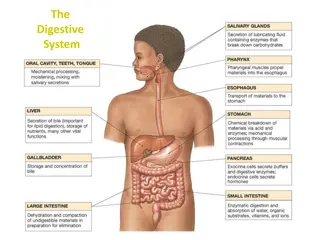
The human digestive system is a complex pathway that starts from the mouth and ends at the anus, involving various organs like the mouth, pharynx, esophagus, and stomach. Each part plays a crucial role in breaking down food particles, absorbing nutrients, and eliminating waste products. From chewing in the mouth to peristalsis in the esophagus and gastric motility in the stomach, each step is essential for proper digestion and nutrient absorption.

  • Digestive system
  • Human body
  • Digestion process
  • Gastrointestinal tract
  • Anatomy

Uploaded on Sep 16, 2024 | 0 Views


Download Presentation

Please find below an Image/Link to download the presentation.

The content on the website is provided AS IS for your information and personal use only. It may not be sold, licensed, or shared on other websites without obtaining consent from the author. Download presentation by click this link. If you encounter any issues during the download, it is possible that the publisher has removed the file from their server.

E N D

Presentation Transcript


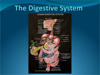
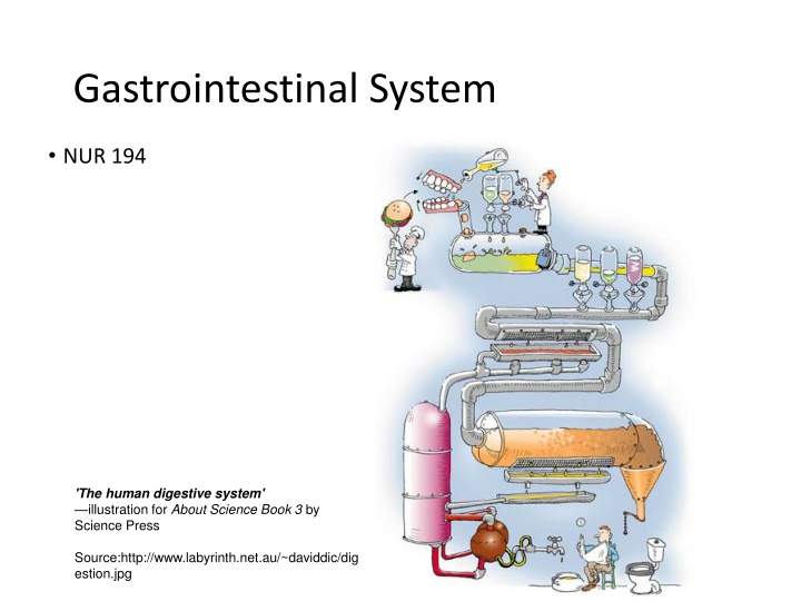
  1. Gastrointestinal System NUR 194 'The human digestive system' illustration for About Science Book 3 by Science Press Source:http://www.labyrinth.net.au/~daviddic/dig estion.jpg

  2. Quick Review: A&P GI tract 25 ft pathway from mouth to anus Function of GI tract: Break down food particles into molecules for digestion Absorb into the bloodstream the small molecules produced by digestion Eliminate undigested and unabsorbed foodstuffs and other waste products from the body

  3. Mouth Food entry Process of digestion begins with chewing (mastication) to break down foods into small particles that can be swallowed and mixed with digestive enzymes Saliva: First secretion that comes in contact with food; contains water and mucus; lubricant for swallowing Contains salivary amylase (enzyme: ptyalin) digests starches Produced in 3 pairs of glands: parotid, submaxillary, and sublingual. Approx. 1L of saliva is produced daily

  4. Pharynx The pharynx or throat extends from the soft palate to the esophagus. It is lined with mucous membrane and contains 3 pairs of organs: the adenoids, the lingual tonsils, and the tonsils.

  5. Esophagus Responsible for food passage from mouth to stomach Distends Smooth muscle contracts in rhythmic sequence (peristalsis) from upper esophagus to stomach to propel food along Upper Esophageal Sphincter (UES) (entry). Closed at rest to prevent air entering the esophagus during respiration. passes through diaphragm at opening called diaphragmatic hiatus Lower esophageal sphincter (LES)relaxes to allow food to enter stomach then closes tightly to prevent reflux of stomach contents back into esophagus. about 24cm or 10 inches long

  6. Stomach Lined with smooth muscle that promotes gastric motility. Expandable pouch, stores and mixes food with secretions; holds approx. 1500ml Parietal cells line wall and secrete hydrochloric acid (HCl) Breaks down food Kills most ingested bacteria Gastric secretions contain pepsin for protein digestion Gastric mucosa secretes intrinsic factor to combine with Vitamin B12 in food to be absorbed in ileum Parietal cells also produce intrinsic factor, which works to facilitate the absorption of Vit B12. Absence of the intrinsic factor causes pernicious anemia. Chyme: food particles mixed with gastric secretions

  7. Stomach Inlet: esophagogastric junction surrounded by smooth muscle (lower esophageal sphincter or cardiac sphincter) which contracts and closes off stomach from esophagus 4 anatomic regions: Cardia (entrance) Fundus Body or corpus Pylorus or antrum: outlet Outlet: Pyloric sphincter circular smooth muscle that controls the opening of the stomach to the intestine

  8. 3 phases of gastric secretion Cephalic- begins with sight, smell, and taste of food and is regulated by the vagus nerve. Gastric- begins with the presence of food in the stomach. Intestinal- begins as the chyme passes from the stomach into the duodenum, causing distention. Secretin : a hormone that inhibits further acid production and decreases gastric motility.

  9. Pancreas Gland that lies behind the stomach and extends to the spleen 2 cellular bodies within Pancreas: Exocrine: 80% of the organ and cells secrete enzymes needed for digestion of carbohydrates, fats, and proteins (trypsin, chymotrypsin, amylase and lipase Endocrine- produces glucagon & insulin.

  10. Liver Largest organ of body other than skin Located in RUQ of abdomen. Primary functions: Forms & secretes bile which is necessary for fat breakdown. Bile empties into the duodenum at sphincter of Oddi. If sphincter is closed, the bile is stored in the gallbladder.

  11. Small Intestine Longest segment about 2/3 the total length of GI tract Approx 7000 cm of surface area for secretion and absorption (the process by which nutrients enter bloodstream through intestinal wall --primary fx of small intestine). 3 anatomic parts: Duodenum (upper) Sphincter of Oddi Jejunum (middle) Ileum (lower) Cecum: junction between small and large intestine; ileocecal valve controls passage of contents into large intestine & prevents reflux of bacteria into small intestine. Appendix located at junction of small and large intestine

  12. Small intestine The common bile duct and pancreatic duct join to form the ampulla of Vater, emptying into the duodenum at the duodenal papilla. This papillary opening is surrounded by muscle known as the sphincter of Oddi. Sphincter of Oddi leads to the jejunum.

  13. Small Intestine Duodenum: contains intestinal glands & receives secretions from pancreas, liver, and gallbladder that aid in digestion (contain enzymes and bile) Intestinal glands: secrete mucus, electrolytes, and enzymes: Coat cells and protect mucosa from HCl Hormones, neuroregulators, and local regulators control rate of secretion production; influence GI motility Pancreas: Secretions have alkaline pH due to high bicarbonate concentration; neutralizes acid entering from stomach Digestive enzymes: Trypsin (protein digestion); Amylase (starch); lipase (fats) Liver: secretes bile (emulsify fats making them easier to digest and absorb) know all functions of the liver Chapter 55 Gallbladder: Bile storage Two types of contractions- stimulated by presence of chyme Segmentation: mixing waves; churns Intestinal peristalsis: propels contents of small intestine to colon Vitamins and minerals: absorbed essentially unchanged Absorption begins in jejunum diffusion and active transport

  14. Large Intestine Functions: movement, continuation of absorption and beginning of elimination Ileocecal valve allows passage of residual waste material from terminal ileum into proximal colon restricts reflux of contents back into ileum, Bacteria is major component of large intestine 2 colonic secretions: an electrolyte solution (bicarb to neutralize) and mucus (protects mucosa and provides adherence of fecal mass) Waste materials reach and distend rectum in about 12 hours

  15. Large Intestine Components: Begins at cecum. Ascending segment (R-side of abdomen) Transverse (right to left of upper abdomen) Descending segment (L side of abdomen) Terminal portion: sigmoid and anus Anus -- striated muscles form internal and external anal sphincters

  16. Waste Products Feces 75% fluid, 25% solid materials: foodstuffs, inorganic materials, water and bacteria Brown color comes from breakdown of bile Chemicals formed by bacteria are largely responsible for odor Approx 150 ml of gases are contained in GI tract: methane; hydrogen sulfide; and ammonia either absorbed into portal circulation and detoxified by liver, or expelled through rectum as flatus Process of elimination: Stimulation of rectum causes rectal distention which reflexively initiates contraction of rectal muscles causing the internal anal sphincter to relax (autonomic nervous system). The external anal sphincter is under conscious control of the cerebral cortex once allowed to relax will allow contents to be expelled. Contraction of the abdominal muscles (bearing down or straining) facilitates the emptying of the colon. Average frequency of defecation: once a day, but varies among individuals

  17. GI Changes with Aging Stomach: Decreased gastric HCL -Atrophic gastritis due to bacterial overgrowth Encourage bland foods high in vitamins & iron Assess for epigastric pain Large Intestine: Decreased peristalsis Constipation & Impaction High fiber diet Increased fluid intake (1500ml) if not contraindicated Encourage activity as tolerated Pancreas: Pancreatic duct change Steatorrhea: excess fat in stool. Small frequent feedings Assess for diarrhea Liver: Decreased enzyme activity & cholesterol synthesis Depresses drug metabolism leading to accumulation of drug and possible toxicity Assess for adverse effects of drugs Know Chart 55-1

  18. Assessment Take a complete history; focus on symptoms common to GI dysfunction Read Gordon s functional health patterns-Chart 55-2 Family & Genetic HX * Ex: Familial adenomatous Polyposos(FAP)-rpedisposition to colon cancer Previous hx of GI disease Past and current medication use. Including OTC & herbal products Previous or current treatments Previous surgeries Nutritional/Dietary hx: Food Allergies Tobacco and alcohol use Changes in appetite or eating patterns Anorexia (loss of appetite for food); dysphagia (difficulty swallowing); N/V; dyspepsia (indigestion/heartburn) Unexplained weight gain or loss Stool characteristics Socioeconomic, psychosocial, spiritual, or cultural factors Cultural: food restrictions, use of ingredients. Fasting 80-90% African Americans are lactose intolerant. Lack of enzyme lactase which converts dairy to glucose and galactose. (s/s: bloating, cramping, diarrhea) PE: Abdominal girth; tenderness/guarding; rectal exam; pelvic exam Diagnostics: CBC, urinalysis, abdominal x-rays, EKG, pregnancy test for the female

  19. PQRST P: precipitating or palliative Q: quality or quantity R: region or radiation S: severity scale T: timing

  20. Symptoms Common to GI Dysfunction Pain: can be major symptom; assess character, frequency, location, pattern, time of pain be aware of common sites of referred pain Indigestion: Upper abdominal discomfort or distress; most common symptom; Peristaltic activity may be contributing; BMs may or may not relieve pain. Fatty foods often perpetrator remain in stomach longer. Coarse vegetables and highly seasoned foods may cause distress.

  21. Symptoms Intestinal Gas: Accumulation of gas may leading to belching (expulsion from stomach thorough mouth) or flatus (from rectum) Complaint of bloating, distension, or full of gas Excessive flatulence may be a symptom of gallbladder disease or food intolerance. Nausea and Vomiting: Nausea : usually triggered by odors, activity, or food intake Emesis may vary in color and content; may contain undigested food particles or blood (hematemesis). Blood: If bright red signal of hemorrhage; if dark or has coffee-ground appearance it has been subjected to gastric enzymes

  22. Symptoms Change in Bowel Habits and Stool Characteristics May signal colon disease Diarrhea: abnormal increase in frequency or liquidity of stool Constipation: decrease in frequency of stools; stools that are hard, dry, or of smaller volume than normal

  23. Symptoms Stool Characteristics color and appearance Color: can be affected by foods or meds Blood: can present in several ways Tarry-black: blood shed in upper GI tract Bright red: blood enters in lower portion of GI Dark red: passing rapidly through GI tract Streaking of blood on surface of stool/toilet tissue: may be lower rectal or anal bleeding Common abnormalities of stool: Bulky, greasy, foamy; foul in odor; gray with silvery sheen Light gray or clay-colored caused by absence of urobilin Mucus threads or pus visible on gross inspection Small, dry, rock-hard masses called scybala may be streaked with blood from rectal trauma during passage Loose, watery stool that may or may not be streaked with blood

  24. Physical Assessment Examination includes: Mouth: tongue, buccal mucosa, teeth, gums note any ulcerations, nodules, swelling, inflammation; color changes Abdomen Rectum - anal and perineal areas: rash or excoriation; fissures or fistulas; hemorrhoids. Digital exam: masses or tender areas Performance of the exam must be followed in this order so intestinal activity & bowel sounds are not increased prior to exam. Inspection Auscultation Percussion Palpation

  25. Inspection Skin changes May result from GI Tract disorders such as liver and biliary obstructions Scars trauma, previous surgeries Contour and symmetry Bulging Distension Peristaltic waves Cullen s sign

  26. Auscultation Bowel Sounds all 4 quads using the diaphragm of the stethoscope (best for high-pitch and gurgling): Increased bowel sounds-borborygmus Character Location Frequency Normal sound is heard every 5 to 20 seconds Hypoactive one or two sounds heard in 2 minute period Hyperactive: 5 to 6 sounds heard in less than 30 seconds Absent: No sounds heard in 3 to 5 minutes BRUITS

  27. Percussion and Palpation Percussion: Tympany (high pitched) or dullness Palpation: 2 types: Light: detects large masses & areas of tenderness. Rebound tenderness (Blumberg s sign-used to further evaluate areas of pain). : exert pressure over an area and release quickly is there pain on withdrawal of pressure? If so, report to health care provider. Note any areas of tenderness or guarding for further evaluation during deep palpation. If rigidity noted, indicative of peritoneal inflammation. Deep: identify masses in 4 quads. Current practice is that only MD or APN s perform deep palpation. Used to determine size & shape of organs & masses.

  28. Diagnostics Blood Tests: CBC, PT, Carcinoembryonic antigen (CEA and the CA19-9); total calcium, Potassium, Albumin levels AST, ALT, amylase and lipase bilirubin, ammonia. Serum Cholesterol and Triglycerides Urine Tests: amylase; urobilinogon Stool Tests ( FOBT)-Fecal Occult Blood Test , FIT, Stool samples for ova/ parasites; stool test for clostridium difficile Know: Labs: Chart 55-3 for normal ranges

  29. Diagnostics contd Abdominal Ultrasound Plain films of the abdomen Upper GI Study Lower GI study PTC CT

  30. Endoscopy Endoscopy: direct visualization of GI tract with a flexible fiberoptic endoscope. Used to evaluate bleeding, ulceration, inflammation, tumors, and cancer of esophagus, stomach, biliary system, or bowel. May obtain specimans for biopsy or studies (H.pylori). Requires Informed consent by patient prior to procedure. EGD- examines esophagus, stomach & duodenum. May be used to dilate an esophageal stricture (during procedure) ERCP- includes exam of liver, gallbladder, bile ducts, and pancreas to identify cause & location of obstruction(s). Uses contrast so x-rays may be obtained. May obtain biopsies, or remove gallstones.Lasts 30 minutes to 2 hours. Reqiures Iv access for moderate sedation drugs and contrast.

  31. Colonoscopy Colonoscopy: endoscopic exam of entire large bowel.. Recommended every 10 years for all persons age 50+. More frequently if family hx of colon cancer. Evaluates for causes of chronic diarrhea, source of GI bleeding. Prep: Clear liquid diet 12-24 hours prior to procedure. NPO 6-8 hours prior may have water. Avoid aspirin, NSAIDS, and anticoagulants (with MD order) for several days Diabetic patients should check With MD re: diabetic meds as pt is NPO Night before procedure, Pt drinks Sodium Phosphate (Phospho-Soda) to clean bowel. May be repeated the morning of procedure. May prescribe Go-Lytely as a bowel cleanser the day before as well. Chill drinks to improve taste. Have pt drink quickly to avoid N/V. Watery diarrhea begins in about 1 hour. Additional laxatives or cleansing enemas may be required. Procedure: IV access for moderate sedation. Versed. Air is instilled to prmoote better viewing. Endoscopy passed through rectum to cecum. Atropine Sulfate : available in case of bradycardia from vasovagal response. After-Care: NPO until sedation wear off and pt is fully alert. VS every 15 minutes until pt is stable. Siderails up until Pt fully alert. Monitor for rectal bleeding or pain. May expect abdominal cramping for several hours. Monitor for bowel perforation, hypovolemic shock. Chart 55-4.

  32. Tests cont. Gastric Analysis, Gastric Acid Stimulation Test(Histolog), pH monitoring Ultrasonography, Endoscopic ultrasonography Liver-spleen scan

  33. The nurses role Is to prepare the client, explain the procedure, and provide the necessary post procedure care. Read and know the procedures so you can explain them to the client.

  34. Oral and Esophageal Disorders Chapter 56 Examining the Lips, Mouth, and Gums: Examples of Lip Conditions : lips-ppd-s herpes%25202 Cold Sore Chancre Contact Dermititis

  35. Stomatitis/Candidiasis Stomatitis is a painful ulceration of the oral mucosa that appear as inflammation and denudation of the oral mucosa, known as canker sores Candidiasis is a fungal/yeast infection resulting from an overgrowth of normal flora. Type of secondary stomatitis. Overgrowth may be caused by long term antibiotic use, chemotherapy, radiation, and HIV. White plaque on tongue. Red and painful underneath.

  36. Mouth Candidiasis Hairy Leukoplakia Lichen planus

  37. Mouth and Gums Aphthous Stomatitis (Canker Sore) Periodontitis

  38. Salivary Glands Acute Sialadenitis: Inflammation of salivary glands Causes: dehydration; radiation therapy, malnutrition, improper oral hygiene. Invasion of staph a.; streptococcus viridans;or pneumococcus S/Sx: Pain, swelling, purulent discharge Tx: Antibiotics; massage, hydration, corticosteroids; surgery to drain; analgesics, warm compresses, use of lemon slices & fruit/citrus flavor candy to stimulate saliva Best prevented by good routine oral hygiene. Xerostomia-very dry mouth . Usually caused by exposure of glands to radiation.

  39. Esophagus Chapter 57 Dysphagia: difficulty swallowing Most common symptom of esophageal disease Odynophagia: acute pain on swallowing Motility disorders: Achalasia: rare disorder. absent or ineffective peristalsis; accompanied by the esophageal sphincter that fails to relax during swallowing resulting in sensation of food sticking in lower esophagus. Diffuse spasm: Motor disorder with unknown cause; stress produces contractions of the esophagus

  40. Esophagus Gastroesophageal Reflux Disease (GERD) Caused by an incompetent lower esophageal sphincter, pyloric stenosis, or a motility disorder that allows backflow of gastric or duodenal contents S/Sx: Pyrosis (burning sensation in the esophagus); dyspepsia (indigestion); regurgitation; dysphagia; odynophagia- Chart 57-1 & Table 57-1 Tx: Low-fat diet; avoid caffeine, tobacco, beer, milk; peppermint/spearmint; carbonated beverages; avoid eating 3 hours before bedtime; maintain a healthy weight; avoid tight- fitting clothes; elevated head and/or HOB; surgical intervention for severe cases not responding to medical intervention. Chart 57-2

  41. GERD Tx: Meds: Antacids; Histamine receptor blockers (prevent or block the production of gastric acid): Cimetidine (Tagamet); Famotidine (Pepcid); Nizatidine (Axid); Ranitidine (Zantac) Proton Pump Inhibitors (decrease gastric acid production): Prevacid (lansoprazole), Aciphex (rabeprazole); Prilosec (Omeprazole); Nexium (esomeprazole magnesium) Prokinetic Agents (accelerate gastric emptying): Urecholine (bethanechol); Motililium (domperidone) and Reglan (metroclopramide) Untreated GERD can lead to Barrett s Esophagus a precancerous condition

  42. Esophagus Hiatal Hernia: opening in the diaphragm (through which the esophagus passes) becomes enlarged and the upper part of the stomach moves up into the lower part of the thorax. 2 major types: Sliding HiatalHernia Heartburn, regugitation, chest pain, dyshapgia, belching Paraesophageal Hernia Feels full after eating, breathless after eating, feeling of suffocation, chest pain mimics angina, s/s worse in recumbent position. Diverticulum: (Of esophagus). Herniation of mucosa into surrounding tissue. S/S: dysphagia, reflux, bad breath, nocturnal cough.DX by EGD. Small meals of semisoft foods, elevate HOB. May require surgical removal:

  43. Gastric Disorders-Chapter 58 Gastritis: inflammation of the gastric (stomach) mucosa; membranes become edematous and hyperemic (congested with fluid and blood) and begin to erode. Leads to ulceration and a potential for hemorrhage Acute: lasts hours to a few days; infection with Helicobactor pylori. usually caused by eating contaminated foods; highly seasoned foods; overuse of aspirin or NSAIDs; excessive alcohol intake; bile reflux; radiation therapy; or the ingestion of a strong acid or alkali. S/Sx: Acute: abdominal pain; headache; N&V; anorexia; hiccoughing Tx: Usually self-limiting; mucosa able to repair itself; refrain from food and alcohol until resolved; non-irritating diet during recovery; IV fluids. Acids or alkalis: dilution and neutralization; acids (common antacids), alkali (diluted vinegar or lemon juice)

  44. Gastric Disorders Gastritis: Chronic: repeated exposure to irritating agents; recurring episodes of acute gastritis Causes: Prolonged inflammation-- benign or malignant ulcers; bacteria Helicobacter pylori; autoimmune diseases (pernicious anemia) B12 deficiency; caffeine; medications; alcohol; smoking; reflux of intestinal contents. S/Sx: anorexia; heartburn; belching; N&V; sour taste in the mouth Tx: Modify diet; promote rest; reduce stress; pharmacotherapy antibiotics; proton pump inhibitors; bismuth salts (Pepto-Bismol).

  45. Gastric and Duodenal Ulcers Peptic Ulcer: musocsal lesion of the stomach or duodenum. Peptic Ulcer Disease (PUD): occurs when mucosal defenses are impaired and no longer protect the epithelium from the effects of acid and pepsin. Types of Peptic Ulcers: Gastric: occurs when break in mucosal barrier and HCL acid injuries lining. Delays gastric emptying resulting in regurgitation of duodenal contents. Deep & penetrating ulcers. Duodenal: chronic break in duodenal mucosa and leaves a scar after healing. High gastric acid secretion.. Most common ulcer. Stress Ulcers: acute lesions that occur after an acute medical crisis or trauma. Gastritis may lead to stress ulcers. Review Table 58-2

  46. Gastric and Duodenal Ulcers Stress and anxiety formerly thought to be the cause Caused by infection with gram-negative bacteria Helicobacter pylori Familial tendency may predispose; Blood Type O at greater risk; also associated with chronic pulmonary disease or chronic renal disease Associated with chronic use of NSAIDs, alcohol ingestion, and excessive smoking

  47. Gastric and Duodenal Ulcers S/Sx: May come and go; days to weeks to months and then disappear then reappear often without identifiable cause Dull, gnawing pain; usually relieved with eating Burning sensation in midepigastrium or back Pyrosis (heartburn); belching N&V (vomiting usually is indicative of complication and often follows a bout of pain and bloating; usually consists of undigested food ) Constipation or diarrhea (usually as a result of medication or diet regimen)

  48. Gastric and Duodenal Ulcers Assessment: Pain and epigastric tenderness; abdominal distention Diagnostics: Barium study of Upper GI; Endoscopy; Stool testing for occult blood; biopsy and histology with culture, breath test, or serological testing for H. pylori Tx:Medications:Antibiotics for h. pylori; histamine 2 (H2) receptor antagonists; proton-pump inhibitors for ulcers not associated with h. pylori. Also, lifestyle changes and surgical intervention Surgical Intervention read p.1278

  49. Nursing Considerations Relieving pain: meds; avoid aspirin, caffeine; regular meals; relaxation techniques to manage stress and pain; smoking cessation Reduce anxiety: Answer questions, explain diagnostic tests; administer meds on schedule; encourage family support. Maintain optimal nutrition: Assess for malnutrition and wt loss; teach about importance of complying with med and dietary regimen. Promote self-care teach clients to manage disease and factors that help or aggravate; medication regimen; dietary considerations; smoking cessation

More Related Content

giItT1WQy@!-/#giItT1WQy@!-/#giItT1WQy@!-/#giItT1WQy@!-/#giItT1WQy@!-/#giItT1WQy@!-/#giItT1WQy@!-/#giItT1WQy@!-/#