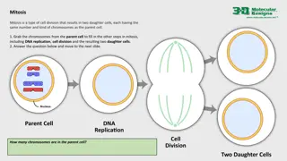
Understanding Human Chromosomes and Genetics in Health and Disease
Human chromosomes play a crucial role in genetics, ranging from heredity to disease. Cytogenetics studies their structure and behavior, essential for diagnostics like prenatal testing and identifying genetic disorders. The coiling and folding of DNA within chromosomes, along with the mitotic cell cycle phases, are key processes in understanding genetics. Karyotyping steps, such as culturing and staining, help analyze chromosomes for abnormalities. This informative guide sheds light on the significance of human cytogenetics and chromosome structure in various medical contexts.

Download Presentation

Please find below an Image/Link to download the presentation.
The content on the website is provided AS IS for your information and personal use only. It may not be sold, licensed, or shared on other websites without obtaining consent from the author. Download presentation by click this link. If you encounter any issues during the download, it is possible that the publisher has removed the file from their server.
E N D
Presentation Transcript
HUMAN CHROMOSOMES Human genetics 442 1 Foundation block RED : Important PINK : F. slides BLUE : M. slides GREEN : Doctors notes GRAY : Extra Editing file Editing file
Eukaryotic cell Not important
GENETICS: Cytogenetics The study of the structure and function of chromosomes and chromosome behaviour during somatic and germline division. Non-Banded Karyotype Banded karyotype High resolution karyotype 1 Molecular genetics The study of the structure and function of genes at a molecular level and how the genes are transferred from generation to generation. 2 Fluorescent in situ hybridization (FISH) Extra note: most of cancers are genetic mutations and most of the treatments are biological
Human Cytogenetics Definition : the study of human chromosomes in health and disease. Chromosome studies are an important laboratory diagnostic procedure in : prenatal diagnosis e.g. diagnosing Trisomy certain patients with mental retardation and multiple birth defect patients with abnormal sexual development some cases of infertility or multiple miscarriages in the study and treatment of patients with malignancies & hematologic disorders CHROMOSOMES 1- carry genetic material heredity: each pair of homologues consists of one paternal and one maternal chromosome, Crossing over occurs in meiosis I (prophase I). 2- 3- The intact set is passed to each daughter cell at every mitosis. EM of human chromosomes
Structure of Chromosomes Orders of DNA coiling and folding: Primary coiling: DNA double helix Secondary coiling: around histones (basic proteins) nucleosomes, Histones are positively charged proteins. Remember: Nucleosomes consist of the histone octamer. (Biochemistry first lecture) Tertiary coiling chromatin fiber Chromatin fibers form long loops on non-histone proteins tighter coils chromosome Mitotic cell cycle Mitotic cell cycle can be divided into two major phases: 1. Interphase - G1 (cell growth) - S (DNA synthesis) - G2 (cell growth) 1. Mitotic phase - Prophase - Metaphase - Anaphase - Telophase Extra Note : Chromosome develop in mitotic phase (prophase)
Karyotype Steps involved: CULTURING HARVESTING Slide-Making Banding Staining Karyotyping Chromosome Analysis Chromosome Preparation from Peripheral Blood Important points Phytohemagglutinin: stimulate T lymphatocytes to divide Colchicine and hypotonic saline: arrest cell division during metaphase Giemsa: stain
Dont forget single complete set of chromosomes. (N=23 for humans) Metaphase chromosomes 1 The 2 sister-chromatids are principally held together at the centromeric region. 2 Each chromosome has a centromere (CEN) region contains the kinetochore. CEN divides the chromosome into two arms: short arm (P arm) long arm (Q arm) 3 4 Each arm terminates in a telomere. Centromeric position arm length 1 The ratio of the lengths of the two arms is constant for each chromosome. This ratio is an important parameter for chromosome identification and allows classification of chromosomes into several basic morphological types: i-metacentric ii-sub-metacentric iii-acrocentric 2 In the human karyotype chromosome pairs 13, 14, 15, 21, 22, Y chromosome are acrocentric. Chromosome pairs 1,3,16,19 are metacentric. Rest chromosome pairs are sub-metacentric, including X chromosome. 3
Chromosomal classification 22 pairs of autosomes, numbered from 1 to 22 by order of decreasing length 1 pair of sex chromosomes: XX in the female XY in the male. Karyotyping Based on: 1-the length. (Each chromosome is unique in length) 2-the position of the centromere. 3-the presence or absence of satellites. Chromosomal satellite is the term given to that part of the end of a chromosome that is separated from the rest of the chromosome by a secondary constriction.
Items in the Description Of Karyotype Normal Karyotypes Abnormal Karyotypes Down Syndrome (47,XY,+ 21) Turner Syndrome (45,X) Banding 1 Certain staining techniques cause the chromosomes to take on a banded appearance, 2 Each arm presenting a sequence of dark and light bands 3 Patterns are specific and repeatable for each chromosome 4 Allowing accurate identification and longitudinal mapping for locating gene positions and characterising structural changes. 5 Patterns, and the nomenclature for defining positional mapping have been standardised
Chromosome Banding Band resolution = estimate of number of light + dark bands per haploid set of chromosomes 400 -850+ G Banding:Treat with trypsin and then with Geimsa Stain. Most common in karyotyping. Adenine&Thymine will appear as dark regions(stained), while Cytosine&Guanine will appear as light regions(not stained). R Banding:Heat and then treat with Geimsa Stain. Unlike G Banding, Adenine&Thymine will apear as light regions(not stained), while Cytosine&Guanine will appear as dark regions(stained). Q Banding:Treat with Quinicrine dye giving rise to fluorescent bands. It requires an ultraviolet fluorescent microscope. C Banding:Staining of the Centromere. Treat with acid followed by alkali prior to G banding The difference is in the stain and how to create it depending on what you are looking for
Fluorescence In-Situ Hybridization (FISH) Banded Karyotype: Normal Banded Karyotypes FISH of interphase nuclei with a chromosome 21 centromeric probe showing 3 signals consistent with trisomy 21 FISH of metaphase with a probe for telomere showing signals at the end of each chromatid A normal G-banded male Karyotype A normal R-banded male Karyotype AT-rich regions stain darker than GC-rich regions GC-rich regions stain darker than AT-rich regions
Take Home Messages The packaging of DNA into chromosomes involves several orders of DNA coiling and folding The normal human karyotype is made up of 46 chromosomes consisting of 22 pairs of autosomes and a pair of sex chromosomes, XX in the female, and XY in the male. Each chromosome consists of a short (p) and a long (q) arm joined at the centromere. Chromosomes are analyzed using cultured cells and specific banding patterns can be identified using special staining techniques. FISH is based on the ability of a single-stranded DNA probe to anneal to its complementary target sequence. It can be used to identify and study genes on chromosomes in metaphase or interphase.
MCQs: 1 .A Molecular cytogenetic technique based on the ability of a single stranded DNA probe to anneal with its complementary target sequence: A) (FISH) B) G. Binding C) Quinidine D) Karyotype Answer key: 1-A. 2-B. 3-A. 4-D. 5-D 2. Which one of the following chemicals is used to arrest cell division during metaphase for karyotyping? C) A) Cell culture media 3. In we treat it with trypsin and then with Geimsa stain. B) Colchicine Phytohemagglutini n D) Trypsin A) G Banding B) R Banding C) C Banding D) Q Banding 4. The study of human chromosome in health and disease: C) Molecular genetics D) Human cytogenetics A) Biochemistry B) Cytogenetics 5. Cytogenetics: A) Non-banded karyotype B) Banded karyotype C) High resolution karyotype D) All of them
MCQs: 6. Treat with heat and then Geimsa stain: A) G Banding B) R Banding C) Q Banding D) C Banding 7. The study of the structure and function of chromosomes and chromosome behavior during somatic and germline division: C) Molecular genetics A) Biochemistry B) Cytogenetics D) Histology 8. The genotype of Turner s syndrome is A) 47,XY,+21 B) 45,X C) 47,XXY D) 46,XX 9. CEN divides the chromosome into two arms: the short arm (p arm) and the long arm (q arm), each arm terminates in a A) Colchicine B) Trypsin C) Telomere D) Centromere 10. Culture media contains Phytohemagglutinin to stimulate to divide. A) B lymphocyte B) T lymphocyte C) Trypsin D) Colchicine
Team members M - team F - team Ali Jabaan Mohammed Alzeer Khaled Arbeed Abdulaziz Alnasser Muhannad Almuadi Saleh Alkholaifi Malik Halees Abdullah Awartani Walaa Almutawa Shahad Almarshad Razan Alsulami Atheer Alahmari Reema Aljubreen Mayssam Aljaloud Jana Alhazmi Layan Alburikan Team leaders Razan Almohanna leader Maram Beyari sub-leader Nawaf Alrefaei leader Ibrahim Alhezam sub-leader & humangenetics442@gmail.com


")