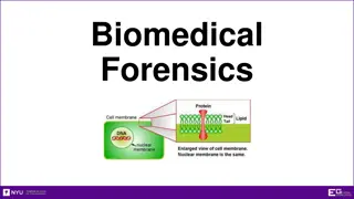
Understanding DNA Cloning with Filamentous Coliphages

Filamentous coliphages M13, f1, and fd are utilized as cloning vectors due to their circular single-stranded DNA molecules and advantages over other vectors. These phages have dimensions of 900 nm x 9 nm and infect bacteria through F pili, releasing up to 1000 phage particles per cell per generation without host-cell lysis. Gene VIII codes for the major structural protein, while Gene III codes for the minor coat protein.

Download Presentation

Please find below an Image/Link to download the presentation.
The content on the website is provided AS IS for your information and personal use only. It may not be sold, licensed, or shared on other websites without obtaining consent from the author. Download presentation by click this link. If you encounter any issues during the download, it is possible that the publisher has removed the file from their server.
E N D
Presentation Transcript
DNA cloning with single stranded DNA vectors M13, f1 and fd are filamentous coliphages containing a circular single-stranded DNA molecule. These coliphages have been developed as cloning vectors, for they have a number of advantages over other vectors, including the other two classes of vector for E. coli, plasmids and phage . This phage particles have dimensions 900 nm 9 nm contain a single-stranded circular DNA molecule (6407 (M13) or 6408 (fd) nucleotides long). The complete nucleotide sequences of fd and M13 are available and they are 97% homologous. Sequencing of f1 DNA indicates that it is very similar to M13 DNA.
M13 PHAGE Lambda Phage
The biology of the filamentous coliphages The phages only infect enteric bacteria harbouring F pili the end of the F pilus is the adsorption site . phage particles bind to F pilus only infects F+, Hfr, F' cells >single-stranded DNA genome enters cell designated as + strand infected cells continue to grow and divide, and extrude virus particles (but at slower rate compared to uninfected cells) Up to 1000 phage particles may be released into the medium per cell per generation Replication of phage DNA does not result in host-cell lysis. The process of phage transfect and release 1. The single-stranded phage DNA enters the cell 2. the single-stranded phage DNA is released.
The single-stranded phage DNA enters the cell the virus discard the capsid proteins the viral DNA passes into the cell converted to a double- stranded replicative form . the decapsidation and replication are tightly coupled. The RF multiplies rapidly inside the cell until about 100 RF molecules.
a viral-encoded protein accumulated. The protein binds to the viral strand and prevents synthesis of the complementary strand. As the DNA passes through the membrane, the DNA-binding protein is stripped off and replaced with capsid protein.
Gene VIII codes for the major structural protein of the bacteriophage particles Gene III codes for the minor coat protein
Replicative form (RF) RF contains + and strands strand is template for mRNA synthesis > for production of new + strands > by rolling circle replication + strands are packaged in phage coat protein > exit cell as phage particle M13 does not kill host > phage particles released without lysing cell membrane > slows growth of host, produces turbid plaques zones of slowed bacterial growth Single-stranded DNA > collected by growing M13 infected cells in culture > cultures centrifuged to pellet bacterial cells > phage remains in supernatant > until precipitated with Ficoll And then; DNA extracted from phage by phenol extraction.
When a normal M13 phage infect E. coli with F plasmid(strain JM101 in our experiment), it will use F pilus to put its genome into cytosol. Then this single strand genome will use host s polymerase to make itself a double strand structure and stay in cytosol like a plasmid. M13 genome contains major 9 genes, we call each gene s product as gp1, gp2, etc. When it wants to produce progeny phage, gp2 will recognize f1 ori and make a single strand break on genome. Then gp5 will form dimer structure and start to package this single strand DNA from package signal on f1 ori, this will help stabilize single strand genome in cytosol. After that, progeny phage will be released from host cell and be coated by gp3, gp6, gp7, gp8, gp9.
M13 Phage vectors A series of vectors (M13 mp series) have been developed from this phage. Wild type phage is not suitable as it contains few unique restriction sites. These vectors have a polylinker with unique restriction enzyme sites in lac Z gene that complements host (e.g. JM 103 or JM 104). Screening of recombinants is done based on formation of blue/white plaques. M13 vectors are used for obtaining sufficient quantity of DNA for sequencing by Sanger's dideoxy chain termination method.
M13 Cloning vectors M13mp18 & M13mp19 (SIZE: 7249bp) M13 phage with lacZ ' containing multiple cloning site; same gene and cloning site as pUC18 & pUC19 Packaging process is not linked to any size constraint of the M13 genome
Important points for cloning vectors >M13 occurs in both single and double stranded forms >RF can be digested with restriction endonucleases inserts can be cloned in, like plasmid + strands from phage particles > convenient source of single-stranded DNA used for sequencing and site-directed mutagenesis > different sized DNA molecules packaged as phage particle phage with inserts > 2 kb replicated slower different sized DNA molecules produce different size phage particles Both RF and single-stranded DNA will transfect competent E. coli cells to yield either plaques or infected colonies.
Why use single-stranded vectors? Sequencing by dideoxy method required single- stranded DNA oligonucleotide-directed mutagenesis required single- stranded DNA. certain probe preparation required single-stranded DNA. In summary: Filamentous phages possess all the advantages of plasmids and producing particles containing single-stranded DNA in an easily obtainable form. Inserts of up 42 Kb have been introduced into M13 genome and packaged (7x genome size) Ideal Insert capacity: 1 - 4 Kb


")
")