Understanding Plasmid Partitioning Mechanisms in Bacteria

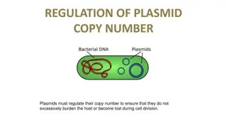
The stable maintenance of low-copy-number plasmids in bacteria relies on partition mechanisms that ensure proper positioning during cell division. Different from high-copy-number plasmids, which rely on random diffusion, low-copy-number plasmids require regulated partitioning mechanisms to prevent dilution from the population. These mechanisms involve genes like par organized on plasmids, a cis-acting centromere-like DNA site, centromere-binding proteins, and motor proteins. The partitioning process involves the formation of a nucleoprotein complex at the centromere-like site, directing plasmid movement to specific locations within the cell.

Download Presentation

Please find below an Image/Link to download the presentation.
The content on the website is provided AS IS for your information and personal use only. It may not be sold, licensed, or shared on other websites without obtaining consent from the author. Download presentation by click this link. If you encounter any issues during the download, it is possible that the publisher has removed the file from their server.
E N D
Presentation Transcript
The stable maintenance of low-copy-number plasmids in bacteria is actively driven by partition mechanisms that are responsible for the positioning of plasmids inside the cell. Partition is a dynamic process; plasmids are moved and positioned inside the cell so that cell division separates at least one copy into each daughter cell. For plasmids present in high copy numbers (50 to 100 copies per cell; e.g., pBR322), random diffusion may be enough to get at least one copy of the plasmid to each daughter cell. random segregation of low-copy-number plasmids (only 2 to 4 copies per cell) would likely mean that, following cell division, one of the daughter cells would not receive a plasmid. The plasmid would eventually be diluted from the population. Consequently, regulated partitioning mechanisms are essential for these plasmids. The mechanism used for partitioning differs depending on the plasmid.
PROPERTIES OF PARTITIONING SYSTEM Partitioning, especially of low-copy-number plasmids such as F factor and P1, usually involves genes called par organized on plasmids as gene cassettes. Plasmid copies are paired around a centromere-like site and then separated in the two daughter cells. Partition systems involve three elements, organized in an auto-regulated operon: 1) A cis-acting centromere-like DNA site 2) Centromere binding proteins (CBP) 3) The motor protein Cis-acting means that, for a plasmid to replicate, the DNA sequence in question must be part of that plasmid. It does not encode a diffusible product. In contrast, a trans-acting replication gene encodes a protein that does diffuse through the cytoplasm. Thus, the gene does not have to reside on the plasmid to help that plasmid replicate.
The centromere-like DNA site is also called partition, or par, site, which are the DNA sites that direct the action of the segregation machinery. The par sites are considered prokaryotic centromeres because they are required in cis for plasmid stability and because they are the assembly sites of the segregation machinery. It often contains one or more inverted repeats which are recognized by multiple CBPs. One partition protein is a site-specific DNA binding protein that recognizes the par site(s), and is often referred to as the centromere-binding protein, or CBP. The second protein is an ATPase or GTPase, which uses the energy of nucleotide binding and hydrolysis to move plasmid DNA inside the cell. Cis-acting centromere-like site and two trans-acting proteins that are usually called Par A and B, form a nucleoprotein complex at the centromere-like site termed the partition complex. This complex recruits the motor protein, which is a nucleotide triphosphatase (NTPase). The NTPase uses energy from NTP binding and hydrolysis to directly or indirectly move and attach plasmids to specific host location (e.g. opposite bacterial cell poles).
Plasmids are intrinsically located at mid-cell where they replicate. After replication, the daughter plasmids move from mid-cell to quarter-cell positions, by partition mechanism ensuring each daughter cell receives a plasmid copy. Partition Complex Recognition and Assembly The first step in plasmid partition is site-specific DNA binding of the CBP to the par site. Binding of one CBP is not sufficient for partition, and all partition complexes contain multiple CBPs bound to their respective par sites. This large structure therefore presents many potential binding sites for the cognate NTPase. The interaction of the CBP/plasmid partition complexes stimulates the NTPase activity of the partner, and this modulation of the ATP binding and hydrolysis cycles is necessary for plasmid dynamics. The CBPs are thought to pair (or group) plasmids together.
The partition systems are divided in three types, based primarily on the type of NTPases Type I : Walker type P-loop ATPase Type II : Actin-like ATPase Type III : tubulin-like GTPase Name of the different elements in the different types Centromere binding protein (CBP) Centromere-like binding site Type Motor protein (NTPase) Other proteins Type I ParA ParB or ParG parS (Ia) or parC (Ib) Type II ParM ParR parC Type III TubZ TubR tubS TubY
Type I partition system This system is also used by most bacteria for chromosome segregation. Type I partition systems are composed of an ATPase which contains Walker motifs and a CBP which is structurally distinct in type Ia and Ib. ATPases and CBP from type Ia are longer than the ones from type Ib, but both CBPs contain an arginine finger in their N- terminal part. ParA proteins from different plasmids and bacterial species show 25 to 30% of sequence identity to the protein ParA of the plasmid P1. The partition of type I system uses a "diffusion-ratchet" mechanism. This mechanism works as follows: Dimers of ParA-ATP dynamically bind to nucleoid DNA ParB bound to parS stimulates the release of ParA from the nucleoid region surrounding the plasmid The plasmid then chases the resulting ParA gradient on the perimeter of the ParA depleted region of the nucleoid The ParA that was released from the nucleoid behind the plasmid's movement redistributes to other regions of the nucleoid after a delay After plasmid replication, the sister copies segregate to opposite cell halves as they chase ParA on the nucleoid in opposite directions
Type Ia The CBP of this type consists in three domains: N-terminal NTPase binding domain Central Helix-Turn-Helix (HTH) domain C-terminal dimer-domain Type Ib The CBP of this type, also known as parG is composed of: N-terminal NTPase binding domain Ribon-Helix-Helix (RHH) domain
Diffusion-ratchet mechanism, as proposed for plasmid P1 type I par system. ParB (red circles) loads onto the plasmid at the centromere-like site parS, forming the partition complex. After plasmid replication, partition complexes develop repulsive interactions. ParB stimulates ParA (dark blue circles) ATPase activity, and ParA-ADP molecules (open blue circles) are then excluded from the nucleoid. The motive force for plasmid movement is directed toward regions of high ParA concentration. Movement of the partition complex is thus constrained to one direction because of the low ParA concentration behind it, and at the nucleoid end, it changes direction. ParA-ADP molecules diffuse randomly, exchange ADP for ATP (light blue circles), and then rebind the nucleoid.
Type II partition system This system is the best understood of the plasmid partition system. It is composed of an actin-like ATPAse, ParM, and a CBP called ParR. The centromere like site, parC contains two sets of five 11 base pair direct repeats separated by the parMR promoter. The amino-acid sequence identity can go down to 15% between ParM and other actin-like ATPase. The mechanism of partition involved here is a pushing mechanism: ParR binds to parC and pairs plasmids which form a nucleoprotein complex, or partition complex The partition complex serves as nucleation point for the polymerization of ParM; ParM-ATP complex inserts at this point and push plasmids apart The insertion leads to hydrolysis of ParM-ATP complex, leading to depolymerization of the filament At cell division, plasmids copies are at each cell extremity, and will end up in future daughter cell The filament of ParM is regulated by the polymerization allowed by the presence the partition complex (ParR-parC), and by the depolymerization controlled by the ATPase activity of ParM.
Pushing mechanism, exemplified by R1 type II par system. Partitioning complexes are formed through specific binding of ParR proteins (red circles) to the centromere-like site parC of newly replicated plasmid molecules, and serve as a nucleation point for ParM-mediated filament formation. Continuous insertion of ParM-ATP motor proteins (blue circles) on the filament ends pushes plasmid molecules apart. Conversion of ParM-ATP to ParM-ADP (open blue circles) leads to destabilization of the filament, thus allowing the entry of another ParM-ATP. At cell division, plasmid molecules localize near opposite cell poles, thus ending in daughter cells.
Type III partition system The type III partition system is the most recently discovered partition system. It is composed of tubulin-like GTPase termed TubZ, and the CBP is termed TubR. Amino-acid sequence identity can go down to 21% for TubZ proteins. The mechanism is similar to a treadmill mechanism: Multiple TubR dimer binds to the centromere-like region stbDRs of the plasmids. Contact between TubR and filament of treadmilling TubZ polymer. TubZ subunits are lost from the - end and are added to the + end. TubR-plasmid complex is pulled along the growing polymer until it reaches the cell pole. Interaction with membrane is likely to trigger the release of the plasmid. The net result being transport of partition complex to the cell pole.
Treadmilling by TubZ in type III partition systems. TubZ forms dynamic filaments, which grow at the plus (+) end by addition of TubZ-GTP and shrink at the minus (-) end by dissociation of TubZ-GDP. TubR associates with the C-terminal tail of TubZ. TubR/plasmid complexes move toward the plus direction as they are handed from one TubZ to the next in the polymer.