mmWave SAR
The world of millimeter wave SAR, including its operating frequencies, system design, and applications such as personal security, ground imaging, automotive imaging, and 3D imaging.
6 views • 15 slides
Evolution of Digital Radiography in Medical Imaging
Explore the evolution of digital radiography in medical imaging, from the invention of CMOS in 1963 to the development of Gadolinium-based flat-panel detectors in 1997. Learn about the advantages and challenges of digital imaging technology and its impact on the field of radiography.
0 views • 102 slides
Automated CT Perfusion Imaging in Acute Ischemic Stroke: Overview
This presentation delves into the significance of automated CT perfusion imaging in diagnosing and treating acute ischemic stroke. It covers essential information such as the burden of stroke in Ontario, the critical aspect of time in stroke treatment, hyperacute stroke treatment goals, and the impo
4 views • 41 slides
Basic Principles of MRI Imaging
MRI, or Magnetic Resonance Imaging, is a high-tech diagnostic imaging tool that uses magnetic fields, specific radio frequencies, and computer systems to produce cross-sectional images of the body. The components of an MRI system include the main magnet, gradient coils, radiofrequency coils, and the
2 views • 49 slides
Radiographic Imaging Methods of the Respiratory System
Radiographic imaging plays a crucial role in the evaluation and diagnosis of thoraco-mediastino-pleuro-pulmonary conditions. Techniques like radioscopy, digital radiography, computer tomography, magnetic resonance imaging, conventional pulmonary angiography, and hybrid imaging methods offer detailed
10 views • 21 slides
SALIVARY GLAND IMAGING
Salivary gland imaging plays a crucial role in diagnosing and monitoring diseases of the major salivary glands, including the parotid, submandibular, and sublingual glands. Different imaging techniques such as computed tomography, MRI, and ultrasound are used to visualize these glands and aid in dif
1 views • 12 slides
Overview of Jaw Cysts: Classification and Clinical Features
A cyst in the jaw is a fluid-filled cavity lined by epithelium that commonly occurs in the jawbones. There are different types of jaw cysts, such as odontogenic cysts and non-odontogenic cysts, each with specific clinical features and radiographic appearances. Understanding the classification and fe
9 views • 13 slides
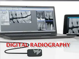
Understanding Digital Radiography in Modern Healthcare
Digital radiography revolutionizes imaging by capturing, displaying, and storing radiographic images digitally, offering benefits such as dose reduction, image manipulation, and electronic transfer. While there are advantages like environmentally friendliness, it also comes with challenges like high
0 views • 25 slides
Understanding Radiographic Changes Due to Tooth Resorption
Tooth resorption can be classified into physiologic and pathologic types, with external and internal resorption processes affecting teeth differently. Physiologic resorption is natural during primary teeth shedding, while pathologic resorption is triggered by abnormal stimuli. External resorption oc
0 views • 13 slides
Veterinary Dental Radiographic Positioning Techniques
Learn about common indications for dental radiographs in veterinary practice and proper patient positioning for accurate imaging. Explore techniques such as parallel view and the bisecting angle method for capturing quality dental X-rays. Enhance your skills in dental radiographic positioning for ef
0 views • 30 slides
Radiographic Imaging Processing Equipment Overview
The chapter delves into processing equipment for radiographic imaging, covering materials selection, manual processor construction, controls like thermostat regulators and inlet valves, and typical manual processing unit details. The content emphasizes factors like material resistance and cost, as w
0 views • 32 slides
Understanding X-Ray Production and Interactions in Radiology
Explore the fundamentals of X-ray production, including the types of ionizing radiation and interactions of photons with matter. Learn about the photoelectric effect, Compton effect, and characteristics of X-ray tubes, essential for radiology professionals and students. Delve into the mechanisms beh
0 views • 17 slides
Troubleshooting Today's X-ray Systems by John DiPasquale
Gain insights into troubleshooting modern X-ray systems with a comprehensive guide presented by John DiPasquale. Explore X-ray system subsystems, including generator systems and imaging systems, with detailed explanations and visuals. Learn about the basics of generator systems, required circuits, a
1 views • 33 slides
Diagnostic Reference Levels in Medical Imaging and Radiation Protection
The concept of Diagnostic Reference Levels (DRLs) is crucial in evaluating the amount of ionizing radiation used in medical imaging procedures. DRLs help determine if radiation levels are appropriate and need optimization. Authorized bodies establish numerical DRL values as advisory guidelines. Loca
1 views • 26 slides
Australian Imaging Biomarkers and Lifestyle Study: Progress and Focus
The Australian Imaging Biomarkers and Lifestyle Study of Ageing (AIBL) commenced in late 2006, with a cohort size and follow-up progress detailed. The study includes assessments and biomarker imaging, such as MRI, amyloid PET, and tau PET scans, with ongoing review cycles and data additions. Current
0 views • 12 slides
Enhancing Stroke Treatment Response Through Advanced Imaging Management Center Strategy
This information details the strategy proposed by the Imaging Management Center to improve stroke treatment response. It emphasizes the importance of timely uploading of brain imaging data within specified timeframes using the AMBRA platform. The process involves modalities like NCCT, CTA, MRI, and
0 views • 6 slides
Understanding Ionizing Radiation in Medical Imaging
Radiation in medical imaging plays a critical role in diagnosis and treatment but also poses risks to patients and healthcare workers. Ionizing radiation, used in various imaging modalities, can cause harm to healthy cells if not managed properly. This article discusses the implications of radiation
0 views • 54 slides
Radiology Techniques for Upper Limb Imaging at Al-Mustaqbal University College
Explore radiographic imaging techniques for upper limbs including fingers and thumbs at the Radiology Techniques Department of Al-Mustaqbal University College. Learn about PA, lateral, oblique, and AP views for detailed analysis. Proper positioning and imaging guidelines are provided to ensure accur
0 views • 20 slides
Sparse Millimeter-Wave Imaging Using Compressed Sensing and Point Spread Function Calibration
A novel indoor millimeter-wave imaging system based on sparsity estimated compressed sensing and calibrated point spread function is introduced. The system utilizes a unique calibration procedure to process the point spread function acquired from measuring a suspended point scatterer. By estimating
2 views • 26 slides
Portal Vein Imaging Techniques and Anomalies Overview
Portal vein imaging is crucial for evaluating conditions affecting the abdominal part of the gastrointestinal tract. Techniques such as spleno-portography and CT triphasic contrast imaging are used to visualize the portal vein and diagnose anomalies like portal-systemic collaterals and porto-systemi
4 views • 7 slides
Radiographic Evaluation of Olecranon Apophyseal Ossification System Guide
Comprehensive radiographic evaluation guide for the olecranon apophyseal ossification system, detailing stages from no ossification to complete fusion. Provides visual references and descriptions for each stage to aid in interpretation.
0 views • 11 slides
Introduction to Radiology: Imaging Modalities and Techniques
Radiology is a medical specialty that utilizes various imaging modalities such as X-Ray, MRI, CT, and Ultrasound to diagnose and treat patients. This field involves the supervision, performance, and interpretation of imaging studies, with findings reported to referring physicians. Radiology also inv
0 views • 10 slides
Evaluation of Success in Endodontic Treatment
Endodontic treatment success is crucial for patient well-being. Various criteria, including clinical, radiographic, and histological, are used to assess outcomes. Clinical signs like absence of pain and normal tooth mobility indicate successful treatment. Radiographic criteria involve checking for n
1 views • 44 slides
Understanding Filtration in Radiography
Filtration is a crucial process in radiography that involves eliminating undesirable low-energy photons to enhance image quality and reduce radiation dose to patients. This process involves using filters made of materials like aluminum to selectively absorb photons. Measurement of filtration is expr
0 views • 63 slides
Understanding Radiographic Faults and Artefacts in Veterinary Imaging
Learn about radiographic faults and artefacts in veterinary radiography, including technical errors like motion artefacts, dark radiographs, gray non-contrast images, and light radiographs, as well as processing errors such as dark spots and white spots on radiographs. Discover the causes of these e
0 views • 12 slides
Advanced Microscopy Techniques in EUV Lithography: SHARP Overview
SHARP utilizes Fresnel zone plate lenses to achieve diffraction-limited quality in EUV lithography, offering a range of NA values and image magnifications. The system allows emulation of mask-side imaging conditions with hundreds of lenses available. Coherence control and engineering are provided th
1 views • 18 slides
Insights into Multi-View Imaging System Optimization
Delve into the simulation and calibration of a multi-view imaging system using differentiable ray tracing and gradient-based optimization. Explore the challenges of ambiguity in results and the impact of angular offset on imaging accuracy. Discover how the system handles errors and maintains precise
0 views • 6 slides
Understanding Radiographic Testing: History, Science, and Radiation Safety
Exploring the history and science behind Radiographic Testing (RT), from the discovery of X-rays to the development of methods using gamma radiation. Learn about the terminology, natural radiation, and the characteristics of radiation in various forms. Understand the importance of safety measures in
0 views • 47 slides
Understanding PET-CT Imaging in Adrenal Tumors at Tehran University
Tehran University of Medical Sciences' Research Center for Nuclear Medicine specializes in PET-CT imaging for adrenal tumors. Dr. Arman Hassanzadeh-Rad, MD, explains the principles of PET imaging, hybrid imaging combining PET and CT, F-18 FDG use, SUV measurement, and more. PET-CT allows for both at
0 views • 21 slides
Simulation Study of Photoacoustic Imaging for Bragg Peak Reconstruction
This study explores the use of simulation in Photoacoustic Imaging for reconstructing the Bragg peak in proton therapy. It discusses the concept of Photoacoustic Imaging, the generation of acoustic waves from pulsed electromagnetic radiation, and the experimental setups for characterizing proton dos
0 views • 13 slides
Overview of Radiography and Ultrasound Imaging Techniques
This content provides a comprehensive overview of conventional radiography, digital radiography, and ultrasound imaging techniques. It covers equipment details, safety issues, advantages vs. limitations, as well as the interpretation, magnification, and integration capabilities of these imaging moda
0 views • 28 slides
Understanding Magnetic Resonance Imaging (MRI)
Magnetic Resonance Imaging (MRI) is an imaging technique based on nuclear magnetic resonance principles. It was first developed in the 1970s by Paul Lauterbur and Peter Mansfield. MRI uses the interaction between protons in the body and magnetic fields to create detailed images. This technology has
0 views • 77 slides
Comprehensive Radiological Imaging Review in Abdominal and Adrenal Pathologies
This extensive radiological imaging review covers a wide range of topics in abdominal and adrenal pathologies, discussing differential diagnoses, imaging modalities such as CT and USG, specific syndromes like prune belly syndrome, and conditions like retroperitoneal fibrosis. It delves into various
0 views • 39 slides
Introduction to Forensic Radiology: Techniques and Applications
Forensic radiology, as pioneered by Dr. Nashwa Mahmoud Mostafa, involves the use of radiographic techniques to assist in legal investigations and post-mortem examinations. Beginning with the historical overview of X-rays in forensic science, the field has evolved to utilize advanced imaging technolo
0 views • 68 slides
Understanding Elbow Joint Disorders in Veterinary Medicine
The elbow joint is a true diathrodial joint formed by the articulation of various structures. Disorders such as elbow luxation, un-united anconeal process, and fragmented coronoid process can lead to lameness and other clinical signs in animals. Causes include osteochondritis dissecans and trauma, w
0 views • 16 slides
Comprehensive Overview of Radiology in Pediatric Dentistry at Semmelweis University Budapest
Radiology plays a crucial role in diagnosing and treating diseases within the body, especially in pediatric dentistry at Semmelweis University Budapest. From the historical evolution of radiography to the modern radiologic diagnostic imaging techniques used in dentistry, this department utilizes a r
1 views • 40 slides
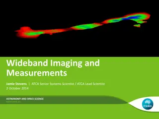
Wideband Imaging and Measurements in Astronomy
Wideband imaging and measurements play a crucial role in astronomy and space science. This involves utilizing wide bandwidths to make accurate measurements and create images for continuum mapping and spectral-line observations. Increasing bandwidth enables lower thermal noise levels, quicker identif
0 views • 32 slides
Overview of DICOM WG21 Multi-Energy Imaging Supplement
The DICOM WG21 Multi-Energy Imaging Supplement aims to address the challenges and opportunities in multi-energy imaging technologies, providing a comprehensive overview of imaging techniques, use cases, objectives, and potential clinical applications. The supplement discusses the definition of multi
0 views • 33 slides
Understanding Fluoroscopy in Veterinary Surgery and Radiology
Fluoroscopy is a dynamic radiographic examination primarily used by radiologists in the Department of Surgery and Radiology at the College of Veterinary Science and Animal Husbandry, DUVASU. This imaging technology, discovered in 1896, involves X-ray tubes and image receptors mounted on a C-arm for
0 views • 51 slides
Understanding Initial Asymptotic Acoustic RTM Imaging Results in Salt Model
Acquire insights into the initial asymptotic acoustic RTM imaging results for a salt model in Xinglu Lin, San Antonio. This study delves into the concept of Reverse Time Migration (RTM), showcasing the methodology, workflow, and imaging conditions involved in this innovative seismic imaging techniqu
0 views • 22 slides


