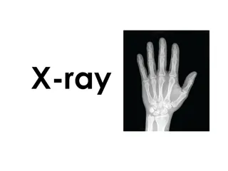
Troubleshooting Today's X-ray Systems by John DiPasquale
Gain insights into troubleshooting modern X-ray systems with a comprehensive guide presented by John DiPasquale. Explore X-ray system subsystems, including generator systems and imaging systems, with detailed explanations and visuals. Learn about the basics of generator systems, required circuits, and specialty circuits for controlling x-ray lengths and radiographic processes.
Download Presentation

Please find below an Image/Link to download the presentation.
The content on the website is provided AS IS for your information and personal use only. It may not be sold, licensed, or shared on other websites without obtaining consent from the author.If you encounter any issues during the download, it is possible that the publisher has removed the file from their server.
You are allowed to download the files provided on this website for personal or commercial use, subject to the condition that they are used lawfully. All files are the property of their respective owners.
The content on the website is provided AS IS for your information and personal use only. It may not be sold, licensed, or shared on other websites without obtaining consent from the author.
E N D
Presentation Transcript
Troubleshooting Todays X-ray Systems Presented by John DiPasquale
A little about me Got my start in electronics while in the USAF Aircraft electrician 4 yrs. BMET 18 yrs. o Technician, Manager, Instructor and Regional Manager After military service 8 years in various imaging and biomedical capacities including asset management for both in- house and 3rd party contract positions Left industry to teach HS Mathematics in the Connecticut Vocational Technical HS System Came back to industry in 2006 o 1st OEM experience with Hologic o Technical trainer on both analog / digital imaging systems and support systems for their mammography lines Moved to Wisconsin in early 2013 and began career with TP in December of that year. Education & Credentials Masters degree in Education Bachelors degree in Electronics Engineering and Business Administration AAS in Instructor and Biomedical Equipment Technologies ICC CBET since 1994 USAF Master Instructor 2
What we are going to cover What we are going to cover X-ray System s Subsystems Troubleshooting 3
X X- -ray Systems Subsystems ray System s Subsystems Generator Systems Imaging Systems 4
Generator Systems The Basics 5
Generator Systems(2) Required Circuits PHT / SHT Controls length of x- ray Develops kV Split Secondary Phase shifting Regulated Older days Step up transformer, 1:500 ratio AC voltages & line frequency Currently Rectifier & frequency controlled inverter circuits on primary side Rectified secondary PFT / SFT Develops mA Direct & indirectly heated cathodes Negative side of tube Split regulation Filament preheat & boost mA amount during x-ray Older days Step down transformer, 200:1 AC voltages & line frequency Currently Rectifier & frequency controlled inverter circuits on primary side Rectified secondary Power Line to transformer 230VAC to 480VAC Single or three phase 6
Generator Systems (3) Specialty Circuits Timer circuits Controls the length of the x-ray Radiographic (high dose x-rays) 2 timers Simultaneous start Primary (5 seconds maximum radiographic mode) Backup (energizes after ~6 seconds) Fluoroscopic (low dose x-rays) 5 minute & resettable Rotor circuits Phase shift caused by capacitor start motor circuit Speeds Low ( 4500 RPM) High ( 10K RPM) Start circuits AC Pulsed DC Brake circuits DC Temp electro magnet Interlock Circuits Monitors all conditions set by law or the operator Techniques Environmental Prevents exposure start Unless all conditions are satisfied Types: Relay logic And gate logic Microprocessor controlled today 7
Generator Systems (4) Generator Types Voltage Waveforms Single Phase Half wave Full wave Three Phase 6 pulse 12 pulse High Frequency Most common today Pulsating DC Constant Potential Generally found in portable systems if used 8
Imaging Systems Radiographic Imaging Systems Types & Components Film Systems Components 3D Top & Tube Stands Tube & Collimator positioning X, Y, & Z axis movements & rotations Table / Wall Stand Phototimers Bucky/Grids Film Direct Screen type - Single/double emulsion Cassettes Paper Screen Processor Manual Automatic Viewing Light Boxes Hot lights Film Storage Rooms Size Temp & Humidity controlled CR Systems Components 3D Top & Tube Stands Table / Wall Stand Cassettes Reader Computer Monitors PACS Digital Systems Components 3D Top & Tube Stands Tube & Collimator positioning X, Y, & Z axis movements & rotations Table / Wall Stand Phototimers Bucky/Grids Detectors Direct Scintillation layer type Flash circuits Types Computers Processing software Image Storage Monitors Technologist Physician PACS Storage & Retrieval Tube & Collimator positioning X, Y, & Z axis movements & rotations Phototimers Bucky/Grids Intensifying screen Plate Laser PMT / camera Light - flashing Fixed Wireless Processing software Image Storage Technologist Physician Storage & Retrieval 13
Imaging Systems (2) Radiographic Imaging Key Terminology SID Source to Image Distance SOD Source to Object Distance OID Object to Image Distance Magnification of Image HVL Half Value Layer How much aluminum is needed to cut dose in half Used to measure changes in inherent filtration Focal Spot Actual Physical area bombarded by electrons Effective Geometric derived value of central ray used in determining size Filaments are rated by this. 14
Imaging Systems (3) Fluoroscopic Imaging Systems Types & Components Film Systems Components Spot film device Holds I.I. device Phototimer Film Electro-mechanical device Image Intensifier Tube Converts x-rays to light energy & brightens image Light Sensor Part of auto kV circuit PMT Mirror Optics Directs image to different viewing /recording devices Pick up devices Camera tubes Monitors Monitors CR Systems Components Spot film device Holds I.I. device Phototimer CR cassette Electro-mechanical device Image Intensifier Tube Converts x-rays to light energy & brightens image Light Sensor Part of auto kV circuit PMT / PDA Mirror Optics Directs image to different viewing /recording devices Pick up devices Camera tubes CCD cameras Digital Systems Components Spot Film Devices Houses detector Electronic image capture Detectors Direct Scintillation layer type Flash circuits Types Light Sensor Part of auto kV circuit PDA Pick up devices CCD cameras Computers Processing software Image Storage Monitors Fixed Portable 15
Imaging Systems (4) Fluoroscopic Imaging Key facts Generally real time exams Typically uses an under-table tube and radiation is shooting upward Exam dependent May require use of contrast media x-ray altering media that either positively or negatively affects the image depending on what is used to make something standout X-ray tube & Imaging Device mounted on C-arms to maintain SID 16
Troubleshooting Todays X Troubleshooting Today s X- -ray Systems ray Systems General Info & Thoughts Fluoroscopic Radiographic Preventive Items 17
Troubleshooting General Information & Thoughts Discreet devices evolution Individual components Single purpose PCBs Multiple purpose PCBs 4 key checks Signal in Signal out Power Reference Earth Ground (Protective) Circuit AC DC Floating 18
Troubleshooting (2) General Information & Thoughts (cont.) Analysis questions to ask yourself What was it told to do? Did it do it? Another set of questions Proper squiggly in? Proper squiggly out? Size & shape 19
Troubleshooting (3) General Information & Thoughts (cont.) X-ray machines are VERY logical These systems must have things happen in a sequential order It is a building process Figure out what is missing in the process and focus on that Types of logic used over the years Relay And/Or Gates Microprocessors & programs Microprocessor looks for information (feedback) Time period responses Understand the limitations of the test equipment you are using Example: Oscilloscope vs DMM Both read voltage Both are connected in parallel to circuit Use circuit reference NOT ground Difference is the time based response Oscilloscope Voltage at an instant in time Time per division DMM RMS voltage over time Sampling rate 20
Troubleshooting (4) General Information & Thoughts (cont.) Understand the components PRIMARY function in the system Generator Produce kV & mA Control exposure length Control Panel / Operator Console Sets techniques OGP Manual Environment Table Patient height May have top adjustments Motorized Floating May or may not tilt 90/90 90/(15-20) 2 key signals for generator Houses Bucky/Grid assemblies - movement Understand the components PRIMARY function in the system (cont.) Wall Stand May or may not - tilt or be motorized Houses Bucky/Grid assemblies Similar inputs as table Stand Movements (Tube & Detector) 3D Movements X, Y, & Z axis Position Movements & Brakes & Tube positioning detents Collimator Light field represents x-ray field May be manual / automatic Automatic PBL / ACSS associated acronym CAN system Typically 4 blades w/ 2 motors May have IRIS function 8 blades w/ 4 motors (Fluoro) X-ray Tube Produces the x-rays Houses the rotor 21
Troubleshooting (5) General Information & Thoughts (cont.) The unwrittenrule of troubleshooting If you don t know what you are looking for stay out of the machine!!! Justifying what the machine tells you will only lead you down the rosy red path to nowhere & probably give you a headache from beating your head up against the wall 22
Troubleshooting (6) Radiographic Systems Suggestions Get as much information from the end user as possible describing what is wrong with the system or about the problem. Do not be afraid to ask questions and get clarification if you aren t sure what you are being told, especially if the message isn t clear cut Table won t go down The image isn t clear, blurry, has lines in it Allow the system to help you identify what may be happening and causing your issue Example: Generate a table top x-ray Generate a table x-ray Generate a photo-timed x-ray Know the differences on how the system is expected to perform and focus on those areas. What was added/removed by the environment change 23
Troubleshooting (7) Radiographic Systems Suggestions (cont.) If in the generator Figure out what s missing If kV is missing All interlocks satisfied Half split methodology Generator vs Imaging System Items needed Intensifying screen kV meter mA/mAs meter Generate an x-ray Use either a 2 or 3 point technique Screen glows x-rays coming out kV meter indicates selected kV mA/mAs meter indicates amount of selected mAs If all are good, problem will generally lie in the imaging system, NOT the generator 2 key external interlock signals Cassette present Bucky movement Are items reset Error codes, information available If mA/mAs missing Filaments open Proper filament regulation during standby & boost Proper regulation for mA during x-ray Are the power supplies proper and working 24
Troubleshooting (8) Radiographic Systems Suggestions (cont.) Film Based Film Processor biggest culprit Compare sample against daily QC results for processor If not within range processor needs attention Generally speaking Chemical strength Temperature issues Optical Density (OD) not proper Dark Room Safe light Chemical fumes Storage cabinet Cassettes Poor contact Light leaks If problem is in the Imaging System, then the type of Imaging System determines what needs to be checked/done Film Based CR Based Detector Based 25
Troubleshooting (9) Radiographic Systems Suggestions (cont.) CR Based S-curve value Must be minimum value for reader to work Similar in nature to OD value used in film but generally higher Reader Not clearing plate Flash deteriorating Ghosted image (double exposure) Defective laser Defective pickup device Computer Data transfer Corrupt program Defective computer Monitor SMPTE adjustment Defective display 26
Troubleshooting (10) Radiographic Systems Suggestions (cont.) Detector based Detector files Detector Correct & current files Latest mapping/calibration files Power supply Flash deteriorated or not working Defective pixels Bad cables (fiber optic) Wireless Defective battery Not paired Computer Data transfer Corrupt program Defective computer or PCB Files corrupt, missing or incorrect Monitor SMPTE adjustment Defective display 27
Troubleshooting (11) Fluoroscopic Systems Suggestions On the Imaging System side one has to keep in mind this is live imaging not a single shot Image Intensifier Tube Biggest factor are the voltages to the acceleration plates High voltage Requires a HV probe for your meter Some systems have representative low voltage test points for this Use a mirror to check the phosphor output of the tube Digital Detector High Voltage to Detector Use mirror to check the phosphor output of the detector All previous items discussed still apply Half split methodology Generator info Two key additional checks to add on the generator side though Max R output of the tube ensuring the generator is doing its part Ensure you measure it according to manufacturer s specifications Use the probe sensor indicator to set distance Use different items of thickness & density in the beam Verify the auto kV circuit is working 28
Troubleshooting (12) Fluoroscopic Systems Suggestions (cont.) Pickup devices Types Collimator Fluoro typically has an 8 blade collimator effectively creating a octagonal (circular) input to the I.I. Smaller the field, the larger the magnification of the item being examined. Most systems have 3 different levels of magnification Most common problems Motors Calibration PMT / PDA Typically positioned on the output side of the I.I. / detector Used to sense the amount of light coming out of the I.I. / detector Part of the auto kV adjustment circuit used to ensure the correct amount of light is being sent to the pickup device. Alters penetration of the x-rays by changing kV Changes output light back to normal value Video tubes CCD cameras Takes the light from the I.I./detector and sends it off to the computer for processing as digital data Video tubes output degrades over time CCD cameras Require defect maps for computer processing Bad pixels Camera Iris not working correctly Mirror Optics Directs the output of the I.I./detector to different devices for the purpose determined by the radiologist. Mirror movement Light out 29
Troubleshooting (13) Fluoroscopic Systems Suggestions (cont.) Spot Film Devices (C-arm) Older days Electro-mechanical nightmares Movement controls for adjusting the c-arm mechanism during the exam Positions film for a radiographic exposure based on programming pattern 1x1, 2x1 (L/H), 4x1, 6x1, 9x1, 12x1, 16x1 Temporarily shifted operation of the system from fluoroscopy to radiography and then back again when completed Records what Doctor saw during an exam Higher mAs & dose to patient Phototimed x-ray Collimator leaves Iris mode goes to rad mode, then back to Iris mode Brakes c-arm during rad exposure Spot Film Devices (C-arm) (cont.) Newer devices Movement control for positioning the c- arm over the patient during the exam Does an image capture of the fluoroscopic image that is seen by the doctor Multiple screen systems can show both the live fluoro exam and the most recent image capture Lower radiation to patient as the fluoro image is what is captured Less mechanical devices for FE s to concern themselves with More remote movement controls for operator s to use creating multiple ways of controlling the movements of the components. All images processed by computer on the system 30
Troubleshooting (14) Fluoroscopic Systems Suggestions (cont.) Monitors Calibrations SMPTE Patterns VGA/HDMI outputs Color / Monochrome images Use separate monitors Set color for B&W (monochrome) if desired Power supplies Proper pixelization Doctor vs technologist monitors Computers Input connections Fiber Optic Cables used with Imaging Keyboards Mouse Power supplies HDDs Damaged Corrupt files Maps & Calibration files Video cards Image processing 31
Troubleshooting (15) Suggested Preventive Items for the Field Engineer When performing service Do a backup before you begin Perform the work necessary Do a backup after the service is done Update info left on site For those of you servicing multiple sites Sharing information between all engineers responsible for the site Do NOT solely rely on the internet for access to this information Cloning computer hard drives Suggested programs to use Norton Ghost CloneZilla Frequency After initially installed Changes to programming Any software upgrade Annual PM Backups At the conclusion of a PMI Things to consider Generator System calibrations Computer information 3D Top / Floor Stand Table Collimator Wall Stand 32
ContactInformation My information: John DiPasquale, ext. 8480 jdipasquale@technicalprospects.com Technical Prospects 1000 County Road CB Appleton, WI 54919 Main #: (920) 757-6583 Toll Free #: (877) 604-6583 Fax #: (920) 757-6591 www.technicalprospects.com Parts/Sales: Customer Service Reps, ext. 2101 parts@technicalprospects.com Training Information: Linda Fuerst, ext. 8202 training@technicalprospects.com 33
