Understanding LacZ - Galactosidase Enzyme: Structure, Function, and Applications
LacZ - galactosidase enzyme, encoded by the lacZ gene in the lac operon, plays a crucial role in cleaving lactose, transgalactosylation, and regulating gene expression. Its application in labs using X-gal as a substrate allows for easy detection of active enzyme through color changes. This versatile enzyme serves as a valuable tool in genetic, cellular, and molecular biology research.
Download Presentation

Please find below an Image/Link to download the presentation.
The content on the website is provided AS IS for your information and personal use only. It may not be sold, licensed, or shared on other websites without obtaining consent from the author. Download presentation by click this link. If you encounter any issues during the download, it is possible that the publisher has removed the file from their server.
E N D
Presentation Transcript
LacZ -galactosidase : Structure and function Dr. Susan A. Ibrahim
Lac operon made up of three specific lac genes; there are lacZ, lacY and lacA and they can be found in the structural genes of the operon. The lacZ gene, encoding the enzyme galactosidase ( -gal), is a large (120 kDa, 1024 amino acids protein that forms a tetramer. It is a ubiquitous reagent applied to the study of problems in genetics, cell and molecular biology, development, and the recently emerging fields of genomics and proteomics. Because -gal activity is readily assessed invitro or invivo, in hosts as evolutionarily diverse as bacteria, yeast, and mammals, its induction has become a standard means of assaying clonal insertion, transcriptional activation, protein expression, and protein interaction.
-Galactosidase has three enzymatic activities (Fig. 1). - First, it can cleave the disaccharide lactose to form glucose and galactose, which can then enter glycolysis. - Second, the enzyme can catalyze the transgalactosylation of lactose to allolactose. - Third, the allolactose can be cleaved to the monosaccharides. It is allolactose that binds to lacZ repressor and creates the positive feedback loop that regulates the amount of -galactosidase in the cell. Fig. 1: Schematic summarizing the roles of -galactosidase in the cell. The enzyme can hydrolyze lactose to galactose plus glucose, it can transgalactosylate to form allolactose, and it can hydrolyze allolactose. The presence of lactose results in the synthesis of allolactose, which binds to the lac repressor and reduces its affinity for the lac operon. This in turn allows the synthesis of -galactosidase, the product of the lacZ gene.
Application of lacZ -galactosidase in the lab -galactosidase is best recognized for its reaction with X-gal (5-bromo-4- chloro-3-indoyl-b-D-galactopyranoside), a soluble colorless compound consisting of galactose linked to a substituted indole. X-gal is an analog of lactose, and therefore may be hydrolyzed by the - galactosidase enzyme which cleaves the -glycosidic bond in D-lactose. X-gal, when cleaved by -galactosidase, yields galactose and 5-bromo-4- chloro-3-hydroxyindole (1). The latter then spontaneously dimerizes and is oxidized into 5,5'-dibromo-4,4'-dichloro-indigo (2), an intensely blue product which is insoluble. X-gal itself is colorless, so the presence of blue-colored product may therefore be used as a test for the presence of active -galactosidase. This also allows for bacterial -galactosidase (so called lacZ ) to be used as a reporter in various applications.
On growth medium containing X-gal, colonies of E. coli that have an active -galactosidase become blue because of this reaction. The blue color of a crystal of -galactosidase exposed to X-gal confirms that the enzyme in the crystal is catalytically competent.
White-blue screen Some strains of E. coli have a modified lacZ gene, one that lacks the segment referred to as lacZ and coding for the a-peptide portion of -galactosidase. These mutants can synthesize the enzyme only when they harbor a plasmid, such as pUC8, that carries the missing lacZ segment of the gene. Most plasmid vectors carry a short segment of lacZ gene that contains coding information for the first 146 amino acids of -galactosidase. The host E. coli strains used are competent cells containing lacZ M15 deletion mutation. When the plasmid vector is taken up by such cells due to -complementation process, a functional -galactosidase enzyme is produced. A cloning experiment with pUC8 involves selection of transform on ampicillin agar followed by screening for -galactosidase activity to identify recombinants. Cells that harbor a normal pUC8 plasmid are ampR and able to synthesize -galactosidase (Fig. 2a); recombinants are also ampR, but unable to make galactosidase (Fig.2b).
Fig.2: The cloning vector pUC8: (a) the normal vector molecule; (b) a recombinant molecule containing an extra piece of DNA inserted into the BamHI site.
Screening for -galactosidase presence or absence is in fact quite easy. Rather than assay for lactose being split to glucose and galactose, we test for a slightly different reaction that is also catalyzed by -galactosidase. This involves a lactose analog called X-gal, which is broken down by -galactosidase to a product that is colored deep blue. If X-gal (plus an inducer isopropylthiogalactoside, IPTG) is added to the agar, along with ampicillin, then non-recombinant colonies, the cells of which synthesize -galactosidase, will be colored blue, whereas recombinants with a disrupted lacZ gene and unable to make -galactosidase, will be white. This system, which is called Lac selection, is summarized in Fig.3. * Note that both ampicillin resistance and the presence or absence of -galactosidase are tested for on a single agar plate. of the enzyme such as
Fig.3: The rationale behind insertional inactivation of the lacZ gene carried by pUC8. (a) The bacterial and plasmid genes complement each other to produce a functional - galactosidase molecule. (b) Recombinants are screened by plating onto agar containing X-gal and IPTG.
Activity of Individual -Galactosidase Molecules Craig and coworkers have developed a novel procedure to measure the activity of single -galactosidase molecules. It depends on the conversion of the weakly fluorescent substrate resorufin -D-galactopyranoside to the highly fluorescent product resorufin. Individual -galactosidase molecules produce several thousand-product molecules per minute, and using a typical incubation of 15 min, the amount of product can be measured with an estimated error of about 15%. Various -galactosidase protocols for bacteria have been described, adapting some of the Miller method. However, these methods include many of the drawbacks inherent to the original method and remain labor intensive. One of the challenges in further speeding up this assay is the cell permeabilization stage, which is required for the o-nitrophenyl -D- galactopyranoside (ONPG) substrate to enter the cell and interact with - galactosidase. This typically requires the transfer of cultures due to the fact that permeabilization is normally performed using chloroform/sodium dodecyl sulfate (SDS) or toluene, which can interfere with the optical density readings in standard microtiter plates. Deep well nonreactive polypropylene blocks have been suggested; however, the organic solvents were reported to be difficult to manipulate using multichannel pipettes.
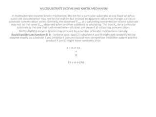
The enzyme's function in the cell is to cleave lactose to glucose and galactose so that they can be used as carbon/energy sources. The synthetic compound o- nitrophenyl- D-galactoside (ONPG) is also recognized as a substrate and cleaved to yield galactose and o-nitrophenol, which has a yellow color. When ONPG is in excess over the enzyme in a reaction, the production of o-nitrophenol per unit time is proportional to the concentration of - Galactosidase; thus, the production of yellow color can be used to determine enzyme.
For example, an investigator may fuse a promoter to the lacZ gene and use -Gal levels as a read out for promoter activity under various conditions. In 1972, Jeffrey Miller published "Experiments in Molecular Genetics" which contained a protocol for determining the amount of -Gal with ONPG. Because of this, ONPG/ -Gal assays are referred to as "Miller" assays, and a standardized amount of -Gal activity is a "Miller Unit". 1 Miller unit=1000 x (Abs420) / (t x v x Abs600) where: Abs420 is the absorbance of the yellow o-nitrophenol, t = reaction time in minutes, v = volume of culture assayed in milliliters, Abs600 reflects cell density.
*Note that this value is different for each spectrophotometer used and should be calibrated by plating dilutions of known Abs600 cultures to determine the colony forming units per Abs600. Also note that because of this, and numerous other possible reasons, although one person s Miller Unit should be equivalent to someone else s, this is most likely not always the case. It is therefore critical that you include positive and negative controls, in parallel to your experimental, whenever doing -Gal assays.
