Gel Electrophoresis for Assessing Nucleic Acid Quality
Gel electrophoresis is a crucial technique for assessing the quality and yield of nucleic acids such as DNA and RNA. It separates DNA fragments based on size, allowing researchers to determine the integrity of the nucleic acids. By running a small amount of sample on a gel and using DNA-binding dyes, damaged or fragmented DNA can be detected. Different dyes like Ethidium bromide, Gel red, and SYBR safe are commonly used to visualize DNA and RNA bands.

Download Presentation

Please find below an Image/Link to download the presentation.
The content on the website is provided AS IS for your information and personal use only. It may not be sold, licensed, or shared on other websites without obtaining consent from the author. Download presentation by click this link. If you encounter any issues during the download, it is possible that the publisher has removed the file from their server.
E N D
Presentation Transcript
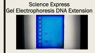
Assessing the quality and yield of nucleic acids PART 2: gel electrophoresis The spectrophotometer cannot tell you if your DNA or RNA are intact, undamaged Vigorous shaking, pipetting up and down, and vortexing, can damage DNA, breaking it into smaller pieces To be sure it is not damaged, you can run a small amount, 100-200 ng, on a gel 1
Agarose gel electrophoresis for DNA: Gel electrophoresis separates DNA fragments (and RNA too) according to their size. DNA samples are loaded into wells (indentations) at one end of a gel, and an electric current is applied to pull them through the gel. DNA fragments are negatively charged, so they move towards the positive electrode (which is red in most systems). https://www.sciencedirect.com/topics/bioche mistry-genetics-and-molecular- biology/agarose-gel-electrophoresis 2
Agarose gel electrophoresis for DNA: DNA fragments move different distances through a gel based on size When we speak of the size of a DNA fragment, we mean the length in base pairs Agarose is like a mesh with pores- smaller fragments can move through the pores more easily while longer fragments take longer A ladder is used to determine the sizes of the DNA fragments in each lane https://www.sciencedirect.com/topics/bioche mistry-genetics-and-molecular- biology/agarose-gel-electrophoresis 3
DNA runs through a gel in linear pieces proportionately to length RNA must be made linear to maintain the relationship between length and distance migrated When a gel is stained with a DNA-binding dye, the DNA fragments show up as bands, each representing a group of same-sized DNA fragments (same for RNA) https://commons.wikimedia.org/w/index.php?title=File_talk:Gel_Electrophoresis.svg& action=edit&redlink=1 4
Dyes used to visualize DNA and RNA in gels (just a few common ones- there are others) Ethidium bromide: (UV) Fluoresces when bound to DNA with low background High sensitivity A known mutagen Gel red: (UV) A safer form of ethidium, modified so that it cannot penetrate cell membranes, therefore should not be toxic (according to manufacturer) As sensitive as ethidium bromide (some claim it is more sensitive) More expensive than ethidium bromide SYBR safe: (blue light or UV) Safest of all these, because not toxic and UV is not needed Less sensitive than Gel red Not as pricey as Gel red 5
Intact vs degraded DNA We use a gel of 0.7- 1.5% for examining the length of DNA fragments and extent of DNA degradation. Good quality DNA should migrate as a high molecular weight band, 20-40 kilobase pairs, kb), with little or no evidence of smearing. ladder of DNA fragments with known lengths provide references to estimate the length of your DNA fragments smears of partially degraded DNA
Separation of RNA according to size by denaturing formaldehyde agarose gel electrophoresis Method: Incubate RNA in the presence of formaldehyde and formamide at 55-65oC for a few minutes. (it is bad for you, use the fume hood) RNA denatures/ secondary structures are removed Carry out electrophoresis in agarose gel containing formaldehyde RNA remains denatured during electrophoresis RNases are denatured, inhibiting RNA degradation RNA molecules will migrate according to their molecular weight rather than ability to form secondary and tertiary structures *Nucleic acids must be LINEAR in order to migrate by size in a gel < < Caution: 1. formaldehyde is a strong fixative/irritant 2. buffer warms up during electrophoresis fumes can fix your corneas! 7
The integrity of ribosomal RNA can be used as an indicator of mRNA quality Ribosomal RNA (rRNA) makes up more than 80% of total RNA samples. Total RNA preps should display two prominent rRNA bands after gel electrophoresis. 25S 18S These correspond to the 25S and 18S rRNAs, in plants, 28S and 18s rRNAs in animals Good quality RNA will have: - No/little evidence of smearing - 25S/18S or 28S/18S ratio ~ 2 mRNA is present but in very small amounts, but will typically be intact if ribosomal RNAs are intact 8
Ribosomal RNA Sizes vary with organism Don t memorize you would need to know what sizes you are expecting to see for your research organism Svedberg (S) units Species rRNA 18S 28S 18S 28S 18S 28S 16S 18S 23S 25S 18S 26S 16S 23S Size (kb) 1.9 5.0 1.9 4.7 2.0 4.1 1.5 1.9 2.9 3.7 2.0 3.8 1.5 2.9 Human Mouse Drosophila Tobacco Leaf Yeast E. coli What are the 16S and 23S rRNAs in plant cells? Where do they come from? 9