Overview of Congenital Heart Diseases
Congenital heart diseases are abnormalities in the heart's structure present at birth. Factors like viral infections, genetic conditions, and maternal health contribute to these conditions. There are non-cyanotic and cyanotic classifications based on shunting patterns. Examples include atrial septal defects, patent ductus arteriosus, and Fallot's tetralogy. Understanding the etiology and classification of these defects is crucial for diagnosis and treatment.

Download Presentation

Please find below an Image/Link to download the presentation.
The content on the website is provided AS IS for your information and personal use only. It may not be sold, licensed, or shared on other websites without obtaining consent from the author. Download presentation by click this link. If you encounter any issues during the download, it is possible that the publisher has removed the file from their server.
E N D
Presentation Transcript
CONGENITAL HEART DISEASES Dr Eman MS Muhammad
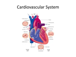
Little is known about the causation of congenital abnormalities of the heart. Rubella infection of the mother in the first 3 months of pregnancy is associated with serious abnormalities in 10-20% of infants of which heart diseases constitutes about 50%. A high proportion of congenital abnormalities of the heart results from defects in the formation of the septa in the developing hearts. At an early stage of development the heart consists of 3 chambers, an atria, a ventricle and the aortic bulb.
Division of each of these into two takes place separately: The ventricular septum grows upwards from the apex. There is ultimately a small aperture at the base. The aortic bulb undergoes division into two nearly equal parts. The two vessels formed must rotate spirally in order to establish their normal continuity with the ventricle. The septum of the bulb ultimately fuses with the up- growing ventricular septum. The last portion to close is the membranous part of the septum.
Etiology: The real cause is unknown, but the following factors are blamed: 1. Viral infection of the mother in the first 3 months by: German measles (rubella) or Coxsachie virus. 2. Certain medications or drugs; tertatogenic drugs such as thalidomide, cortisone. 3. Alcohol and/or tobacco. 4. Nutritional and vitamin deficiencies in pregnancy. 5. Maternal diabetes. 6. Down syndrome, Turner syndrome, and Marfan syndrome. 7. Syphilis.
Classifications: Non-cyanotic group: I. A. Left to right shunt: a) Atrial septal defect (ASD). b) Ventricular septal defect (VSD). c) Patent ductus arteriosus (PDA).
B. No shunt: a) Pulmonary stenosis. b) Aortic stenosis. c) Coarctation of the aorta.
II. Cyanotic group: (right to left shunt) A. Fallot s trilogy. B. Fallot s tetralogy. C. Eisenmenger s syndrome. D. Transposition of great vessels.
Fallots Tetralogy The commonest anomaly of the cyanotic group is the tetralogy of Fallot. It is composed of: a. Obstruction of the outflow tract of the RV, usually from stenosis of the pulmonary valve, or obstruction of the infandibular part of the RV.
b. This results in right ventricular hypertrophy and the pressure in this chamber is raised. c. Some of the un-oxygenated blood in the chamber is shunted into the aorta through a high interventricular septal defect. d. The aorta partially overrides the septal defect, and so in addition to receiving oxygenated blood from the LV, it also receives venous blood from the RV.
In extreme cases, the the pulmonary orifice and artery may be atretic, and blood reaches the lungs from the aorta through the PDA. In about 25% of cases of Fallot there is right aortic arch.
Effects: Right to left shunting of blood, decreased pulmonary blood flow, and increased aortic blood volume. The clinical severity depends on the degree of pulmonary stenosis. Secondary polycythemia and clubbing of figures due to hypoxia. Increased risk of infective endocarditis, and systemic embolization.
Fallots Trilogy Fallot s Trilogy is composed of: a) Pulmonary stenosis. b) Right ventricular hypertrophy. c) Atrial septal defect.
Eisenmengers Syndrome Eisenmenger's syndrome, ES is defined as a long-standing left-to-right cardiac shunt caused by a congenital heart defect (typically by a VSD, ASD, or less commonly, PDA) causes pulmonary hypertension and eventual reversal of the shunt into a cyanotic right-to-left shunt.
Transposition of great vessels A curious anomaly results from failure of the proximal aorta and pulmonary artery to undergo the spiral rotation necessary for establishment of their correct relationships with the ventricles. In consequence, the aorta arises from the RV, and the pulmonary artery from the LV. Such condition alone is incompatible with extra-uterine life.
Sometimes it may be compensated for a time by persistence of the PDA, patent foramen oval, ASD, and/or VSD. The greater the volume of the shunt, the better the admixture of the arterial blood and venous blood and the less marked the cyanosis.
Patent Ductus Arteriosus (PDA) There is a persistent communication between the descending thoracic aorta and the pulmonary artery that results from failure of normal physiologic closure of the fetal ductus. It may be the only abnormality present. It may coexist with other anomalies. It shunt of the blood from the aorta to the pulmonary artery.
The LV its output to compensate for the shunted blood so it undergoes hypertrophy and dilatation. The increased strain on the RV causes its hypertrophy and dilatation. Failure to close the ducts leads to HF. There is also risk of endocarditis of the ductus. Ductus is usually present immediately beyond the origin of left subclavian artery. Such a patient may have a cyanotic tinge in the nail-beds of the toes, but not in those of the hands.
Atrial Septal Defect (ASD) This is the commonest congenital malformations of the heart. Even when the defect is large, it appears to have little effect on the circulation. The blood flows from the left to the right atrium (RA). The RA undergoes hypertrophy and dilatation. Hypertrophy and dilatation of the RV with loud systolic murmur.
The pulmonary artery is dilated and atherosclerotic, while the aorta is small. The overworked RV fails off, this causes in the blood supply to the lung.
Ventricular Septal Defect (VSD) A high septal defect isfrequently part of other congenital anomaly such as Fallot s tetralogy. There is two types: Small defect: Rare and occurs in the muscular part of the septum. Cardiac hypertrophy does not occur (Roger s disease). Big defect: More common and occurs in the membranous septum just below the aortic valve.
Blood is shunted from the LV to the RV to the pulmonary artery causing slight enlargement. SIE may develop on the endocardial surface of the RV opposite the defect. Clinically it produces thrill and a loud systolic murmur.
Coarctation (stenosis) of the aorta) All degrees of narrowing up to the degree of atresia may occur. Infantile type: Slight narrowing of the aorta between the left subclavian artery and the ductus arteriosus (proximal to the ducutus) which remains patent. It is common and occurs predominantly in males. The blood flow through the ductus from the pulmonary artery to the aorta. This defect is usually incompatible with life.
Adult type: The narrowing is distal to the ductus which is closed. With severe narrowing opening of the collateral from the carotid and subclavian arteries links the aorta above and below the narrowed segment. Blood pressure increased in the arms, head and neck and decreased blood pressure in the legs. So the pulse in the legs is poor compared with that in the arms. Death results from LSHF, aortic aneurysm or dissection, cerebral hemorrhage or SIE.
Complications of congenital heart diseases: 1. Cyanosis: develops early in life in Fallot s tetralogy. 2. Predisposition to SIE. 3. Heart failure. 4. Pulmonary hypertension.
TUMOURS OF THE HEART Primary Tumors: They are extremely rare and include myxoma, fibroma,lipoma, rhabdomyoma, angiosarcoma and rhabdomyosarcoma. Secondary Tumors: They occur in about5%of patients dying of cancer. The commonest are bronchial carcinoma, breast carcinoma, malignant melanoma, lymphoma and leukemia.
Thank you Thank you


")


")


")


")


 of the aorta)")