Club Foot (Congenital Talipes Equinovarus): A Comprehensive Overview
Club foot, also known as Congenital Talipes Equinovarus, is a condition characterized by a tri-planar deformity of the foot. This article covers the aetiology, clinical features, diagnosis, treatment options, and patient management strategies for club foot. It discusses the gene variations, environmental factors, malpositioning in utero, and other considerations that contribute to the development of this condition. The pathology, prognosis, complications, operative treatments, and old methods of treatment are also addressed.

Download Presentation

Please find below an Image/Link to download the presentation.
The content on the website is provided AS IS for your information and personal use only. It may not be sold, licensed, or shared on other websites without obtaining consent from the author. Download presentation by click this link. If you encounter any issues during the download, it is possible that the publisher has removed the file from their server.
E N D
Presentation Transcript
Objectives Introduction Aetiology Diagnosis Treatment Old methods of treatment Prognosis & Out come Complications if not treated Operative treatment / surgeries Patient management Conclusion
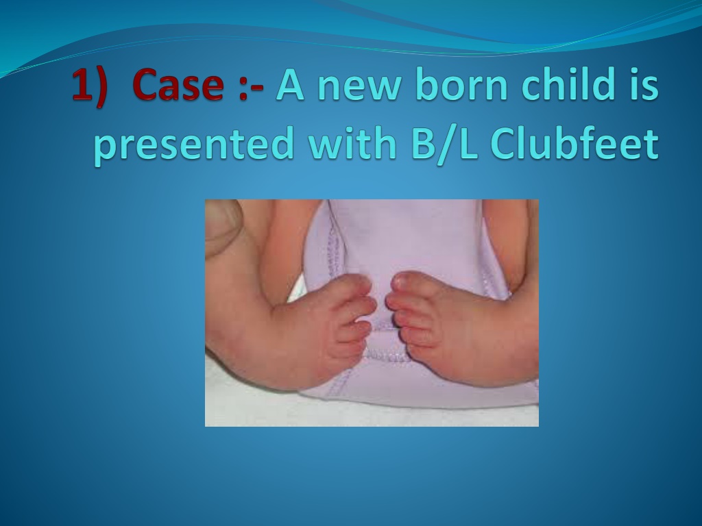
Introduction Club foot - Congenital Talipes Equinovarus In this condition, the foot is deformed in three planes called Tri- planar deformity. Hind foot is in equinus (i.e pointing downwards) and varus (i.e tilted towards the mid-line) positions. Mid foot is in cavus (i.e high arched) adducted and supinated positions. Fore foot is adducted and supinated. 1. 2. 3. The condition is more prevalent in boys and affects 1:1000 live births. Both limbs are affected in up to 50% of cases. Similar deformity is seen in neurological disorders such as myelomenigocele and arthrogryposis.
Pathology The talonavicular joint is subluxed with the navicular bone is displaced medially and plantarwards. The calcaneofibular ligament and posterior tibial tendon sheath are shortened and thickened and contain contractile myofibroblasts. The gastrocsoleus and posterior tibial muscle are smaller and thinner than normal with reduced myofibrils. The vascular supply via the dorsalis pedis may be altered. There could be regional nerve abnormalities as well.
Clinical Features Foot is turned and twisted inwards. Sole faces posteriomedially. Heel is small and high and deep creases appear posteriorly and medially. Small calf. Small foot. In normal babies, the foot can be dorsiflexed and everted until the toes almost touch the front of the leg. However, in clubbed foot babies this manoeuvre meets with varying degree of resistance. This condition may be associated with other congenital disorders such as congenital hip dislocation and spina bifida.
Aetiology 1)Gene variation A variation in the gene that processes folate 2)Environmental factors During pregnancy > Use of drugs > Infections > Smoking > Other negative exposures to the fetus
3)Malpositioning of foot in uterus If the fetus foot was twisted or cramped 4)Spina Bifida Damaged nerves in the open spine > Effect on the development of the legs & feet
5)Other considerations Abnormalities of > Muscles > Joints > Ligaments > Tendens Lesions > Vascular > Joint Malformed bones
Risk factors 1.Boyes are affected more frequently than girls 2.If one parent has a clubfoot 3.If previous children has a clubfoot
History taking Mother had oligohydroamniosis Intrauterine compression (common in positional telipes) First degree relatives Ethnic group (maoris have high risk) Any accident or trauma
Examination Do the routine new born examination (the foot can be fully dorsi flexed to touch the front of the lower leg in a normal neonate but unable to perform in club foot child) Check the weakness of the foot(floppiness, delayed motor milestones, unseady abnormal gait, fatigability) Gross features Varus Entire foot is inverted and supinated Mid foot adduction Fore foot supination and adduction Heel is rotated,inverted and planter flextion Affected foot is shorter Claf muscle is thinner than normal Tight achilles tendon Foot is kidney bean shape
Investigation Antenataly by ultrasound Check for occult spinal problems by radiography or MRI Radio graphical findings Neck of the talus is deformed medially Navicular is subluxed medially Plantar wards on the head of the talus Loss of alignment
Indications for surgery If other treatments fail If the clubfoot is rigid Older children or adults Recurrence 30% of cases - manipulation and casting is successful BUT 70 % Need surgical repair
At what age usually the surgery perform ? Should perform early as possible . Because immature joints, bones and ligaments will facilitate to gain the normal position of the foot . But the foot also should grow a little larger to facilitate surgery when the child is about to walk 10 11 months
Surgical procedure STR soft tissue release (ligaments, tendons) wedge osteotomy - of the lateral column and the calcaneum - relapsing club foot Fusion or arthrodesis - 2 or more bones are fused together. Metal pins or plates may be used to hold the bones together for a while
After surgery ? The foot will be casted and kept elevated, ice packs used to reduce swelling and pain, medications for pain. The skin around the cast and the toes will be checked frequently for the first 48 hours to make sure that the circulation, movement, and feeling are maintained. Cast will be kept about 12 weeks. It will be changed at least 2 or 3 times how to take care of the cast Physical therapy Dennis Browne Night Splint (DBNS) is given to the child, to be worn when not walking, for a period of about 2 years.
Complications Due to anesthesia - Breathing problems , Reactions to medicines Risks from any surgery Bleeding, Infection Possible problems from clubfoot surgery are - Foot swelling , Problems with blood flow to the foot , Damage to nerves in the foot foot may be stiffer than normal foot and calf may be smaller than normal the rest of their life.
CLUB FOOT Outcome and Prognosis
Approximately 50% of clubfeet in newborns can be corrected non operatively. Ponseti reports an 99% success rate using his technique (including an Achilles tenotomy). Others report success rates of 10-35%. Most series report 75-90% satisfactory results of operative treatment (appearance and function of the foot). The amount of motion in the joints of the foot and ankle correlates with the degree of patient satisfaction
Satisfactory results were obtained in 81% of cases, and the range of ankle movement was a major factor in determining the functional result, which again was influenced by the degree of talar dome flattening (suggesting that the primary bone deformity present at birth dictates the eventual result of treatment). Forty-four percent of patients had no dorsiflexion beyond neutral, and 38% of patients required further surgery (nearly two thirds of these were bony procedures). Recurrence rates of deformity were reported at around 25%, with a range of 10-50%. Menelaus reported a 38% recurrence rate
The best results were obtained with children older than 3-4 months with a foot large enough to perform the surgery without compromise (longer than 8 cm, as specified by Simons). The age at operation is directly related to the result. Less than satisfactory results may be associated with overcorrection, which occurs in approximately 15% of cases. Previous surgery seems to have a deleterious effect on the result.
As small infants with operated clubfeet have grown into heavy adults, they have been prone to painful stiff feet, despite good correction. Deitz and Cooper published a 30-year follow-up study of patients treated with the Ponseti method.These cases had comparatively pain-free supple feet. The Ponseti method is gaining mainstream acceptance as evidenced by the emergence of Ponseti clubfeet centers at major teaching hospitals across the United States.
Of the patients who have been monitored long term, those who are heavy and those in jobs involving long periods on their feet (especially performing manual labor) were found to be more likely to have painful feet.This correlated with the trend seen in the general population at large.
Underdevelopment of the calf muscles Plantagrade foot Affected foot become smaller and shorter Tarsalbone assume an abnormal shape Soft tissue underdevelopment at the medial side Hyperplasia of the lower leg muscles Stiff joints
Conclusion A clubfoot, or congenital talipes equinovarus is a congenital deformity involving one foot or both feet. Most cases are idiopathic and not associated with other conditions. Babies should be referred early for treatment. Current best treatment is by casting and bracing according to the Ponseti method. Results are better with manipulative methods than with surgical release. Recurrences can occur and are normally caused by non-compliance with bracing. The standard treatment of clubfoot has changed greatly in the past 10 years. Previously, extensive surgery was common in children born with this condition. The publication of long term evidence of good outcomes with more minimally invasive methods, such as the Ponseti technique, has led surgeons worldwide to change their approach. Ponseti treatment consists of sequential plasters and prolonged bracing, with minor surgical procedures.


Malpositioning of foot in uterus")
Other considerations")