Infectious Diseases 4/5th Semester Classes: Approach & Prevention

Infectious Diseases class focuses on various topics including Antibiotic Stewardship Practices, Community-Acquired Infections, Gram-Positive and Gram-Negative Bacteria, Spirochetal Diseases, Diseases Caused by Bacterial and Viral Infections, HIV/AIDS, Fungal and Parasitic Infections. The class also covers Diphtheria, its etiopathogenesis, clinical manifestations, and risk factors for outbreaks. Understanding the transmission, virulence factors, and pathologic findings of Diphtheria is crucial for effective prevention strategies.

Download Presentation

Please find below an Image/Link to download the presentation.
The content on the website is provided AS IS for your information and personal use only. It may not be sold, licensed, or shared on other websites without obtaining consent from the author. Download presentation by click this link. If you encounter any issues during the download, it is possible that the publisher has removed the file from their server.
E N D
Presentation Transcript
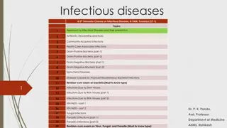
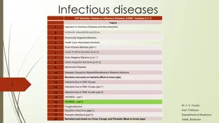
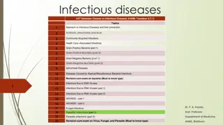
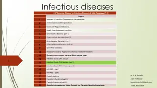
Infectious diseases 4/5thSemester Classes on Infectious Diseases, 8-9AM, Tuesdays (LT-1) Topics Approach to Infectious Diseases and their prevention 1 Antibiotic stewardship practices 2 Community-Acquired Infections 3 Health Care Associated Infections 4 Gram-Positive Bacteria (part-1) 5 Gram-Positive Bacteria (part-2) 6 Gram-Negative Bacteria (part-1) 7 Gram-Negative Bacteria (part-2) 8 Spirochetal Diseases 9 Diseases Caused by Atypical/Miscellaneous Bacterial Infections 10 Revision-cum-exam on bacteria (Must to know type) 11 Infections Due to DNA Viruses 12 1 Infections Due to RNA Viruses (part 1) 13 Infections Due to RNA Viruses (part 2) 14 HIV/AIDS part 1 15 HIV/AIDS part 2 16 Dr. P. K. Panda, 17 18 Fungal Infections Parasitic Infections (part 1) Asst. Professor Department of Medicine 19 Parasitic Infections (part 2) 20 Revision-cum-exam on Virus, Fungal, and Parasite (Must to know type) AIIMS, Rishikesh
DIPHTHERIA Nasopharyngeal and skin infection caused by Corynebacterium diphtheriae Toxigenic strains most frequently cause pharyngeal diphtheria, nontoxigenic strains commonly cause cutaneous disease Cutaneous diphtheria is usually a secondary infection after primary skin lesions due to trauma, allergy, or autoimmunity In tropical latitudes, cutaneous diphtheria is more common than respiratory diphtheria In temperate climate, Pharyngeal one is most common during winter months Risk factors for diphtheria outbreaks include older age, lack of vaccination, alcoholism, low socioeconomic status, crowded living conditions Carriers are defined as individuals who have positive cultures for C. diphtheriae and who either are asymptomatic or have symptoms but lack pseudomembranes
ETIOPATHOGENESIS Unencapsulated, nonmotile, and nonsporulating with club-shaped bacillary appearance and typically form palisades, known as Chinese characters The specific laboratory media using tellurite, colistin, or nalidixic acid Phenotypes:- nontoxigenic (tox ) and toxigenic (tox+) using corynebacteriophages Under iron-limiting conditions leads to the optimal expression Transmitted via the aerosol route, with only human reservoirs, IP-2 5 days Diphtheria toxin is the primary virulence factor and is taken up in the bloodstream, from which it is distributed to all organ systems in the body In vitro, the toxin may be separated into two chains by digestion with serine proteases: the N-terminal A fragment and the C-terminal B fragment Delivery of the A fragment into the eukaryotic cell cytosol results in irreversible inhibition of protein synthesis by NAD+-dependent ADP-ribosylation of elongation factor 2 Pathologic findings of diphtheria include mucosal ulcers with a pseudomembranous coating composed of an inner band of fibrin and a luminal band of neutrophils
CLINICAL MANIFESTATIONS Should be considered when a patient has severe pharyngitis, particularly when there is difficulty swallowing, respiratory compromise, or signs of systemic disease (e.g., myocarditis or generalized weakness) Clinical diagnosis of diphtheria is IF sore throat; pseudomembranous lesions; and low- grade fever Most commonly, the pseudomembranous lesion is located in the tonsillopharyngeal region It is gray or whitish, sharply demarcated, tightly adherent to the underlying tissues (Unlike streptococcal pharyngitis) Massive swelling of the tonsils and may present with bull-neck diphtheria Polyneuropathy (3 5 weeks after) and myocarditis are late toxic manifestations Cranial nerve dysfunction typically precedes other neuropathy and typically reversible in patients who survive the acute phase Other complications include pneumonia, renal failure, encephalitis, cerebral infarction, pulmonary embolism, and serum sickness from antitoxin therapy Cutaneous Diphtheria IS characterized by punchedout ulcerative lesions with necrotic sloughing or pseudomembrane formation
Diagnosis The diagnosis of diphtheria is based on clinical signs and symptoms plus laboratory confirmation Laboratory diagnosis of diphtheria is based either on cultivation or on the demonstration of local lesions with characteristic histopathology A diagnosis of cutaneous diphtheria requires laboratory confirmation since the lesions are not characteristic and are indistinguishable from other dermatoses
Treatment Patients in whom diphtheria is suspected should be hospitalized in respiratory isolation rooms, with close monitoring of cardiac and respiratory function Once a clinical diagnosis of diphtheria is made, diphtheria antitoxin should be administered as rapidly as possible after a test dose Antibiotics are used in the management of diphtheria primarily to prevent transmission to susceptible contacts Procaine penicillin G, 600,000 U IM q12h until the patient can swallow comfortably; then oral penicillin V, 125 250 mg qid to complete a 14-day course (or Erythromycin, 500 mg IV q6h, rifampin and clindamycin) For patients in whom the organism is not eradicated after a 14-day course of erythromycin or penicillin, an additional 10-day course followed by repeat culture is recommended Patients who recover from diphtheria should receive the appropriate vaccine if diphtheria antitoxin titer of <0.01 U/mL
Prognosis and prevention Risk factors for death include bullneck diphtheria; myocarditis with ventricular tachycardia; atrial fibrillation; complete heart block; an age of >60 years or <6 months; alcoholism; extensive pseudomembrane elongation; laryngeal, tracheal, or bronchial involvement Interval between the onset of local disease and the administration of antitoxin DTaP is currently recommended for children up to the age of 7 It is recommended that all adults receive a single dose of Tdap if they have not received it previously, regardless of the interval since the last dose of Td (tetanus and reduced-dose diphtheria toxoids, adsorbed); after that decennial Td booster Close contacts of diphtheria patients should undergo throat culture and After samples for throat culture obtained, antimicrobial prophylaxis should be considered for all contacts (7 10 days of oral erythromycin or one dose of IM benzathine penicillin G, 1.2 million units for persons 6 years of age)
OTHER CORYNEBACTERIAL INFECTIONS Nondiphtherial corynebacteria, referred to as diphtheroids or coryneforms, are frequently considered colonizers or contaminants; however, they have been associated with invasive disease, particularly in immunocompromised patients Humans are the natural reservoirs for several coryneforms, including C. xerosis, C. pseudodiphtheriticum, C. striatum, C. minutissimum, C. jeikeium, C. urealyticum, and Arcanobacterium haemolyticum Animal reservoirs are responsible for carriage of Arcanobacterium pyogenes, C. ulcerans, and C. pseudotuberculosis Soil is the natural reservoir for Rhodococcus equi
Listeria monocytogenes Infections L. monocytogenes is of interest not only to clinicians but also to basic scientists as a model intracellular pathogen that is used to study A food-borne pathogen that can cause serious infections, particularly in pregnant women and immunocompromised individuals Broad host range of reservoirs, facultatively anaerobic, nonsporulating, motile, grows over a broad temperature range Commonly found in processed and unprocessed foods of animal and plant origin Like other enteric pathogens, L. monocytogenes induces its own internalization by cells that are not normally phagocytic Weakly -hemolytic (an essential determinant of its pathogenicity, listeriolysin O Expresses a surface protein, ActA, an essential actin-based motility system (Other pathogens like Shigella, Mycobacterium, Rickettsia, and Burkholderia species use a related pathogenic strategy that allows cell-to-cell spread) There is an inverse relationship between toxicity and virulence i.e., the more cytotoxic the strain, the less virulent it is in animals
Symptoms/diagnosis/treatment Symptoms overlap greatly with those of other infections It presents with several clinical syndromes (meningitis and septicemia are most common) Listerial gastroenteritis typically develops within 48 h of ingestion of a large inoculum of bacteria A nonspecific flulike illness with fever is a common presentation in pregnant women F/B myalgias, arthralgias, backache, and headache Granulomatosis infantiseptica is an overwhelming fetal infection with miliary microabscesses and granulomas It should be considered in all older or chronically ill adults with aseptic meningitis (frequently subacute) The diagnosis is typically made by culture of blood, cerebrospinal fluid (CSF), or amniotic fluid Ampicillin is the drug of choice with/out Gentamycin
They are pleomorphic, gram variable, mostly motile (except C. perfringens, C. ramosum, and C. innocuum, with variable oxygen tolerance Produce more protein toxins than any other bacterial genus, including neurotoxins, enterotoxins, cytotoxins, collagenases, permeases, necrotizing toxins, lipases, lecithinases, hemolysins, proteinases, hyaluronidases, DNases, ADPribosyltransferases, and neuraminidases C. tetani and C. botulinum, cause specific clinical disease by elaborating single toxin while, C. perfringens and C. septicum cause aggressive necrotizing infections due to multiple toxins Botulinum and tetanus neurotoxins are the most potent (LD- 0.2 10 ng/kg)and Epsilon toxin of C. perfringens is the most lethal one (used as agent of bioterrorism) Widespread in nature, forming endospores, Commensals in the intestinal tract of humans and animals, in the female genital tract, and on the oral mucosa (not all commensal clostridia are toxigenic)
The acute onset of hypertonia or painful muscular contractions (usually of the muscles of the jaw and neck) and generalized muscle spasms without other apparent medical cause CDC Maternal tetanus is defined by the WHO as tetanus occurring during pregnancy or within 6 weeks after the conclusion of pregnancy (whether wit The spores or bacteria enter the body through abrasions, wounds, or (in the case of neonates) the umbilical stump.h birth, miscarriage, or abortion) In 20 30% of cases of tetanus, no puncture entry wound is found Higher risks in old age, Injection drug users, and incomplete vaccination Only those bacteria producing tetanus toxin (tetanospasmin) can cause tetanus Although closely related to the botulinum toxins in structure and mode of action, tetanus toxin undergoes retrograde transport into the central nervous system and thus produces different clinical effects Unlike other components of the endosomal contents, which undergo acidification following internalization, tetanus toxin is transported in a carefully regulated pH- neutral environment that prevents an acid induced conformational change Prevents transmitter release and effectively blocks inhibitory interneuron discharge
CLINICAL MANIFESTATIONS Broadly divided into generalized (including neonatal) and local Muscles of the face and jaw often are affected first The incubation period and the period of onset are of particular significance; shorter times are associated with worse outcome The commonest initial symptoms are trismus (lockjaw), muscle pain and stiffness, back pain, and difficulty swallowing Autonomic disturbance is maximal during the second week of severe tetanus, and death occurs due to cardiovascular events
DIAGNOSIS The diagnosis of tetanus is based on clinical findings Culture of C. tetani from a wound provides supportive evidence Serum anti-tetanus immunoglobulin G may also be measured in a sample taken before the administration of antitoxin or immunoglobulin; levels >0.1 IU/mL are deemed protective and do not support the diagnosis of tetanus D/D- strychnine poisoning and dystonic reactions to antidopaminergic drugs Episodic nature
TREATMENT Management strategies aim to neutralize remaining unbound toxin and support vital functions until the effects of the toxin have worn off Patients should be nursed in calm, quiet environments because light and noise can trigger spasms Entry wound should be identified, cleaned, and debrided of necrotic material Metronidazole (400mg rectally or 500 mg IV every 6 h for 7 days) is the preferred antibiotic; Or penicillin, although this may exacerbate spasms TIG is the preparation of choice (less likely to be associated with anaphylactoid reactions); 3000 5000 IU as a single IM dose, a portion should be injected around the wound; nowadays 50 1500 IU administered intrathecally also Equine derived antitoxin (available widely), 10,000 20,000U, after testing for hypersensitivity Spasms are controlled by heavy sedation with benzodiazepines Cardiovascular stability (difficult to treat) is improved by increasing sedation with IV magnesium sulfate, morphine, or other sedatives Patients must be given a full primary course of immunization
Prevention Good wound care and immunization Individuals sustaining tetanus-prone wounds should be immunized if their vaccination status is incomplete or unknown or if their last booster was given >10 years earlier Patients sustaining wounds not classified as clean or minor should also undergo passive immunization with TIG
Botulism Ingestion of botulinum toxin in contaminated food (food-borne botulism) Wound botulism Infant botulism Adult intestinal colonization botulism Botulism symptoms have been reported in patients receiving injections of botulinum toxin for cosmetic or therapeutic purposes (iatrogenic botulism; Moreover, botulism was reported after inhalation of botulinum toxin in a laboratory setting Intentional Botulism
Pathogenesis Consumption of home-preserved fish or vegetable products main source Seven serologically distinct confirmed serotypes of botulinum toxin (A through G); Human cases associated with serotypes A, B, E, and F Four recognized species of clostridia: Clostridium botulinum, Clostridium argentinense, Clostridium baratii, and Clostridium butyricum Neurotoxin enters the vascular system and is transported to peripheral cholinergic nerve terminals, including neuromuscular junctions, postganglionic parasympathetic nerve endings, and peripheral ganglia with Inhibition of acetylcholine release
CLINICAL MANIFESTATIONS All forms of botulism manifest as a relatively distinct clinical syndrome of symmetric cranial-nerve palsies followed by descending bilateral flaccid paralysis of voluntary muscles, which may progress to respiratory compromise and death Weakness descends, often rapidly, from the head to involve the neck, arms, thorax, and legs; occasionally, weakness is asymmetric In some instances, unfortunately, the severe ptosis, expressionless face, and weak phonation of patients with botulism have been interpreted as signs of mental status changes due to alcohol intoxication, drug overdose, encephalitis, or meningitis a conclusion that delays an accurate diagnosis
DIAGNOSIS Diagnosed primarily on clinical grounds, with laboratory confirmation by specific tests that identify botulinum toxin in clinical and food samples (mouse bioassay, BEST METHOD) The temporal occurrence of two or more cases with symptoms compatible with botulism is essentially pathognomonic because other illnesses that resemble botulism do not usually occur in clusters Food history and the names of contacts who may have shared foods should be obtained before illness progresses Ascertainment of the patient s behavioral history related to injection drug use is critical A history of recent abdominal surgery or antibiotic use may be important in the diagnosis of adult intestinal colonization botulism D/D- Guillain-Barre syndrome (GBS), myasthenia gravis, stroke syndromes, Eaton-Lambert syndrome, tick paralysis poisoning by tetrodotoxin, shellfish, or a host of rarer agents and antimicrobial drug associated paralysis
TREATMENT Meticulous intensive care and immediate administration of botulinum antitoxin (Botulism Antitoxin Heptavalent OR Botulism Immune Globulin Intravenous) Persons of all ages (including infants) in whom botulism is suspected should be hospitalized immediately Suspect wounds and abscesses should be cleaned, debrided, and drained promptly. The role of penicillin and metronidazole in treatment and decolonization is unclear
POLYMICROBIAL INFECTIONS INVOLVING CLOSTRIDIA In the absence of devitalized tissue, the presence of clostridia does not necessarily lead to infection (CLOSTRIDIAL WOUND CONTAMINATION) It may appear in association with non-spore-forming anaerobes and facultative or aerobic organisms Often associated with severe local inflammation but may lack the characteristic systemic signs of toxicity and rapid progression seen in other clostridial infections The treatment of mixed aerobic/anaerobic infection of the abdomen, perineum, or gynecologic organsshould be based on Gram s staining, culture, and antibiotic sensitivity information Ampicillin or ampicillin/sulbactam combined with either clindamycin or metronidazole is DOC
ENTERIC CLOSTRIDIAL INFECTIONS Illness results from the ingestion of food containing at least ~1 Billion viable vegetative cells, which sporulate and produceenterotoxin The diarrhea develops within 7 30 h of ingestion is generally mild and self-limiting; however, in the very young, the elderly, and the immunocompromised, symptoms are more severe and occasionally fatal Enterotoxin-producing C. perfringens type A has been implicated and has been considered to play a role in antibiotic-associated diarrhea without pseudomembrane Enteritis necroticans (gas gangrene of the bowel) is caused by toxin and toxin (Pathogenic) producing strains of C. perfringens type C Necrotizing enterocolitis, associated with C. perfringens type A, in previously healthy adults AND low-birth-weight (premature) infants hospitalized in neonatal intensive care units, affects ileum in contrast to jejunum in enteritis necroticans They should undergo nasogastric suction and receive IV fluids; Pyrantel is given by mouth, and the bowel is rested by fasting; Benzylpenicillin (1 mU) IV every 4 h, and the patient is observed for complications requiring surgery Surgical indications; gas in the peritoneal cavity, absent bowel sounds, rebound tenderness, abdominal rigidity; however, the mortality rate ranges from 35% to 100%
CLOSTRIDIAL BACTEREMIA By C. perfringens and C. tertium mostly, rarely C. septicum C. tertium can be found in patients with malignancy or acute pancreatitis, with or without neutropenic enterocolitis; and is resistant to metronidazole, clindamycin, and cephalosporins Positive C. septicum patients have some association with gastrointestinal anomaly or underlying malignancy and in addition, with neutropenia of any origin and, more specifically, with neutropenic enterocolitis Anaerobic blood cultures and Gram s stain interpretation remain the best diagnostic tests
CLOSTRIDIAL SKIN AND SOFT-TISSUE INFECTIONS Necrotizing clostridial soft-tissue infections; Severe pain, crepitus, brawny induration with rapid progression to skin sloughing, violaceous bullae, and marked tachycardia are characteristics Traumatic gas gangrene: conditions predisposing include crush-type injury, laceration of large or medium-sized arteries, and open fractures of long bones that are contaminated with soil or bits of clothing containing the bacterial spores with IP- 6 h <4 days Diagnosis is not difficult because the infection always begins at the site of significant trauma, is associated with gas in the tissue, and is rapidly progressive Non- inflammatory nature Spontaneous (nontraumatic) gas gangrene: occurs via hematogenous seeding of normal muscle with histotoxic from a gastrointestinal tract portal of entry The first symptom may be confusion followed by the abrupt onset of excruciating pain in the absence of trauma, along with fever, should heighten suspicion THEY should undergo aggressive diagnostic studies to rule out gastrointestinal pathology Surgical exploration, Gram s staining, blood culture, and histopathologic examination are main stay of diagnosis and treatment