Understanding the Autonomic Nervous System in Anatomy Lecture

In this anatomy lecture by Prof. Ahmed Fathalla Ibrahim from King Saud University, students will learn about the autonomic nervous system. The objectives include defining the system, describing its structure, and tracing neurons in both sympathetic and parasympathetic systems. The lecture also covers the main effects of sympathetic and parasympathetic actions, explaining the role of nerve cells in maintaining homeostasis. Detailed information on the sympathetic nervous system, including its structure and pathways, is provided to enhance understanding.

Download Presentation

Please find below an Image/Link to download the presentation.
The content on the website is provided AS IS for your information and personal use only. It may not be sold, licensed, or shared on other websites without obtaining consent from the author. Download presentation by click this link. If you encounter any issues during the download, it is possible that the publisher has removed the file from their server.
E N D
Presentation Transcript
Prof. Ahmed Fathalla Ibrahim Professor of Anatomy College of Medicine King Saud University E-mail: ahmedfathala@gmail.com
OBJECTIVES At the end of the lecture, students should: Define the autonomic nervous system. Describe the structure of autonomic nervous system Trace the preganglionic & postganglionic neurons in both sympathetic & parasympathetic nervous system. Enumerate in brief the main effects of sympathetic & parasympathetic system
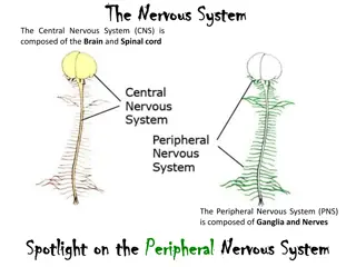
DEFINITION Nerve cells located in both central & peripheral nervous system that are concerned with innervation of involuntary structures: viscera, smooth & cardiac muscles, glands. Function: maintains homeostasis of internal environment. Regulation: by hypothalamus.
STRUCTURE OF AUTONOMIC NERVOUS SYSTEM
SYMPATHETIC NERVOUS SYSTEM Cells of lateral horn of spinal cord (T1 L3) Short axon Cells of sympathetic chain Cells of plexuses surrounding abdominal aorta (Coeliac, superior & inferior mesenteric) Long axon
SYMPATHETIC NERVOUS SYSTEM Preganglionic sympathetic neurons: cells of the lateral horn of spinal cord in all thoracic + upper 3 lumbar segments. Preganglionic axons leave the spinal cord, join corresponding spinal nerves & reach the sympathetic chain (via the white ramus communicans). They either: 1. Synapse with cells of paravertebral ganglia located in sympathetic chain (postganglionic neurons are cells of paravertebral ganglia: postganglionic axons leave the sympathetic chain & join again the spinal nerve (via grey ramus communicans) to supply structures in head & thorax + blood vessels & sweat glands .
SYMPATHETIC NERVOUS SYSTEM 2. Leave the sympathetic chain (without synapse) to reach coeliac & mesenteric plexuses (around branches of abdominal aorta) to synapse with their cells. Postganglionic neurons are cells of coeliac & mesenteric plexuses. Postganglionic axons supply abdominal & pelvic viscera.
PARAVERTEBRAL GANGLIA They are interconnected to form 2 sympathetic chains, one on each side of vertebral column. Number of ganglia: 1. Three ganglia in cervical part of chain 2. Eleven to twelve ganglia in thoracic part 3. Four in lumbar & sacral parts. The chains end into a common ganglion impar in front of coccyx
PARASYMPATHETIC NERVOUS SYSTEM Cranial: cells in brain stem: nuclei of 3rd, 7th 9th & 10th Sacral: cells in S2 S4 segments of spinal cord Long axon Cranial: cells of ciliary, pterygopalatine, submandibular, otic & peripheral ganglia Sacral: cells of peripheral ganglia Short axon Nucleus: group of neurons inside CNS Ganglion: group of neurons outside CNS
PARASYMPATHETIC NERVOUS SYSTEM Preganglionic parasympathetic neurons: 1. Cells located in brain stem: Preganglionic axons leave the brain stem, join 3rd, 7th, 9th & 10th cranial nerves & reach ciliary, pterygopalatine, submandibular, otic & peripheral ganglia (Postganglionic neurons are cells of those ganglia). Postganglionic axons supply structures in head, thorax & abdomen. 2. Cells located in 2nd, 3rd & 4th sacral segments of spinal cord. Preganglionic axons leave the spinal cord, join corresponding sacral spinal nerves to reach peripheral ganglia in pelvis where they synapse. Postganglionic neurons are cells of peripheral ganglia. Postganglionic axons supply pelvic viscera.
QUESTION 1 At which one of the following sites are located preganglionic neurons of the sympathetic nervous system ? 1. Brain stem 2. Thoracic segments of spinal cord 3. Sacral segments of spinal cord 4. Sympathetic chain
QUESTION 2 Regarding the parasympathetic nervous system, which one of the following statements is correct? 1. Its preganglionic axons are short. 2. It supplies sweat glands. 3. Its preganglionic neurons are located in the sacral segments of spinal cord. 4. Its postganglionic neurons are located in the coeliac & mesenteric plexuses.
THANK YOU THANK YOU