Understanding the Nervous System: A Comprehensive Overview
The nervous system consists of the central nervous system (CNS) and peripheral nervous system (PNS), with neurons as its basic unit. Neurons function as sensory, associative, and motor types, carrying impulses within the body. Parts of a neuron include the cell body, dendrites, axon, and terminal end fibers. Nerve fibers, neurolemma, and nuclei play essential roles in nerve function and communication. This detailed guide explores the structure and functionality of the nervous system.

Download Presentation

Please find below an Image/Link to download the presentation.
The content on the website is provided AS IS for your information and personal use only. It may not be sold, licensed, or shared on other websites without obtaining consent from the author. Download presentation by click this link. If you encounter any issues during the download, it is possible that the publisher has removed the file from their server.
E N D
Presentation Transcript
Chapter 13 The Nervous (n r-vuhs) System
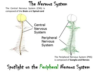
The Nervous (nr-vuhs) System The two major divisions of the nervous system central nervous system = CNS(sehn-trahl n r- vuhs sihs-tehm) = portion of the nervous system that consists of the brain and spinal cord. peripheral nervous system = PNS (pehr-ihf- r- ahl n r-vuhs sihs-tehm) = portion of the nervous system that consists of the cranial and spinal nerves, autonomic nervous system, and ganglia (gahng-gl -ah).
The Nervous (nr-vuhs) System neuron (n -rohn) = basic unit of the nervous system (Figure ).
The Nervous (nr-vuhs) System three types of neurons based on their function: sensory neurons (sehn-s r- n -rohnz) or afferent (ahf-f r-ahnt) or ascending tracts = nerves that carry sensory impulses toward the CNS. associative neurons (ahs- -sh -ah-tihv n -rohnz) or connecting neurons = nerves that carry impulses from one neuron to another motor neurons (m -t r n -rohnz) or efferent ( - f r-ahnt) or descending tracts = nerves that carry impulses away from the CNS and toward the muscles and glands
Parts of the Neuron parts of a neuron = cell body, dendrites (dehn-dr ts), axon, and terminal end fibers. soma (s -mah) = cell body of neuron dendrites (dehn-dr ts) = root like structures that receive impulses and conduct them toward the cell body.
Parts of the Neuron dendr/o = combining form for dendrite axon (ahcks-ohn) = a single process that extends away from the cell body and conducts impulses away from the cell body. ax/o = combining form means axis or main stem (stalk)
Parts of the Neuron terminal end fibers = the branching fibers that lead the impulse away from the axon and toward the synapse. nerve fibers = dendrites and axons
Parts of the Neuron nerves or nerve trunks =Bundles of nerve fibers bound together by specialized tissues neurolemma (n - r lehm-ah) or neurilemma (n -rih- lehm-ah) = tube like membrane covered nerve fibers
Parts of the Neuron nuclei (n -kl - ) = grouped neuron cell bodies within the CNS ganglia (gahng-gl - ah) = grouped neuron cell bodies outside the CNS (Figure).
The Gap synapse = space between two neurons or between a neuron and receptor synaps/o and synapt/o = combining forms for synapse neurotransmitter (n -r - trahnz-miht- r)= chemical substance released into the synaptic space that allows the signal to move from one neuron to another.
Supporting Role neuroglia (n -rohg-l -ah), or glial (gl -ahl) cells or astrocytes (ahs-tr -s tz), microglia (m -kr gl - ah), oligodendrocytes (ohl-ih-g -dehnd-r -s tz), and Schwann (shwahn) cells = supportive cells of the nervous system. Glial cells = astrocytes (ahs-tr -s tz), microglia (m -kr gl - ah), oligodendrocytes (ohl-ih-g - dehnd-r -s tz), and Schwann (shwahn) cells. gli/o = combining form means glue
Supporting Role astr/o = means star. astrocytes = star-shaped, cover the capillary surface of the brain, and help form the blood brain barrier in the CNS. micro- = means small. microglia = small phagocytic cells that help fight infection in the CNS.
Supporting Role oligo- = means few, dendr/o = means branching, -cyte = means cell. oligodendrocytes = cells with few branches that hold the nerve fibers together and help form myelin in the CNS. Schwann cells (named taken from a German anatomist) help form myelin in the PNS.
Surrounding Structures myelin (m -eh-lihn) or myelin sheath = a protective covering over some nerve cells. myelinated (m -lihn- t-ehd) nerves or white matter = nerves are surrounded by myelin Cross section of the brain shows the white matter and gray matter.
Surrounding Structures nonmyelinated (m -lihn- t-ehd) nerves or gray matter = nerves areare surrounded by myelin. nodes of Ranvier (n dz of rohn- v - ) = gaps that interrupted myelin at regular intervals along the length of a fiber. nerve = one or more bundles of impulse-carrying fibersthatconnect the CNS to other parts of the body. neur/i and neur/o = combining forms for nerve or nerve tissue
Terms used in reference to nerves tract (trahckt) = a group of nerve fibers located in the CNS. ascending tracts = groupsof nerve fibers located in the CNS that going up or cranially and carry nerve impulses toward the brain descending tracts = groupsof nerve fibers located in the CNS that going down or caudally and carry nerveimpulses away from the brain.
Terms used in reference to nerves ganglion (gahng-gl -ohn)= a knot like mass of neuron cell bodies located outside the CNS. ganglia less often ganglions = more than one ganglion. gangli/o and ganglion/o = combining forms mean knot like masses of nerve cell bodies located outside the CNS.
Terms used in reference to nerves plexus (plehck-suhs)= network of intersecting (crossover) nerves or vessels. plexi (plehck-s ) = networks of intersecting nerves or vessels. plex/o = combining form for a plexus, or network. innervation (ihn-n r-v -shuhn)= the supply or stimulation of a body part through the action of nerves.
Terms used in reference to nerves receptors (r -sehp-t rz) = sensory organs that receive external stimulation and transmit that information to the sensory neurons. nociceptive (n -sih-sehp-tihv) receptors = pain receptors proprioceptive (pr -pr - -sehp-tihv) receptors = spatial (relating to space or a space) orientation or perception (understanding or feeling or receipt) of movement receptors.
Terms used in reference to nerves stimulus (stihm-yoo-luhs) = something that excites or activates. stimuli (stihm-yoo-l ) = multiple excitations or activations. impulse (ihm-puhlz) = a wave of excitation (stimulation) transmitted through nervous tissue. reflex (r -flehcks) = automatic, involuntary response to change.
Central Nervous System or cerebrospinal system or CNS encephal/o = combining form for brain myel/o = combining form for spinal cord. remember, myel/o also means bone marrow.)
Membrane meninx (meh-nihncks) = connective tissue encased the brain and spinal cord (Figure). meninges (meh-nihn-j z) = plural form of meninx. mening/o and meningi/o = combining forms for the layers of connective tissue enclosing the CNS.
Membrane three layers of the meninges are as follows: dura mater (doo-rah mah- t r) or pachymeninx (pahck- -meh-nihcks) = thick, tough, outermost layer of the meninges. dur/o = combining form for dura mater. ura = means tough pachy- = a prefixmeans thick.
Membrane arachnoid (ah-rahck-noyd) membrane = second layer of the meninges. arachn/o = means spider and the arachnoid membrane resembles a spider web. pia mater (p -ah mah-t r) = third and deepest layer of the meninges. pia = means soft or tender
Membrane leptomeninges (lehp-t -meh- nihn-j z) = pia mater and arachnoid membranes (Figure). epidural (ehpih- doo-rahl)= means located above or superficial to the dura mater. epi- = a prefix meaning above or upon.
Membrane subdural (suhb-doo-rahl) space = area located below (deep to) the dura mater and above (superficial to) the arachnoid membrane. sub- = aprefix means below. subarachnoid (suhb-ah- rahck-noyd) space = area located below (deep to) the arachnoid membrane and above (superficial to) the pia mater which contains cerebrospinal fluid.
Fluid cerebrospinal fluid (s r- - br -sp n-ahl fl -ihd) or CSF = clear, colorless ultrafiltrate fluid that nourishes, cools, and cushions the CNS. cavities = ventricles of the brain choroid plexus (k r-oyd plehck-suhs) =vascular folds of the pia materin the ventricles secrete CSF.
Brain brain = enlarged and highly developed portion of the CNS cranium (kr -n -uhm) = portion of the skull that encloses and protects the brain crani/o = combining formfor skull. intracranial (ihn-trah-kr -n -ahl) = means within the cranium (skull). encephal/o = combining form forbrain
The Brain Divisions 1- cerebrum(s r- - bruhm) = the largest part of the brain, is responsible for receiving and processing stimuli, initiating voluntary movement, and storing information. cerebr/o = combining form for cerebrum
The Brain Divisions gyri (j -r ) = elevated portions of the cerebral cortex convolut/o = combining formmeans coiled gyr/o = combining form means folding. sulci (suhl-k ) or fissures= grooves of the cerebral cortex. sulc/o = combining formmeans groove.
The Brain Divisions ventricles (vehntrih-kuhlz) = small cavities of the brain. ependyma (eh-pehn-dih- mah) = membrane lined the ventricles of the brain and central canal of the spinal cord.
The Brain Divisions 2- cerebellum (sehr-eh-behl-uhm) = second largest part of the brain, that coordinates muscle activity for smooth movement. cerebell/o = combiningformfor cerebellum. vermis (v r-mihs) = inner portion of cerebellum (because it is wormlike) right and left cerebellar hemispheres=other portions of cerebellum
The Brain Divisions 3- brainstem (br n-stehm)= stalk like portion of the brain that connects the cerebral hemispheres with the spinal cord. brainstem consists of the pons (bridge) medulla oblongata midbrain, and interbrain. , , interbrain contains structures such as the pituitary gland, hypothalamus, and thalamus.
The Brain Divisions midbrain contains structures responsible for visual and auditory reflexes, posture, and muscle control. pons (pohnz) = bridge at the base of the brain that allows nerves to cross over so that one side of the brain controls the opposite side of the body. medulla oblongata (meh-duhl-ah ohb- lohng-gahtah) = cranial continuation (continue) of the spinal cord that controls basic life functions.
The Brain Divisions hippocampus (hihp- -kahm-puhs) =portion of the limbic (means border) system (visceral brain) like a seahorse in shape that involves memory. The arbor vitae or tree of life of the cerebellum = tree like outlines seen on sagittal views of the cerebellum.
The Brain Divisions Table : Brain Dvisions Based on Location Brain Part Division Some Components forebrain; prosencephalon telencephalon cerebral cortex and olfactory (pr s-ehn-sehf-ah-lohn) (t l-ehn-sehf-ah-lohn) brain, limbic (lihm-bihck) system (affects emotion and behavior); limbic means border diencephalon halamus, epithalamus, and (d -ehn-sehf-ah-lohn) hypothalamus limbic system optic chiasm (crossing of the vision nerves); chiasm means crossing midbrain; mesencephalon vision and hearing bodies, mesencephalon posture, and muscle (m z-ehn-sehf-ah-lohn) control; limbic system hindbrain; metencephalon cerebellum and pons rhombencephalon (meht-ehn-sehf-ah-lohn (rohmb-ehn-sehf-ah-lohn) myelencephalon medulla oblongata (m -lehn-sehf-ah-lohn)
SpinalCord spinal cord = continuation of the medulla oblongata of the brainstem. foramen magnum (f r- -mehn mahg- nuhm) = opening in the occipital bone which spinal cord passes through it. foramen = means passage or hole magnum = means great.
SpinalCord myel/o = combining form for spinal cord and bone marrow. intumescence (ihn-too-meh-sehns) = normal or abnormal swelling. cervical intumescence = swelling occurs in the area of C6 T2. lumbosacral intumescence = swelling occurs in the area of the L4 caudal segment.
SpinalCord conus medullaris (k -nuhs mehd-yoo-lahrihs) = spinal cord at the level of the cranial lumbar vertebrae which is cone-shaped. conus = means cone. cauda equina (kaw-dah -kw - nah)= collection of spinal roots at the caudal part of the spinal cordincludes the conus medullaris to the caudal vertebrae.
SpinalCord filum terminale (f -luhm t r- mih-nahl) = thread like tapering section of the cauda equina filum = means thread like ( ) structure. tapering = means cone
Cushions (Discs) intervertebral discs = discs located between the vertebrae (Figure) nucleus pulposus (n -kl -uhs puhlp - suhs) = gelatinous center of the intervertebral disc annulus fibrosis (ahn- yoo-luhs f -br -sihs) = fibrous outer layer of the intervertebral disc Intervertebral disc. (a) Normal intervertebral disc. (b) Ruptured disc.
Peripheral Nervous System(PNS) peripheral nervous system consists of cranial and spinal nerves, the autonomic nervous system, and the ganglia. 1- cranial nerves: Twelve pairs of cranial nerves originate from the undersurface of the brain. cranial nerves generally are named for the area or function they serve and are represented by Roman numerals (Table).
Table : Cranial Nerves Cranial Nerve Name Function I olfactory conducts sensory impulses from the nose to the brain (smell) II optic conducts sensory impulses from the eyes to the brain (vision) III oculomotor sends motor impulses to the external eye muscles and to some internal eye muscles IV trochlear sends motor impulses to one external eye muscle V trigeminal has three branches: 1- ophthalmic = sensory to cornea; 2- maxillary = motor to upper jaw; 3- mandibular = motor to lower jaw VI abducent motor innervation to two muscles of the eye VII facial motor to facial muscles, salivary glands, and lacrimal glands and taste sensation to anterior two-thirds of tongue
Table : Cranial Nerves (continue) Cranial Nerve Name Function VIII acoustic or vestibulocochlear two branches: cochlear = sense of - hearing; vestibular = sense of balance IX glossopharyngeal motor to the parotid glands and sensation to caudal third of tongue, and sensory to the pharyngeal mucosa pharyngeal muscles, taste X vagus sensory to part of the pharynx and abdominal viscera; motor for swallowing and voice production larynx and parts of thoracic and XI accessory accessory motor to shoulder muscles XII hypoglossal motor to the muscles that control tongue movement
Peripheral Nervous System(PNS) 2- Spinal Nerves: spinal nerves ( ) arise from the spinal cord. Spinal nerves have dorsal and ventral roots. plexus = network of intersecting nerves contain several spinal nerves which join togetherto form a single peripheral nerve. Each appendage is innervated by a plexus.
Peripheral Nervous System(PNS) Each forelimb is suppliedfrom nerves that arise from the brachial (br -k -ahl) plexus (C6 T2) brachial = means the arm. each hindlimb is supplied from nerves that arise from the lumbosacral plexus (L4 S3). lumbosacral =means the loin and sacrum.
Peripheral Nervous System(PNS) Spinal nerves are named for where theyarise from the spinal cord. C represents cervical, Trepresents thoracic, L represents lumbar, S represents sacral, and Co and Cy represent coccygeal (or Cd represents caudal). These letter abbreviations are followed by numbers to represent the vertebral area from which the nerve exits the spinal cord. C4 represents cervical spinal nerve 4, T11 represents thoracic spinal nerve 11, and so on.
Autonomic Nervous System (ANS) autonomic nervous system (aw-t -nah-mihck n r-vuhs sihs- tehm), or ANS, is the part of the peripheral nervous system that innervates smooth muscle, cardiac muscle, and glands. The two divisions of the autonomic nervous system are the sympathetic nervous system (sihm-pah-theh-tihck n r-vuhs sihstehm) and the parasympathetic nervous system (pahr- ahsihm-pah-theh-tihck n r-vuhs sihs-tehm). The two divisions of the autonomic nervous system work together to maintain homeostasis within the body. Homeostasis (h -m - st - sihs) = process of maintaining a stable internal body environment.
Autonomic Nervous System (ANS) sympathetic and stress response; respiratory rate,and blood flow to muscles decrease gastrointestinal function; pupil dilation provides emergency increase heart rate, parasympathetic returns body to returns heart rate, normal after stressful response;and blood flow maintainsto normal levels; normal bodyreturns normal functiongastrointestinal function; constricts pupil size to normal respiratory rate,

 System")
 System")
 System")


")
")

")
")
")
")
")
")