Global Standards and Development of AI in Radiology

AI systems in radiology have shown promising results, outperforming radiologists in certain cases. However, challenges remain in testing these systems comprehensively across diverse scenarios. The formation of TG-Radiology aims to establish global standards for evaluating and benchmarking AI radiological systems, with a focus on data collection for assessing their applications. The timeline highlights key milestones, from the creation of TG-Radiology to current participant statistics and contributors to the development of Test-Driven Development (TDD).

Download Presentation

Please find below an Image/Link to download the presentation.
The content on the website is provided AS IS for your information and personal use only. It may not be sold, licensed, or shared on other websites without obtaining consent from the author. Download presentation by click this link. If you encounter any issues during the download, it is possible that the publisher has removed the file from their server.
E N D
Presentation Transcript
FGAI4H-I-023-A03 E-meeting, 7-8 May 2020 Source: TG-Radiology Topic Driver Title: Att.3 Presentation (TG-Radiology) Purpose: Discussion Contact: Darlington Ahiale Akogo Founder, Director of AI minoHealth AI Labs This PPT contains a presentation of I-023-A01. E-mail: darlington@gudra- studio.com Abstract:
Topic Group on AI for Radiology Darlington Ahiale Akogo Topic Driver Founder, Director of AI minoHealth AI Labs darlington@gudra-studio.com
AI in Radiology - Overview AI systems in radiology have achieved impressive results. They outperformed radiologists in some cases. Challenge in properly testing AI radiological systems and ensuring they work in all edge and diverse cases radiologists encounter. Study analysed 31,587 AI healthcare studies, only 69 studies provided enough data enabling calculation of test accuracy. Only 25 studies further did out-of-sample external validations. And further, only 14 of such studies compared the models performances to that of radiologists. (Liu et al., 2019) Standardisation and benchmarking is required.
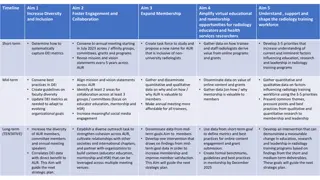
Objectives TG-Radiology is dedicated to the development of global standards for the evaluation and benchmarking of AI radiological systems. We are also collecting diverse data towards the assessment of AI radiological applications.
Timeline 21-24 Jan 2020 - TG-Radiology was officially created at Meeting H, Brasilia 30 Jan - Call for Participants published 25 Feb - First Zoom meeting Use Case Collection 26 Mar - First TDD submission Further work on TDD Updated TDD for 9th Meeting
Participants 17 participants currently From 5 continents; Africa (6), North America (5), South America (2), Europe (1), Asia (3) 11 organisations; AI providers (3), other digital health providers (2), health centers (2), universities (4)
Current Contributors to TDD (10 participants) Xavier Lewis-Palmer, minoHealth AI Labs, GUDRA Issah Abubakari Samori, minoHealth AI Labs, GUDRA Benjamin Dabo Sarkodie, Euracare Advanced Diagnostic Center Andrew Murchison, Oxford University Hospitals NHS Foundation Trust Alessandro Sabatelli, Braid.Health Daniel Hasegan, Braid.Health Pierre Padilla-Huamantinco, Universidad Peruana Cayetano Heredia Judy Wawira Gichoya, Emory University School of Medicine Saul Calderon Ramirez, De Montfort University (Metrics deliverable) Vincent Appiah, minoHealth AI Labs, GUDRA
Topic Description Document Topic Description Overview of Radiology, Challenges facing Radiology, Overview of AI in Radiology, Challenges Facing Overview AI in Radiology, Impact of Benchmarking Existing AI Solutions Use Case Descriptors (9): Condition, Medical imaging modality, AI task/problem description (eg. Image Classification, Image Segmentation), General algorithm description (if shareable), Project goal and current stage (if shareable), Input structure and format, Output structure and format, Evaluation metrics, Explainability and Interpretability framework
Topic Description Document Existing AI Solutions Use Cases from minoHealth AI Labs and Braid.Health 29 conditions: incl, breast cancer, pneumonia, hernia, arthritis X-Ray/body part classification: 56 parts: incl, cervix, wrist, lumbar, spine 3+ modalities: Chest X-Ray (22), Foot X-Ray (6), Mammography (1), other X-Ray (56)
Topic Description Document Existing AI Solutions Algorithms used: Convolutional Neural Networks, Transfer Learning, Bayesian Optimization Input structure and format: 2D image, PNG/JPEG (converted from DICOM) Evaluation metrics: Accuracy Score, ROC curve & Area Under Curve ROC Score, Specificity and Sensitivity
Topic Description Document AI Input Data Structure Image Conversion Considerations: Table with 3 approaches and their Advantages and Disadvantages Approaches: Integrating an automated conversion programme into AI Software, Using a separate software, Using an online tool Scores & Metrics Overview on 3: ROC curve and AUC score, F1 Score, Patient Level Accuracy & Image Level Accuracy (Metrics deliverable - Saul Calderon Ramirez)
Topic Description Document Benchmarking Methodology and Architecture Precision Evaluation framework and platform A radiograph-agnostic benchmarking platform and framework Assess how well an AI system performs across different demographics (Location, Gender, Age and more)
Next Steps Collecting additional metrics & scores, developing guidelines AI Input Data Structure: Overview/Guidelines of various ideal input & modalities (based on use cases) AI output Data Structure: Overview/Guidelines of various outputs (based on use cases) Meeting on Guidelines for AI4Radiology COVID-19 Solutions (27th May - TBD)