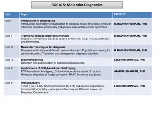
Introduction to Forensic Radiology: Techniques and Applications
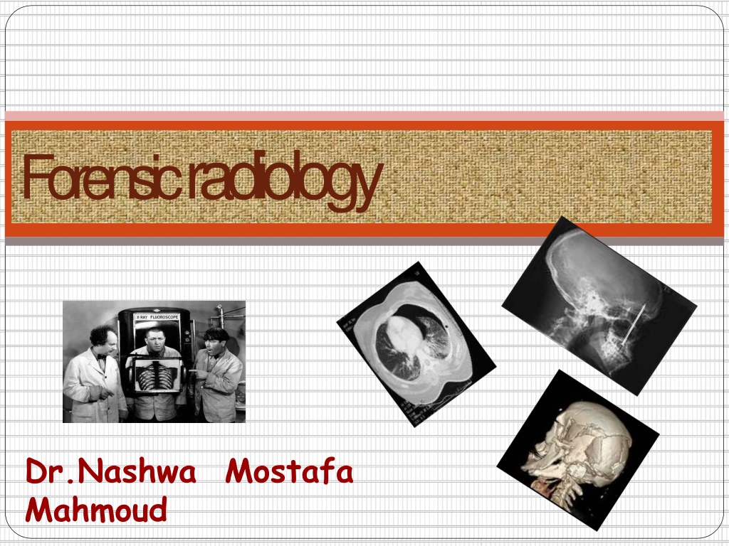
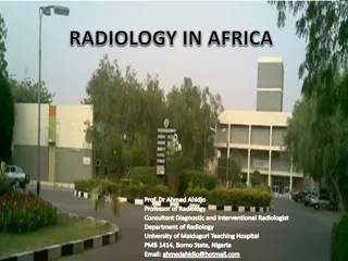
Forensic radiology, as pioneered by Dr. Nashwa Mahmoud Mostafa, involves the use of radiographic techniques to assist in legal investigations and post-mortem examinations. Beginning with the historical overview of X-rays in forensic science, the field has evolved to utilize advanced imaging technologies like CT scans and fluoroscopy. Common techniques include traditional X-rays, fluoroscopy for real-time imaging, and computed axial tomography for detailed cross-sectional analysis. Learn about the scope and significance of forensic radiology in this comprehensive overview.
- Forensic Radiology
- Imaging Techniques
- Legal Investigations
- Dr. Nashwa Mahmoud Mostafa
- Medical Imaging

Download Presentation

Please find below an Image/Link to download the presentation.
The content on the website is provided AS IS for your information and personal use only. It may not be sold, licensed, or shared on other websites without obtaining consent from the author. Download presentation by click this link. If you encounter any issues during the download, it is possible that the publisher has removed the file from their server.
E N D
Presentation Transcript
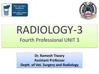
Forensic radiology Dr.Nashwa Mahmoud Mostafa
Outlines: Def Historical overview Common techniques Scope of Forensic radiology
Forensic radiology Definition: Radiography is the creation of radiographs by exposing a photographic film or other image receptor to X-rays. It is thus an examination of the structure of materials by non-destructive methods. Forensic radiography is the creation of radiographs for the purpose of assisting with legal investigations.
History The first time an X-ray was used for a forensic purpose was shortly after the technology was invented. In 1895, Wilhelm Roentgen discovered X-rays and just a few months later, a bullet lodged in the leg of a gunshot victim was shown in an X-ray and the evidence was used in court to successfully prosecute the accused for attempted murder. In addition to living subjects, forensic radiology is commonly used just before autopsies. The science has developed over the years to include CAT scan, MRI and ultrasound technologies.
C o m m o n techniques: 1. x-ray (roentgen ray): an energy form of ionizing radiation from which may be produced fluorescent or photographic images films . forensic radiology depended almost exclusively on the x- ray and the static image captured on the radiograph. Chest roentgenogram, radiograph, Or X-ray film.
C o m m o n techniques: 2. Fluoroscopy :electronically enhanced and directly visualized x-ray in real-time motion, cine- photographed, videotaped, or digitized and stored on magnetic tape or disks for replay A modern fluoroscope with an image intensifier connected to a television camera. The televised image (arrow) can be seen without darkening the room.
C o m m o n techniques: Dental x-ray machine Mortuary X-Ray Room
C o m m o n techniques: 3. Computed axial tomography : CAT Scan as computed tomography or CT scans passed through the body over multiple diametric pathways in the axial or cross-sectional plane resulting in a series of images of cross- axial sectional slices , a much higher differentiation between body tissues than conventional x-ray . in interventricular septum CT slice of heart showing tumor (arrows)
C o m m o n techniques: 4. Magnetic resonance imaging (MRI) : Utilizes strong magnetic fields to generate electromagnetic signals from elements and compounds found in body fluids and tissues . Can obtain multiplanar, multidirectional, sectional images or slices (MR scans) MRI of cardiac tumor (arrows) shown in the previous figure
C o m m o n techniques: 5. Ultrasound or ultrasonography : Sound waves generated outside the body by transponders are reflected back from internal structural interfaces to be recaptured and converted into real-time or static images. The image is a sonogram US of the same heart showing a tumor (arrows) in interventricular septum
C o m m o n techniques: 6-Angiography : Vertebral angiography in case of subarachnoid hemorrhage In case of cerebral embolism occlusion of the right middle cerebral artery by lead pellet embolus (arrow). right carotid arteriogram shows
S C O P E O F F O R E N S I C R A D I O L O G Y Radiography can speak The situations in which forensic radiology can be applied to resolve legal matters are many and varied: 1. Determination of Identity 2. Evaluation & documentation of Injury or cause of Death 3. Criminal Litigation 4. Civil Litigation 5. Recent advances ; Virtopsy. 6. Education & Research
I. Identification Radiological techniques allow forensic personal identification of: the ripped, lacerated corpses, charred or carbonized corpses macerated, putrefied or skeletonized corpses in mass disasters, transportation injuries, airchrach, bomb explosions
I. Identification I.Deductive (general or reconstructive) identification: Sex: radiographs of skull, pelvis and sternum. Age: Appearance of ossific centers Union of epiphysial plates Calcification of laryngeal and costal cartilages Skull radiographs for examination of fontanels, sutures and teeth. Race; negro skull,
I. Identification II. Comparative Identification: Depends on comparing antemortem to postmortem X- Rays of a person. Comparison includes: Normal structures: Comparison of skull bones, sinuses especially frontal sinuses regarding the size and shape ,Sella tursica & others Other bones; ribs, hip, dental, chest and vertebral areas Abnormal structures: congenital anomalies of bones, deformities and/or fractures, metallic prosthesis.. Dental radiographs: comparing root shapes, teeth fillings and abnormal teeth eruptions. Personal objects & Jewells:
I. Identification 1. The frontal sinus The frontal sinus is a triangular, pyramidal air cavity in between the tables of the frontal bone highly variable nature, even among identical twins. stable structure during adult life Its resiliency :It has very strong walls and preserved intact in human remains. Head & paranasal sinus radiographs are taken commonly for diagnostic purposes and almost everybody has one in his/her health folder.
I. Identification 1. The frontal sinus X-ray Comparison : matching of unique features; of external and internal bony anatomy (several-fold, curvatures, trabuclae, septae) can be made by superimposition or coding systems. Comparison of frontal sinuses between ante-mortem (AM) and post- mortem (PM) skull films showing duplication of distinctive pattern of air cells,
I. Identification 1. The frontal sinus Frontal sinus CT is a more precise than conventional radiographs; avoiding the superimposition of structures beyond the plane of interest the images can be easily manipulated and internal points that should be evaluated can be shown by images segmentation. Appearance of Several Frontal Sinuses in CTs
I. Identification 1. The frontal sinus Frontal sinus CT is a more precise than conventional radiographs; allowing the visualization of small differences of density & thickening Craniometric points; precisely located and measurements more accurately performed than on conventional radiographs. Volumes and areas can be determined. CT skull using a bonewindow . C; bony thickening of inner table of frontal bone on CT D; The topogram preliminary to the CT scan, showing craniometric points,
I. Identification 2. Dental radiography Partially skeletonized, badly decomposed remains of a female body. Characteristic dental features: comparing root shapes, teeth fillings , abnormal teeth eruptions, artificial teeth fillings, dental sutures or teeth archades Comparison of A : AM dental radiograph, with B : PM one of disarticulated mandible. There is a perfect match of both the restoration in the molar and the broken- off drill bit tip.
I. Identification 2. Dental radiography A : PM radiograph of mandibular fragment compared with B : AM bitewing radiograph. The root canal work and restorations are identical.bit tip.
I. Identification 2. Dental radiography A : PM facial roentgenogram shows unique restorations and a wire suture in the orbital floor. B : AM panoramic dental examination shows identical findings.
Dismembered trunk of a female found in a sewer. I. Identification 3. Any characteristic bony features B A : PM radiograph shows peculiar beak-like calcification of the 1st costochondral junction, cx rib bilaterally (arrows); B : AM chest with identical calcifications (arrow). A
I. Identification A: PM and B: AM X-ray of the forearm of a air crash victim with plate and screws fixation devices in place. A: PM and B: AM X ray of an air crash victim who had undergone hip replacement surgery.
I. Identification 4. Personal objects & Jewells Radiograph of severely burned remains on which no personal effects were evident on external examination. A wristwatch and ring, clearly seen on the radiograph, were not found on initial autopsy. When recovered, both items were instrumental in identifying the victim.
II.Radiological detection & documentation of injury O R the cause of death Radiological examinations play significant role: in differential diagnosis of non- accidental fractures from accidental fractures in determination of radiological evidence of physical abuse, Torture medical malpractice cases localization and type of bullets or shots remained within body. Pre autopsy ; to diagnose air embolism, cerebral air embolism, barotrauma, pneumothorax or pneumopericardium,
A. Radiology of injury II.Radiological detection & documentation of injury O R the cause of death I. Soft tissue injury: Swelling, edema or hemorrhage: Subdural hematoma around frontal lobe (arrows) on CT.
A. Radiology of injury II.Radiological detection & documentation of injury O R the cause of death I. Soft tissue injury: Laceration of an organ: knife wound to the heart (arrow) shown by MRI
A. Radiology of injury II.Radiological detection & documentation of injury O R the cause of death I. Soft tissue injury: Abnormal collection of air in the chest pneumothorax: Right-sided pneumothorax (arrow) on plain CXR
A. Radiology of injury II.Radiological detection & documentation of injury O R the cause of death I. Soft tissue injury: Abnormal collection of air in the chest pneumothorax: Left-sided pneumothorax (arrow) on CT scan of the chest with chest tube in place
A. Radiology of injury II.Radiological detection & documentation of injury O R the cause of death I. Soft tissue injury: Pneumopericardium >> the dark halo of air surrounding the heart (arrows). There also is pneumomediastinum outlining the inferior border of the thymus (open arrows).
A. Radiology of injury II. Fractures: In child abuse II.Radiological detection & documentation of injury O R the cause of death Battered child cases: A. Multiple regional fractures skull
A. Radiology of injury II. Fractures: In child abuse II.Radiological detection & documentation of injury O R the cause of death Battered child cases: A. Multiple regional fractures skull Skull fissures on CT Skull fissures on plain XR
A. Radiology of injury II. Fractures: In child abuse II.Radiological detection & documentation of injury O R the cause of death Battered child cases: Multiple regional fractures Long bones metaphyseal fractures (avulsion and dislocation of epiphyseal ends): Typical bucket-handle metaphyseal fracture of the distal humerus on plain XR.
A. Radiology of injury II. Fractures: II.Radiological detection & documentation of injury O R the cause of death In child abuse Rib fractures. A : typical healed posterior fracture from AP compression. B : healed lateral rib fractures.
A. Radiology of injury II. Fractures: II.Radiological detection & documentation of injury O R the cause of death In child abuse Rib fractures. D : (beads of string) with multiple bilateral healing fractures (note hazy callus surrounding ribs).
A. Radiology of injury II. Fractures: In child abuse II.Radiological detection & documentation of injury O R the cause of death 2. Battered child cases: B. Fractures of different ages: a new rib fracture (arrow) through one of the old, healed fractur
A. Radiology of injury ` II. Fractures: In domestic abuse II.Radiological detection & documentation of injury O R the cause of death D : panorex study shows fractures through the left mandibular angle and right mentalis, separation of teeth at fracture site depressed fracture of the left zygomatic arch (arrows) hand showing new (arrows), healing (open arrows), and healed (curved arrows) fractures with residual deformity, and dislocation
A. Radiology of injury II. Fractures: In transporation fractures II.Radiological detection & documentation of injury O R the cause of death typical bumper fracture in an adult pedestrian hit from the right.
A. Radiology of injury II.Radiological detection & documentation of injury O R the cause of death III. Foreign bsot adbibei nsg: by glass piece
II.Radiological detection & documentation of injury O R the cause of death III. Foreign bodies: Retained instruments after surgery: clamp Retained instruments after surgery: curved needle (arrow)
II.Radiological detection & documentation of injury O R the cause of death III. Foreign bodies: A bottle was driven into the victim s face. The cap stayed behind as the bottle was withdrawn.
II.Radiological detection & documentation of injury O R the cause of death III. Foreign odies: bIn charred mutilated bodies A woman was found burned beyond recognition after a house fire. The remains were radiographed in order to try to match them with the occupant s ante-mortem chest film. Showing; B revealed several coils of a wire ligature around the victim s neck. C positive radiological identification.
II.Radiological detection & documentation of injury O R the cause of death III. Foreign bodies: In transportation injuries This middle-aged man was sent for a chest film ( A ) because of suspected heart disease. A round mass in the left lung prompted a tomogram ( B and C ) which defined the mass in frontal and lateral projections.
II.Radiological detection & documentation of injury O R the cause of death III. Foreign bodies: At surgery ( D ) a gearshift knob encapsulated in fibrous scar was removed. The man had been in an automobile accident 22 years earlier!
II.Radiological detection & documentation of injury O R the cause of death IV. Firearm injuries wounds: In the location of the bullet Reveal whether there are bullets of a different caliber (in cases where multiple weapons are involved). The number of bullets is also important and must be correlated with the entrance and exit wounds . May also reveal information about the angle and direction of fire. Small metallic fragments produced when a bullet strikes bone may lead directly to the bullet and clearly indicate the bullet s path The radiographs may reveal clues as to the type of weapon . Shots; leave a characteristic Radiology role in gunshot bullets: Fragmented bullet within the head and neck areas of a gunshot wound victim.
II.Radiological detection & documentation of injury O R the cause of death IV. Firearm injuries wounds: Radiology role in gunshot The radiographs may reveal clues as to the type of weapon . Shots; leave a characteristic leadsnowstorm X-rays may be the first indication that a crime has been committed when decomposed bodies are dbi su lcl eotvse: rAedb.ullet traversed the posterior elements of the C-1 vertebra (small arrows), impacted on the posterior body of C-2 (open arrows), then dropped in the spinal canal before coming to rest at the C-5 level (large arrow).
II.Radiological detection & documentation of injury O R the cause of death IV. Firearm injuries bullets: A : frontal and B : lateral view of the skull show a left temporal wound of entry (arrowheads). There are scattered bone and bullet fragments throughout. The bullet bounced off the sella (open arrow). The jacket (short arrow) separated, and the bullet (long arrow) came to rest against the right parietal bone posteriorly.
II.Radiological detection & documentation of injury O R the cause of death IV. Firearm injuries bullets: the characteristic sharp projections of the jacket ,exposed as the bullet mushrooms(dumdamized bullets)
II.Radiological detection & documentation of injury O R the cause of death IV. Firearm injuries bullets: CT reveals bullet deep in posterior costophrenic sulcus (star pattern). Bullet obscured on routine chest film by density of full- thickness liver. A : bullet fired into the base of the skull cut a groove in the occipital bone (large arrow) scattering fragments (small arrows) into the posterior fossa as shown on CT examination.