Comprehensive Overview of Radiology in Pediatric Dentistry at Semmelweis University Budapest
Radiology plays a crucial role in diagnosing and treating diseases within the body, especially in pediatric dentistry at Semmelweis University Budapest. From the historical evolution of radiography to the modern radiologic diagnostic imaging techniques used in dentistry, this department utilizes a range of imaging technologies to ensure accurate diagnosis and treatment. Dental radiographic techniques, including intraoral and extraoral methods, are essential in this field. Explore the specialized devices and procedures involved in special radiologic examinations of the maxillary structures.

Download Presentation

Please find below an Image/Link to download the presentation.
The content on the website is provided AS IS for your information and personal use only. It may not be sold, licensed, or shared on other websites without obtaining consent from the author. Download presentation by click this link. If you encounter any issues during the download, it is possible that the publisher has removed the file from their server.
E N D
Presentation Transcript
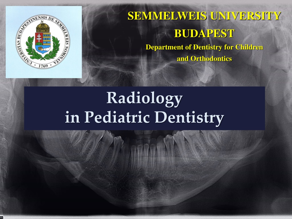
SEMMELWEISUNIVERSITY BUDAPEST Department of Dentistry for Children and Orthodontics Radiology in Pediatric Dentistry
RADIOLOGY the medical speciality, that uses imaging to diagnose and treat diseases seen within the body; Imaging techniques used by radiologists: X-ray radiography Ultrasound Computer tomography (CT) Nuclear medicine Positron emission tomography (PET) Magnetic resonance imaging (MRI) Interventional radiology: the performance of (usually minimal invasive) medical procedures with the guidance of imaging technologies
History Radiographs (Roentgenographs): X-ray: W. C. R ntgen, 8th November, 1895; 3 Studies: 1986; First Nobel Prize in Physics, 1901; Wilhelm Conrad R ntgen (1845-1923)
History Professor K llicker (anatomist) named it roentgen-ray, after its discoverer Rudolf Albert von K llciker (1807-1905) in 1896 the first dental x-ray photograph was taken (5-9-25 minutes exposure time!!!) in 1897 Iszlay was the first, who took dental radiography in Hungary in 1905 Charles G. Barkla, Liverpool the characteristic X-ray .
Dental Radiographic Techniques Dental examinations are the most frequent type of radiological procedure, and account for 21% of the total on a global scale; Individual doses are small but collective doses can not be ignored due to the high volume of procedures. Intraoral techniques (the film is in the mouth) Extraoral techniques (the film is out of the mouth)
Radiologic diagnostic imaging Conventional radiological diagnostics of the maxillary structures device types: - Standard for dental offices Analogue Digital Small device types of for intraoral images, lateral view.
Radiologic diagnostic imaging Special radiologic examination of the maxillary structures Devices: 2 m high device for extraoral techniques like panoramic images (OPG); = ca. 12 hours under fresh air on a field (camp) or 1 hour in the high mountains
Digital radiography = a form of X-ray imaging, using digital X-ray sensors instead of traditional photographic film RIS Radiologic Information System PACS Digital Archiving System Digital image capture devices: Flat Panel Detectors High-density Line-scan Detectors
Radiologic diagnostic imaging Modern scanning imaging methods: virtula slices Computer Tomography (CT) 1972 Godfrey N. Housnfield X-ray free techniques Nuclear Spin Tomography Magnetic Resonance Tomography (MRT) 1973 Paul Lauterbur Ultrasound
Intraoral techniques Periapical (lateral) radiography Bite-wing (crown) radiography Occlusal radiography
General rules of creating pictures: The position of the head The position of the film The position of the tube Two of the fundamental rules of radiography are: 1) the central beam should pass through the area to be examined; 2) the radiographic receptor should be placed in a position to record the image with the least amount of image distortion. Three types of intraoral radiologic examinations commonly used in dental practice periapical, bitewing (interproximal), and occlusal examinations depend on the operator s adherence to these two rules even though specific techniques, processes, and indications differ widely among them.
The rules of the tube- positioning Right angle (the central beam is perpendicular on the line of the bisecting-angle Orthoradial direction of the central beam Apical central beam
1. Periapical radiography The purpose of the intraoral periapical examination is to obtain a view of the entire tooth and its surrounding structures; Two exposure techniques may be employed for periapical radiography: the paralleling technique; bisecting angle technique.
1. Periapical radiography isometric (the same measures) orthoradial projection;
1. Periapical radiography Parallel technique Bisecting angle technique: the length of the tooth is isometric, but the picture is distorted The aim: to exam the apex and the bone around the apex
Paralleling technique is the preferred method; provides less image distortion and reduces excess radiation to the patient; should always be attempted before other techniques. Bisecting angle technique can be employed for patients unable to accommodate the positioning of the paralleling technique; candidates may include those with low palatal vaults and children; disadvantages to the bisecting technique include image distortion and excess radiation due to increased angulations involving the eye and thyroid glands. . 1. Periapical radiography
The bisecting angle technique The bisecting-the-angle or bisecting angle technique is based on the principle of aiming the central ray of the x- ray beam at right angles to an imaginary line which bisects the angle formed by the longitudinal axis of the tooth and the plane of the receptor.
Films for periapical radiography Usually: 30 mm x 40 mm For children: 22mm x 35mm (there is 1-2 mm difference between sizes of different products)
2. Bitewing radiography introduced by Dr. Raper, 1925. focus on the clinical crowns of both the maxillary and mandibular teeth. do not show the apices of the tooth and cannot be used to diagnose in this area. detection of interproximal caries in the early stages of development, before it is clinically apparent. B reveal the size of the pulp chamber and the relative extent to which proximal caries have penetrated. a useful adjunct to evaluating periodontal conditions. They offer a good view of the septal alveolar crest, and in addition, permit changes in bone height to be accurately assessed by comparison with adjacent teeth.
2. Bitewing radiography (interproximal radiography) Bitewing radiographic images are of particular value in detecting interproximal caries in the early stages of development, before it is clinically visible. For this reason horizontal angulation must be accurately projected following the direction of the interproximal contacts and no overlapping contacts are present on the radiographic image. Bitewing radiographic images are also useful in evaluation of the alveolar crests for detection of early periodontal disease.
Films for bitewing radiography Wing-films Size of the film: 30 mm x 40 mm or 27 mm x 54 mm
Occlusal radiography The patient bites for the film Film size: 75 mm x 57,5 mm Upper occlusal radiography Aim: to exam the area of incisors
Occlusal radiography Lower occlusal radiography The aim: to diagnose of the sublingual or submandibular salivary calculus, the structure of the bone at the mental area, and to exam the lower incisors
Extraoral techniques Panoramix radiography Panoramic radiography (orthopantomography) Cephalometric X-ray (tele X- ray)
Panoramix radiography It is a magnified picture separately made about the maxilla and mandible The anterior region is of value The molar region is distorted The size of the film: 10 cm x 24 cm
Orthopantomogram (OP) Layered picture (tomography) Both of the jaws The area of the incisors is not so easy to diagnose The size of the film: 15cm x 30cm
Cephalometric X-ray Specially made for orthodontics Diagnose of the sagittal and vertical direction of the growing jaws Realistic size of the skull The size of the film: 18cm x 24cm
Carpal radiography 1. radius, ulna dist. epiphysis 2. carpal bones 3. metatarsus 4. tarsus in childhood, to classify the bone maturation importance to orthodontics and combined cases in oral surgery
CVM (Cervical Vertebral Maturation) Skeletal age determination
Radioactivity radiation bionegativ effect swallowed dose (Gy = J/kg) ekvivalent dose (Sv = Gy x Q = J/kg x Q) max: 50 mSv/ 1 year or 30 mSv/ 13 weeks! ALARA = As Low As Reasonably Achievable
Radiation - Dosis Dental X-ray: 0,02 mSv Rtg Cranium, Thorax: 0,2 mSv Rtg Ripps: 3,0 mSv Rtg Cervix: 5,0 mSv Mammographiy1,0 mSv CT cranium: 2,0 mSv, Thorax: 10,0 mSv, Abdomen: 7,0 mSv Angiography, Cardiac catheter: 10,0 mSv
How to avoid of overdosing proper indication X-ray film with high sensitivity the use of amplifier proper procedure (without failures!) careful work in the darkroom new techniques (rvg)
Radiation protection Protection of the patient: long tube, using of filters (Al) lead-robe (protection of gonads -especially for children!) Protection of the team: min. 1,5 m distance, the best is 3,5 m! X-ray film is prohibited to be held by the doctors or the assistant! using of protection wall X-ray dosimeter
In primary dentition radiographic examination is not necessary in the following cases: dens connatalis dentitio tarda caries pulpitis periostitis focal infection (focus)
Indications of radiographic examination in primary dentition: after pulpotomy; after pulpectomy; uncertain swelling (to exclude the tooth origin); after traumatic injuries; anomalies of mechanism of 2nd dentition Caries detection: approximal, (occlusal)
Indications of radiographic examination in permanent dentition: root canal treatment traumatic injuries teeth in retention supernumerary, missing and malformed teeth pillars before prosthodontic treatment
Advantages of the digital technique Sensor instead of film Lower radiation Easy storage and sending Save the environment The size of the detector: 40 x 20 x 14 mm
CBCT endodontics orthodontics parodontology TMJ-examinations dentoalv. surgery maxillofac. surgery otolaryngology orthopaedics judicial medicine
References: Intraoral Radiographic Techniques Allan G. Farman, BDS, EdS., MBA, PhD; Sandra A. Kolsom, CDA-Emeritus, RDA; ADAA 2014 Council on Education


")