Survey of Structural Properties of Different Classes of Viruses

Viruses are small obligate intracellular parasites that require living hosts for multiplication. They lack ribosomes and metabolic enzymes, relying on host cells for replication. Viruses vary in size, shape, and genome organization, with classification based on morphology, physicochemical properties, genome type, lipids, and carbohydrates. Antigenic and biological properties also influence virus classification. Understanding these characteristics helps in identifying hosts and developing treatments for viral diseases.

Download Presentation

Please find below an Image/Link to download the presentation.
The content on the website is provided AS IS for your information and personal use only. It may not be sold, licensed, or shared on other websites without obtaining consent from the author. Download presentation by click this link. If you encounter any issues during the download, it is possible that the publisher has removed the file from their server.
E N D
Presentation Transcript
BIOCHEMISTRY OF PARASITES BCH 4I4 LECTURE 7-9 Survey of structural properties of different classes of viruses S.O Anadozie (Ph.D.)
VIRUSES A virus is a small filterable and obligate intracellular parasite requiring a living host for its multiplication i.e can reproduce only within a host cell. The term virus was coined by Pasteur from the Latin word for poison . First discovered after the development of a porcelain filter, the Chamberland-Pasteur filter that could remove all bacteria visible in the microscope from any liquid sample. Adolph Meyer in 1886 demonstrated that a disease of tobacco plants could be transferred from a diseased plant to a healthy one via liquid plant extracts. In 1892, Dmitri Ivanowski showed that this disease could be transmitted even after the Chamberland-Pasteur filter had removed all viable bacteria from the extract.
CHARACTERISTICS OF A VIRUS They use a lock and key fit to identify hosts a process known as Simple Viral Reproductive Cycle . They take control of synthetic and genetic machinery inside their host cell. Viruses are composed of a nucleic acid, RNA or DNA, but never both. Do not have ribosomes, enzymes for metabolism and necessary requirements to make proteins Do not grow but replicate or multiply which is done within living cells. Some viruses have a lipid envelope or membrane surrounding a nucleocapsid core. The source of the envelope is from the membranes of the host cell. All viruses have a protein coat or shell that surrounds and protects the nucleic acid core.
CHARACTERISTICS OF A VIRUS Most virions, or single virus particles, are very small, about 20 to 250 nm in diameter. However, some recently discovered viruses from amoebae range up to 1000 nm in diameter. Viruses have a host range. They infect specific cells or tissues of specific hosts, or specific bacteria, or specific plants. Viruses are important agents of human, animal and plant diseases e.g rice yellow mottle virus of rice, foot and mouth disease of domestic animals and measles virus.
CLASSIFICATION OF VIRUS Viruses are classified using a combination of characteristics, these includes: 1. Morphology: size, shape, presence of envelope, number and sequence of proteins. 2. Physicochemical properties: thermal stability, detergent stability, molecular mass. 3. Genome organization and replication: size and type of nucleic acid, strategy of replication, number and position of open reading frames, transcriptional and translational strategies, site of virion assembly and release. 4. Lipids and carbohydrate: content, character.
CLASSIFICATION OF VIRUS (CTD) 5. Antigenic properties: serological relationships. 6. Biological properties: Host range, mode of transmission, pathogenicity, tissue tropisms, geographic distribution. Other forms of classification includes; a. Orders (virales): Groupings of families of viruses that share common characteristics and are distinct from other orders and families. b. Families (-viridae): Groupings of genera of viruses that share common characteristics and are distinct from the member viruses of other families. c. Subfamilies (-virinae): Not used in all families, but allows for more complex hierarchy of taxa. d. Genera (-virus): Groupings of species of viruses that share common characteristics and are distinct from the member viruses of other species. e. Species (virus): A polythetic class of viruses that constitutes a replicating lineage and occupies a particular ecological niche".
COMPONENTS OF VIRUSES AND DEFINITION OF TERMS Capsid: Composed of numerous repeating subunits. Viral genome is thus conserved in that a single protein can be transcribed many times to make the capsid. Capsomere: Clusters of polypeptides which, when completely assembled, form the capsid. They protect the nucleic acid from the environment Nucleocapsid: Genome together with capsid. Virion: An infectious free viral particle, consisting of nucleocapsid, or nucleocapsid plus envelope. N:B. Not all virus particle are infectious. Envelope: Lipid bilayer and proteins usually obtained from host cell. The Envelope spikes binds to the cell surface proteins.
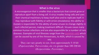
STRUCTURE OF VIRUS All viruses contain the following three components: (A) a nucleic acid genome (viral genomes); (B) a protein capsid that covers the genome. Together this is called the nucleocapsid. In addition, many animal viruses contain a (C) lipid envelope. A: Viral Genomes: The genomes of all known cells are comprised of double stranded DNA, that for viruses can be a single or double stranded DNA or RNA. They vary greatly in size, from approximately 5-10 kb (papovaviridae, Parvoviridae) to greater than 100-200 kb (Herpesviridae, Poxviridae).
STRUCTURE OF VIRUS (Viral genomes ctd) The known structures of viral genomes are: For DNA; - Double Stranded - linear or circular - Single Stranded - linear or circular - Other Structures - gapped circles For RNA; - Double Stranded - linear - Single Stranded - linear : These single stranded genomes can be either + sense, - sense, or ambisense.
STRUCTURE OF VIRUS (Viral genomes ctd) The sense strand can serve directly as mRNA and code for protein, so for these viruses, the viral RNA is infectious. The viral mRNA from (-) strand viruses is not infectious, since it needs to be copied into the (+) strand before it can be translated. In an ambisense virus, part of the genome is the sense strand, and part is the antisense. The genome of some RNA viruses is segmented, meaning that a virus particle contains several different molecules of RNA, like different chromosomes.
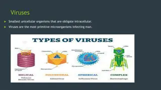
STRUCTURE OF VIRUS B: Protein Capsid (Viral nucleocaspids): Viral genomes are surrounded by protein shells known as capsids. The capsid protein recognises signal (sequence), a special types of packaging on the viral genome. The capsid are made of repeating structural subunits arranged in three major forms of symmetry; 1. Cubic Symmetry (icosahedral symmetry) - composed of proteins (capsomers) subunits arranged in the form of a hollow, quasi spherical structure, with the genome within. The icosahedron is made up of 20 equilateral triangles, forming a symmetric figure.
STRUCTURE OF VIRUS (icosahedral symmetry ctd) Since proteins are not equilateral triangles, each face of an icosahedron contains more than one protein subunit. The simplest icosahedron is made by using 3 identical subunits to form each face which could be a single protein or, more likely, a complex of several polypeptides. Each of the subunits could be a single protein or, more likely, a complex of several polypeptides. Lumps or clusters of virus nucleocapsids on the surface of the particle are often seen under an electron microscope. The protein subunits clustered around an axis of symmetry are called "morphological units" or capsomers.
STRUCTURE OF VIRUS (protein capsid ctd) (2) Helical symmetry - The proteins (capsomers) are arranged in a helix (hollow coil) around the viral RNA, with 3 nt of RNA fitting into a groove in each subunit. Helical capsids can also be more complex, and involve more than one protein subunit. A helix can be defined by two parameters, its amplitude (diameter) and pitch. This structure is very stable, and can be dissociated and re-associated readily by changing ionic strength, pH and temperature. The interactions that hold these molecules together are non-covalent, and involve H-bonds, salt bridges, hydrophobic interactions, and vander Waals forces.
STRUCTURE OF VIRUS (protein capsid ctd) Examples of animal virus with helical nucleocapsids, includes the Orthomyxoviridae (influenza), the Paramyxoviridae (bovine respiratory syncytial virus), and the Rhabdoviridae (rabies). (3) Complex symmetry - Viruses can be complex in shape with no regular symmetry, as in poxvirus. The bacteriophage T4 has three relatively complex virions with its DNA-containing head group and tail fibers that attach to host cells; adenovirus, which uses spikes from its capsid to bind to host cells. The influenza virus uses glycoproteins embedded in its envelope to bind to host cells. The influenza virus also has matrix proteins, internal to the envelope, which help stabilize the virion s shape.
STRUCTURE OF VIRUS (C) Viral Envelope - The nucleocapsid of some animal viruses, is surrounded by a membranes called an envelope which is made up of a lipid bilayer comprised of host- cell lipids. It also contains virally encoded proteins (glycoproteins) which are trans-membrane proteins and serve many purposes, such as binding to receptors on the host cell, playing a role in membrane fusion and cell entry. They can also form channels in the viral membrane. Many enveloped viruses also contain matrix proteins, which are internal proteins that link the nucleocapsid to the envelope. They are very abundant (many copies per virion), and are usually not glycosylated.
STRUCTURE OF VIRUS (viral envelope ctd) Some virions also contain other, non-structural proteins that are used in the viral life cycle and present in low amounts in the virion e.g replicases and transcription factors. Enveloped viruses are formed by budding through cellular membranes, usually the plasma membrane but sometimes an internal membrane such as the ER, golgi, or nucleus. The genome, capsid and matrix occurs on the inside face of the membrane, the envelope glycoproteins cluster in that region of the membrane, and the virus buds out. This ability to bud allows the virus to exit the host cell without lysing, or killing the host. Non-enveloped viruses, and some enveloped viruses, kill the host cell in order to escape.
Host-cell Virus Interactions Productive Infection: Virus successfully completes replication cycle and produces progeny virions. Abortive Infection: Virus enters the host-cell but cannot successfully complete replication. This can result from a non-permissive host-cell, or because the virus is defective. And can become persistent infections. Restrictive Infection: Cells are only transiently permissive, after which they become non-permissive. This results in transformation of the cell. Latent Infection: Virus remains in a cell and does not replicate, as in HSV, VZV, CMV, EBV, and HIV.
TOPIC 8 Viral multiplication mechanism
VIRAL MULTIPLICATION Viruses carry their genome (RNA or DNA) and sometimes functional proteins required for early steps in replication cycle. Viruses depend on host cell machinery to complete replication cycle. Viral genome must be able to produce mRNA; host cell protein- synthesizing machinery may be able to synthesize viral proteins. The virion is disrupted after interaction with the host cell, causing infectivity lost. A process known as the ECLIPSE PERIOD. The yield of infectious virus per cell ranges from 100 - 100,000 particles. Replication cycle varies from 6 - 8 h to about 40 h.
Multiplication of viruses: Basic steps General steps in viral multiplication/replication: In animal virus, differences between naked and enveloped virus is seen. The multiplication cycles includes: Adsorption: Attachment of viruses to host cells. Surface protein on virus attaches to specific receptor(s) on cell surface Penetration: Entry of virions (or their genome) into host cells through receptor-mediated endocytosis. Uncoating: The viral capsid is removed and degraded by viral enzymes or host enzymes releasing the viral genomic nucleic acid. Synthesis: New nucleic acids, capsid proteins, and other viral components- transcription, translation and genome replication. Assembly: Maturation and assembly of newly synthesized viral components into complete virions Release: Departure (pemergian) of new virions from host cells.
Multiplication of viruses: Basic steps Figure 1: Multiplication of viruses
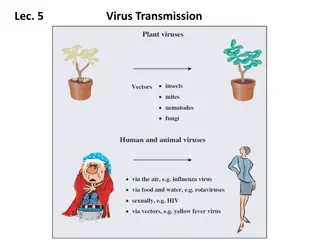
Multiplication of viruses: Basic steps Viruses recognize specific receptors Figure 2 and 3. And then penetrates into the cell Figure 4 Figure 4
DNA VIRUSES With animal DNA viruses, transcription and translation are not coupled, except for poxviruses where transcription occurs in the nucleus and translation in the cytoplasm. The primary transcripts, generated by RNA polymerase II, are larger than the mRNAs found on ribosomes, and in some cases, as much as 30% of the transcribed RNA remains untranslated in the nucleus. The viral messengers, however, like those of animal cells, are monocistronic. Transcription has a temporal organization, with most DNA viruses only a small fraction of the genome is transcribed into early messengers. The synthesis of early proteins is the key initial step in viral DNA replication. After DNA synthesis, the remainder of the genome is transcribed into late messengers.
DNA VIRUSES The complex viruses have immediate early genes, which are expressed in the presence of inhibitors of protein synthesis, and delayed early genes, which require protein synthesis for expression Regulation is carried out by proteins present in the virions, or specified by viral or cellular genes, interacting with regulatory sequences at the 5' end of the genes. These sequences may respond in trans to products produced by other genes and act in cis on the associated genes. Different classes of genes may be transcribed from different DNA strands and therefore in opposite directions e.g. polyomaviruses. The transcripts may undergo post- transcriptional processing so that nonessential intervening sequences are removed.
DNA REPLICATION The mode of replication is semiconservative but the nature of the replicative intermediates depends on the manner of replication. Several methods of replication can be recognized. A. Adenoviruses Adenoviruses show asymmetric replication, which initiates at the 3' end of one of the strands using a protein primer. The growing strand displaces the pre-existing strand of the same polarity and builds a complete duplex molecule and replicates in a similar manner after generating a panhandle structure by pairing the inverted terminal repetitions. B. Herpesviruses Herpesviruses have linear genomes with terminal repeats. On reaching the nucleus, the terminal ends undergo limited exonucleotic digestion and then pair to form circles. Replication is thought to take place via a rolling circle mechanism, where concatemers are formed. .
DNA REPLICATION C. Papovaviruses The DNA of papovaviruses are circular and the replication is bidirectional and symmetrical, via cyclic intermediates D. Parvoviruses The replication of single stranded parvoviruses is initiated when (+) and (-) stranded DNA from different parvovirus particles come together to form a double stranded DNA molecule from which transcription and replication takes place. E. Poxviruses The striking feature of poxvirus DNA is that the two complementary strands are joined. The replicative intermediates, present in the cytoplasm, are special concatemers containing pairs of genomes connected either head to head or tail to tail
DNA REPLICATION F. Hepadnaviruses Hepatitis B virus employs reverse transcription for replication. The genome consists of a partially double-stranded circular DNA with a complete (-) strand and an incomplete (+) strand. Upon entering the cell, the (+) strand is completed and transcribed. RNA transcripts are in turn reverse-transcribed into DNA by a viral enzyme in several steps, following closely the model of retroviruses, including a jump of the nascent positive strand from one direct repeat (DR) to another.
RNA VIRUSES The replication of RNA viral genomes is dictated by the absence of multiple translation units within the same messenger, a characteristic of all animal cell messengers. To overcome this difficulty, 3 main strategies have developed. A. The viral mRNA acts directly as the messenger and is translated monocistronically, followed by cleavage to form different proteins. B. The virion RNA is transcribed to yield various monocistronic mRNAs by initiating transcription at various places. C. The genome itself is a collection of separate RNA fragments that are transcribed into monocistronic mRNAs.
Assignment 1. write on the classes of RNA viruses 2. Replication of Single-stranded RNA viruses 3. List and explain the control mechanism of viral replication
READ UP ON INTERFERON


")


")
")

")
")
")

")