Understanding the Structure and Symmetry of Viruses along with Cultivation Methods
Viruses, defined as obligate intracellular parasitic organisms, exhibit general properties such as small size, filterability, simple structure, and absence of cellular components. They vary widely in size and lack independent metabolism. The cultivation of viruses plays a crucial role in studying their characteristics and behavior.

Download Presentation

Please find below an Image/Link to download the presentation.
The content on the website is provided AS IS for your information and personal use only. It may not be sold, licensed, or shared on other websites without obtaining consent from the author. Download presentation by click this link. If you encounter any issues during the download, it is possible that the publisher has removed the file from their server.
E N D
Presentation Transcript
Structure and Symmetry of Viruses Cultivation of Viruses Mr. S. N. Mendhe Department of Microbiology, Shri Shivaji Science and Arts College, Chikhli
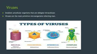
Viruses Viruses Long before the discovery of the microbial world, the term virus was used to denote any agent capable of producing disease. The word is Latin one & originally meant snake venom or poisonous fluid. Now viruses are defined as obligate intracellular parasitic minute organisms. Almost all the types of living organisms are subject to attack by viruses, hence, viruses occur in plants, animals even in bacteria.
General characteristics or General General characteristics or General properties properties Size & visibility Filterability Simple structure Absence of cellular structure No independent metabolism Nucleic acids Crystallization No growth & division
Size Size & & visibility Viruses vary widely in size. The largest among them (e.g., poxviruses) measuring about 300. The smallest viruses (e.g. foot & mouth disease virus ) are about 20nm. On the whole viruses are much smaller than bacteria. Mostly animal viruses, bacteriophages are invisible microscope. As they are too small to be seen under the light microscope, they are also designated as ultramicroscopic. One of the larger viruses such as pox viruses, can be seen under the light microscope visibility : :- - plant under viruses the & light
Filterability Filterability: :- - Iwanowski demonstrated what was thought to be uniquely distinctive property of viruses, the ability when suspended in fluid to pass through the filters that would hold back bacteria. Hence they are also known as filterable viruses.
Simple Simple structure Viruses have very simple structures. The simplest viruses are nucleoprotein particles consisting of genetic material (DNA or RNA) surrounded by protein capsid. In this respect they differ from typical cells which are made up proteins, carbohydrates, lipids & nucleic acids. The most complex viruses contain lipids & carbohydrates. Absence Absence of of cellular cellular structure Viruses do not have cytoplasmic organelles like mitochonria, golgi complex, ribosomes etc. are absent. They do not have limiting cell membrane. structure:- structure: :- - any cytoplasm, thus
No No independent independent metabolism Viruses can not multiply outside a living cell. No virus has been cultivated in cell free medium. Viruses do not have independent metabolism. They are metabolically inactive outside the host cell, because they do not posses enzyme systems & protein synthesis machinery. Viral nucleic acid replicate by utilizing protein synthesis machinery of the host. It codes for the synthesis of a number of viral proteins including subunits or capsomeres of the capsomeres tail proteins & some enzymes concerned with the synthesis or release of virions. metabolism: :- -
Nucleic Nucleic acids Virions have only one nucleic acid, either DNA or RNA. Typical cells have both DNA& RNA. acids: :- - Crystallization Crystallization: :- - Many of the smaller viruses can be crystallized & thus behave like chemicals. No No growth growth & & division power of growth & division. A fully formed virus does not increase in size by addition of new molecules. division:- Viruses do not have
Viral Viral structure structure: :- - Viruses are ultramicroscopic particles containing nucleic acid surrounded by protein, and in some cases, other macromolecular components such as a membrane like envelope. Outside the host cell, the virus particle is also known as a virion. The virion is metabolically inert and does not grow or carry on respiratory or biosynthetic functions. Viral structure:- Certain viruses contain ribonucleic acid (RNA), while other viruses have deoxyribonucleic acid (DNA). The nucleic acid portion of the viruses is known as the genome. The nucleic acid may be single-stranded or double-stranded; it may be linear or a closed loop.
The genome of the virus is surrounded by a protein coat known as a capsid, which is formed from a number of individual protein molecules called capsomeres. Capsomeres are arranged in a definite geometrical pattern around the nucleic acid. A single type of capsomere or several chemically distinct types may make up the capsid. The combination of genome and capsid is called the viral nucleocapsid. Such viruses are known as naked viruses, e.g. Polio virus,Adeno virus. A number of kinds of viruses contain envelopes. An envelope is a membrane like structure that encloses the nucleocapsid and is obtained from a host cell during the replication process. Among the enveloped viruses are those of herpes simplex, chickenpox, mononucleosis. and infectious
Helical / Spiral Symmetry:-The nucleocapsids of viruses are constructed according to certain symmetrical patterns. The virus that causes tobacco mosaic disease, for example, has helical symmetry. In this case, the nucleocapsid is wound like a tightly coiled spiral. The rabies virus also has helical symmetry. Icosahedral Icosahedral Symmetry Symmetry: :- - Other viruses take the shape of an icosahedron, and they are said to have icosahedral symmetry. In an icosahedron, the capsid is composed of 20 faces, each shaped as an equilateral triangle. Among the icosahedral viruses are those that cause yellow fever, polio, and head colds.
The envelope of certain viruses is a lipid bilayer containing glycoproteins embedded in the lipid. The envelope gives a somewhat circular appearance to the virus and does not contribute to the symmetry of the nucleocapsid. Projections from the envelope are known as spikes. The spikes sometimes contain essential elements for attachment of the virus to the host cell.
Binal /Complex Symmetry:- These viruses possess a capsid that is neither purely helical nor purely icosahedral, and that may possess extra structures such as protein tails or a complex outer wall. Some bacteriophages, phage T4, have a complex structure consisting of an icosahedral head bound to a helical tail, which may have a hexagonal base plate with protruding protein tail fibres. such as Enterobacteria
CULTIVATION CULTIVATION OF Viruses are obligate intracellular parasites. They cannot grow in any culture media. There are three methods are used for the cultivation of the viruses: Animal inoculation Tissue culture Embryonated egg inoculation OF VIRUSES VIRUSES: :- -
1. Animal Animal inoculation inoculation: :- - As the name indicates cultivation of viruses occur by using different types of animals. For e.g. Monkeys, rats, rabbits guinea pigs. At the end of incubation period, the animals are slaughtered and washed thoroughly and viruses are obtained from them.
2 2. .Embryonated Embryonated eggs( Prior to the advent of cell culture, animal viruses could be propagated only on whole animals or embryonated chicken eggs. The developing chick embryo, 10 to 14 days after fertilization, provides a variety of differentiated tissues, including the amnion, allantoic cavity, chorion, and yolk sac, which serve as substrates for growth of a wide variety of viruses, including paramyxo viruses, rhabdoviruses, togaviruses, herpes viruses, and pox viruses. The shell surface is first disinfected with iodine and penetrated with a small sterile drill. After inoculation, the drill hole is sealed with paraffin wax and the egg is then incubated at 360C for required period of time. eggs( Chick Chick embryo embryo method) method): :- - orthomyxoviruses,
3. Tissue culture method: 3. Tissue culture method:- - This is the method of choice routinely employed for cultivation of viruses. a) Cell culture: a) Cell culture:- - Tissues, e.g., monkey kidney are broken up with a sterile razor and dissociated into component cells by proteolytic enzyme like trypsin and mechanical shaking. The cells are washed with physiological buffer and then suspended in a buffered nutrient medium in the presence of blood serum or growth medium
which hormones, essential amino acids, vitamins, salt, glucose and factors required for the growth of normal cells. Antibiotics are also added for preventing bacterial contamination. The commonly used medium is Hanks or Earles balanced salt solution. Cell suspension is placed in tissue culture vessel, petri dish, etc and incubated. contains a complex mixture of
Under these conditions, cells will adhere to the surface of the dish, and they will divide and migrate until the surface of the dish is covered with a single layer of cells. This monolayer, sheet of the cells remains viable and is known as cell line or cell culture.
Cell culture:- Primary cell culture:- A primary cell culture is defined as a culture of cells obtained from the original tissue that have been cultivated in vitro for the first time, and that have not been subcultured. These are normal cells freshly taken from the body and culture it in. For Example: Primary cell culture in monkey s kidney, chick embryo eggs and human embryonic kidney is used. It is mainly used for cultivation of viruses for the vaccine production and rarely used for isolation of viruses.
Diploid Diploid cell These are cells of single type that retain their original diploid chromosomal number. They are capable of 50-100 divisions before dying. It is mainly used for isolation of viruses and occasionally for the vaccine production. cell culture culture: :- -
Continuous Continuous cell These are single types of cell usually derived from cancer cells. These cells are capable of an infinite number of divisions, e.g., HeLa cell line, Hep-2 cell line, etc. HeLa cells have been derived from carcinoma of cervix from lady named Heneritta Lacks who had died from cervical cancer. This cell line is also known as HeLa cell line and still maintained in the laboratory. It is mainly used for the isolation of viruses. HeLa cell line (Henritta Lacks) is readily available and easy to propagate serially. cell line line: :- -
Detection of viral growth in cell Detection of viral growth in cell culture culture The viral replication or growth in cell culture is determined methods: 1. Cytopathic effect (CPE): 2. Metabolic inhibition:- 3. Haemadsorption: 4. Immunofluorescence: 5. Interference: by following
1 1. . Cytopathic Cytopathic effect (CPE): Viruses cause morphological changes or cytopathic lesions in cell culture in which they grow. These cytopathic lesions cytopathic effects and can be observed under microscope. Cytopathic effects (CPE) induced by viruses are characteristic, e.g., enteroviuses produce crenation of cells and degeneration of entire cell sheet. effect (CPE): are known as
2. 2. Metobolic Metobolic inhibition: The normal cells produce acid during normal metabolism. However, virus infected cells fail to produce acid due to inhibition of normal cellular metabolism. This can be detected by use of phenol red indicator in cell culture. If the virus grows in cell culture, acid production will be stopped and phenol red will not show colour change. inhibition:- -
3. 3. Haemadsorption Haemadsorption: : In response to multiplication of haemagglutinating viruses in cell culture, adsorption of certain type of erythrocytes on the surface of cells occurs, e.g., Influenza and Parainfluenza viruses. 4. 4. Immunofluorescense Immunofluorescense: : Immunofluorescence (IF) technique is widely used for the rapid diagnosis of viral infections by the detection of virus antigen in clinical specimens, as well as the detection of virus-specific antibody. The technique makes use of a fluorescent- labelled antibody to stain specimens containing specific virus antigens, so that the stained cells fluoresce under UV illumination.
5. Interference: 5. Interference: In this technique, the growth of first virus inhibits the growth of second virus by interference, e.g., non cytopathogenic viruses inhibit cytopathogenic viruses. the growth of