Understanding Orthomyxoviridae: Key Points on Influenza Viruses


ORTHOMYXOVIRIDAE
DR. MOHIT BHATIA
ASSISTANT PROFESSOR
DEPARTMENT OF MICROBIOLOGY
AIIMS, RISHIKESH


LEARNING OBJECTIVES
•
Introduction to Myxoviruses
•
Orthomyxoviridae family
MYXOVIRUSES
•
Group of viruses that bind to mucin receptors on the surface of RBCs (
myxo
in Greek meaning ‘mucin’).
•
Mucin receptors - Clumping of RBCs together to cause hemagglutination.
CLASSIFICATION
ORTHOMYXOVIRIDAE
•
Influenza viruses are the members of Orthomyxoviridae family.
•
Major causes of morbidity and mortality and have been
responsible for several epidemics and pandemics of respiratory
diseases in the last two centuries.
MORPHOLOGY
•
Influenza viruses consist of mainly thee genera- Influenza A, B, and C.
•
Spherical and 80–120 nm in size
•
Helical nucleocapsid (9nm),surrounded by an envelope
•
Viral RNA
comprises of
multiple segments
of negative sense single stranded RNA. Each segment codes for a
specific viral protein having a specific function.
o
Influenza A & B contain
eight segments
of RNA
o
Influenza C contains seven segments of RNA. The segment coding for neuraminidase is absent.
MORPHOLOGY
•
Site of RNA replication
- nucleus
•
Viral proteins
MORPHOLOGY (CONT..)
•
Envelope is lipoprotein in nature.
o
Lipid part is derived from the host cell membrane.
o
Proteins or the peplomers are
virus coded,
10nm long
glycoproteins that are inserted into the lipid envelope.
o
Two peplomers are present-
Hemagglutinin (HA)
Neuraminidase (NA)
MORPHOLOGY (CONT..)
•
Hemagglutinin (HA):
o
Triangular shaped peplomer, binds to mucin or
sialic acid receptors on RBCs, resulting in
clumping of RBCs to cause
hemagglutination.
o
Binds to the receptors on respiratory epithelial
cells, thus facilitating viral entry.
MORPHOLOGY (CONT..)
Neuraminidase (NA):
•
Mushroom-shaped peplomer, present in fewer number than HA.
•
Sialidase enzyme that degrades the sialic acid receptors on RBCs thus helps in:
o
Displaces HA from RBCs resulting in reversal of hemagglutination called
elution.
o
Facilitates release of virus particles from infected cell surfaces during budding process
by preventing self-aggregation of virions to the host cells.
o
NA helps the virus to pass through the mucin layer in the respiratory tract to reach
the target epithelial cells.
ANTIGENIC SUBTYPES AND NOMENCLATURE
•
Genera-
Based on RNP and M proteins, influenza viruses are divided into four genera: A, B and C and D
•
Subtypes-
Based on HA and NA antigens,
•
Influenza A has distinct 18 H subtypes (H1 to H18) and 11 N subtypes (N1-N11),
o
Most of the subtypes infect animals and birds, rarely human major epidemics and pandemics.
o
Example- Six HA (H1, H2, H3, H5, H7 & H9) and two NA (N1&N2) subtypes have been recovered from humans.
•
Influenza B and C viruses though have subtypes; but are not designated.
•
Influenza D virus primarily infects cattle and are not pathogenic to humans.
ANTIGENIC SUBTYPES AND NOMENCLATURE
Standard nomenclature system:
Any influenza isolates should be designated based on the following
information:
•
Influenza Type/ host (indicated only for non-human origin)/ geographic origin/strain number/year of isolation/(HA
NA subtype).
•
Examples:
o
Human strain-
Influenza A/Hong Kong/03/1968(H3N2)
o
Nonhuman strain-
Influenza A/swine/Iowa/15/1930(H1N1)
ANTIGENIC VARIATION
•
Antigenic variation is the unique property of Influenza viruses, which is
due to the result of antigenic changes occurring in HA and NA
peplomers.
o
Antigenic drift
o
Antigenic shift
ANTIGENIC DRIFT
•
Minor change occurring due to
point mutations
in the HA/NA gene.
•
Results in alteration of amino acid sequence of the antigenic sites on HA/NA, such that virus can escape
recognition by the host's immune system.
•
New variant must sustain two or more mutations to become epidemiologically significant.
•
Seen in both Influenza virus type-A&B
•
Results in outbreaks and minor periodic epidemics
•
Antigenic drift occurs more frequently every 2-3 years.
ANTIGENIC SHIFT
•
Abrupt, major drastic, discontinuous variation in the sequence of a viral surface protein (HA/NA)
•
Occurs due to
genetic re-assortment
between genomes of two or more influenza viruses infecting the
same host cells, resulting in a new virus strain, unrelated antigenically to the predecessor strains.
•
Occurs only in Influenza A virus
•
Results in pandemics and major epidemics- e.g. H1N1 pandemics of 2009.
•
Antigenic shift occurs less frequently every 10-20 years.
PATHOGENESIS
PATHOGENESIS
SIALIC ACID RECEPTORS
•
Sialic acid receptors - found on the host cell surfaces are specific for HA antigens
•
α 2-6 sialic acid receptors:
o
Specific for human influenza strains and are found abundantly on human upper respiratory tract epithelium, tracheal
and tracheobronchial cells (mainly ciliated but also non-ciliated cells).
•
α 2-3 sialic acid receptors:
o
Specific for avian influenza strains
o
Found abundantly on bird’s intestinal epithelium.
o
In humans, they are present in very few numbers on lower respiratory tract (trachea, bronchi, alveolar cells) &
rarely nasal mucosa and nosapharynx.
SIALIC ACID RECEPTORS
•
Why pigs are the most common mixing vessels?
o
Both α 2-3 &α 2-6 sialic acid receptors are found on the respiratory epithelium of pigs and swine flu strains have specificity
for both the receptor types.
o
Hence pigs can be infected simultaneously by human, swine and avian strains, thus serving as a mixing vessel.
o
Reassortment between the segments of various strains takes place inside the same
swine cell.
HOST IMMUNE RESPONSE
•
Humoral immunity - predominant
•
Type and subtype-specific and long lasting.
•
Antibodies against HA and NA – protective
•
Antibodies to HA - prevent initiation of infection by inhibiting viral entry
•
Antibodies to NA decrease the severity of the disease and prevent the
transmission of virus to contacts
•
Antibodies against other viral proteins are not protective.
HOST IMMUNE RESPONSE
•
Antibodies against the ribonucleoprotein - useful in typing viral
isolates as influenza A or B or C.
•
All the three types of influenza viruses (i.e. A,B &C) are
antigenically unrelated and there is no cross-protection.
•
Immunity incomplete - reinfection with the same virus can occur.
HOST IMMUNE RESPONSE
•
Original antigenic sin:
o
When a previously infected individual gets a repeated infection with a
different antigenic variant of influenza virus, antibodies are produced against
both the subtypes
o
But predominant response would be against the original strain, a
phenomenon called "original antigenic sin."
•
Components of both cell mediated immunity (e.g. cytotoxic T cells) and innate
immunity (NK cells, interferons) are also important in providing immunity
against influenza infections.
CLINICAL MANIFESTATIONS
•
Incubation period -
18-72 hours
•
Uncomplicated Influenza (Flu syndrome
):
o
Asymptomatic or develop minor upper respiratory
symptoms such chills, headache, and dry cough, followed by
high grade fever, myalgia and anorexia.
o
Self-limiting condition.
COMPLICATIONS
Pneumonia:
•
Secondary bacterial pneumonia - most common
•
Common agents are staphylococci, pneumococci and
Haemophilus influenzae.
•
Primary Influenza pneumonia is rare but leads to more severe complication.
•
Combined viral-bacterial pneumonia- It is three times more common than primary influenza
pneumonia, most common co-infection being
S.aureus.
CLINICAL MANIFESTATIONS
•
Other
pulmonary complications:
o
Worsening of COPD
o
Exacerbation of chronic bronchitis and asthma.
•
Reye's syndrome
CLINICAL MANIFESTATIONS
•
The following are at increased risk of complications:
o
Age- children <2yrs& old age (>65yrs)
o
Pregnancy
o
Underlying chronic lung, cardiac, renal, hepatic, and CNS conditions
o
Low immunity(HIV infected people)
o
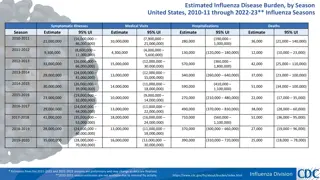
Children have high risk of developing croup, sinusitis, otitis media, high-grade fever, and diarrhoea.
EPIDEMIOLOGY
•
Incidence:
3–5 million cases of severe illness and 2.5–5 lakhs of deaths occur worldwide.
•
Seasonality:
Common during winters. The most common seasonal flu strain varies from
season to season and from place to place (e.g. H3N2 in Pondicherry in 2018)
•
Epidemiological pattern:
Depends upon the nature of antigenic variation that occurs in
the influenza types.
EPIDEMIOLOGY
•
Major influenza outbreaks
:
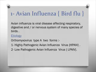
AVIAN FLU
•
Birds - primary reservoir
•
All influenza subtypes (16H types and 9N types) are found in birds and some of the subtypes can be
transmitted to mammals (e.g.; H1, H2, H3, and H5 to humans; H1 and H3 to swine; and H3 and H7 to
horses).
•
Avian flu strains- highly virulent as they possess
PB1F2
protein, which targets host mitochondria and induces
apoptosis.
AVIAN FLU INFECTION IN BIRDS
•
Bird flu strains - highly lethal to chickens and turkeys (but avirulent to ducks) and are the major cause of
economic loss in poultry causing severe mortality in chickens.
•
Avian flu multiplies in intestinal tracts of birds and shed through feces into water.
•
Influenza viruses do not undergo antigenic variation in birds, because of the short life span of birds.
AVIAN FLU INFECTION IN HUMANS
•
Till date, all human pandemic strains have originated by reassortment
between avian and human influenza viruses and the mixing has occurred in
pigs.
•
A/H5N1
is the most common avian flu strain that has been endemic in the
world for the past 15 years.
•
Origin-
It was first reported from Hong Kong in 1997and has spread to various
countries including India within few years.
AVIAN FLU INFECTION IN HUMANS
•
Less morbidity and more mortality
•
Clinical feature
- H5N1 avian flu strains are associated with higher rates of pneumonia and extra-pulmonary
manifestations such as diarrhoea and CNS involvement.
•
Other avian flu strains
that can cause human infections are-
o
A/H7N7(Netherlands)
o
A/H9N2 ( Hong Kong)
o
A/H7N9 (caused an outbreak in China, 2013)
LABORATORY DIAGNOSIS
•
Avian flu strains can be identified by real time reverse transcriptase
PCR detecting specific HA and NA genes.
H1N1 IN INDIA
Seasonal flu:
•
After the pandemic in 2009, A/H1N1 has become a seasonal
influenza strain circulating in India along with the other two
seasonal strains (A/H3N2 and type B).
•
Most cases in a year occur during the winter season (Dec-Jan).
H1N1 IN INDIA (CONT..)
Epidemiological Surveillance for Influenza
•
Integrated Disease Surveillance Program (IDSP) under NCDC,
Government of India has established a network (of 12 regional
centers) for epidemiological surveillance for H1N1 and other
influenza like illness (ILI).
•
Also monitors the changes in the circulating influenza strain (if any).
H1N1 IN INDIA (CONT..)
•
Government of India (NCDC) report for H1N1, 2018:
•
Between 2010- 2017, about 1,15,630 cases and 8,681 deaths due to
H1N1 were reported from India.
•
Varied with geographical regions:
Maharashtra was the worst-hit
state with 23,958 cases and 2,710 deaths, followed by Gujarat and
Rajasthan. Sikkim and Lakshadweep are the only two states/Union
territories with no cases over the past seven years.
•
Varied with year: Year 2015 (maximum), 2017 and 2010
recorded highest number of cases
CLINICAL FEATURES
•
Uncomplicated influenza: Mild upper respiratory tract illness and diarrhea
•
Complicated/severe influenza - high-risk groups - secondary bacterial pneumonia, dehydration, CNS involvement,
and multi-organ failure.
CATEGORIZATION OF SEASONAL INFLUENZA
A/H1N1
•
Guideline on categorization of Seasonal Influenza A/H1N1 cases during screening for home isolation, testing,
treatment and hospitalization (issued by Ministry of Health & Family Welfare, Govt. of India)
GUIDELINE ON CATEGORIZATION
GUIDELINE ON CATEGORIZATION
*
Real time reverse transcriptase PCR can detect and quantify the specific HA and NA genes of H1N1.
**
Oseltamivir (Tamiflu) tablet or Zanamivir (inhalational form)
GISRS
•
Influenza surveillance has been conducted globally through WHO's Global Influenza Surveillance and Response
System (GISRS).
•
Monitors the evolution of influenza viruses and provides recommendations in areas including laboratory
diagnostics, vaccines and treatment.
•
Serves as a global alert mechanism for the emergence of influenza viruses with pandemic potential.
LABORATORY DIAGNOSIS
•
Specimen Collection:
o
Ideal specimens - nasopharyngeal swab or lavage fluid, nasal aspirate or to less extent throat swab
o
Swabs
with a synthetic tip (e.g. polyester or Dacron
swabs) are best for specimen collection
o
Cotton or alginate swabs are unsatisfactory
•
Transport: Swabs are immediately put inside the viral transport media, kept at 4°C during transport up to
4days, thereafter at –70°C.
ISOLATION OF VIRUS
•
Embryonated eggs and primary monkey kidney cell lines have been the methods of choice
for the isolation of influenza viruses in the past.
•
The viral growth in cell line was detected by hemadsorption or hemagglutination test.
•
Because of technical difficulty, isolation is not routinely followed for diagnostics.
DIRECT IMMUNOFLUORESCENCE TEST
•
Viral antigens coated onto epithelial cells can be directly detected in nasal aspirates by using fluorescent
antibodies.
•
Rapid but less sensitive than isolation.
RT-PCR (REVERSE TRANSCRIPTASE
PCR
)
•
Highly sensitive, specific and rapid (turnaround time of <1 day).
•
Also detect the specific type and subtype of influenza virus
REAL-TIME RT-PCR
•
Currently the gold standard
(recommended by Govt of India).
•
Quantitative, higher sensitivity and specificity than RT-PCR with turnaround time of 2–3
hours.
•
Simultaneously detects the three common seasonal flu strains (A/H1N1, A/H3N2 and
type B).
REAL-TIME RT-PCR
•
Result is expressed as the emission of fluorescence during the cycles
ANTIBODY DETECTION
•
Fourfold rise in the antibody titre between acute and convalescent sera is more significant
than a single high titre.
•
HAI test
(Hemagglutination inhibition): Obsolete
•
Neutralization test
- Though it is the most specific & the best predictor of susceptibility
to infection, is time-consuming and difficult to perform.
•
ELISA -
M
ore sensitive than other assays; detects antibodies against HA and/or NA
antigens.
TREATMENT
•
Neuraminidase inhibitors
(such as zanamivir, oseltamivir, and peramivir) can be administered for influenza A
and influenza B infections.
o
Drug of choice for A/H1N1 2009 flu, A/H5N1avian flu and influenza-B.
o
Dosage-
Oseltamivir (Tamiflu 75mg tablets)
Zanamivir (10mg, inhalational form)
o
Schedule-
For treatment- given twice a day for 5 days
For chemoprophylaxis- given once daily. Duration depends on the clinical setting.
TREATMENT
•
Matrix protein M2 inhibitor:
o
Amantadine and rimantadine can be given for some strains of
influenza A infection.
o
Strains of A/H1N1 2009 flu and A/H5N1 avian flu and influenza B
have developed resistance.
GENERAL PREVENTIVE MEASURES
•
Measures of droplet precaution:
o
Strict hand hygiene
to be followed.
o
Isolation room:
Patients should be kept at isolation room or cohorting to be followed.
o
Containment of coughs and sneezes
Respiratory hygiene and cough etiquette
Use of gloves and mask for staff and patient
o
Work restriction:
CDC recommends that people with influenza-like illness remain at home until at least 24 hours
after they are free of fever (100° F) without the use of fever-reducing medications.
VACCINATION PROPHYLAXIS
•
Vaccine strains:
•
Based on WHO recommendations, influenza vaccines are prepared every year.
•
Strains to be included
in the vaccine depend upon the strains isolated in the previous influenza seasons and strains that are anticipated to
circulate in the upcoming season.
•
Formulations
: Most of the influenza vaccines are cocktails containing two type A and one or two type B influenza strains.
o
Trivalent form:
This is the most common form available; comprises of three strains: A/H1N1, A/H3N2 and influenza B strain.
o
Quadrivalent form:
In addition to trivalent form, this contains another type B strain. (Yamagata lineage, 2013).
•
GISRN
: WHO’s Global Influenza Surveillance and Response System (GISRS) reviews the vaccine composition on an annually for both
northern and southern hemisphere.
VACCINATION PROPHYLAXIS
•
For 2018-19 season:
The WHO recommended vaccine:
o
For northern hemisphere is a
Quadrivalent form
vaccine :
o
Type A/H1N1(Michigan lineage, 2015)
o
Type A/H3N2 (Singapore lineage, 2016)
o
Type B (Yamagata lineage, 2013)
o
Type B (Victoria lineage, 2017)
o
For southern hemisphere is a trivalent vaccine: same as that for northern hemisphere except the Type B (Victoria
lineage, 2017).
•
Types:
Both injectable (inactivated) and nasal spray (live attenuated) vaccines are available.
INJECTABLE VACCINES
•
Most widely used vaccines in immunization programmes.
•
Types:
There are three types of injectable vaccines.
o
Inactivated Influenza Vaccine (IIV)
o
Cell Culture-based Inactivated Influenza Vaccine (ccIIV3);
o
Recombinant Influenza Vaccine (RIV)
INJECTABLE VACCINES (CONT..)
•
Schedule:
Single dose administered by intramuscular (IM) route; except for 6 month-8 year of age (2 doses
are required ≥ 4 weeks apart).
•
Timing of vaccination:
Optimally before onset of influenza season, i.e. by end of October.
•
Efficacy:
25–67 % (25% for H3N2, 42% against type B and 67% against 42%). The efficacy is lower if vaccine
virus doesn’t match to currently circulating viruses. Immunity lasts for 6-12 months.
•
Side effects:
Mild reactions can occur in 5% of cases such as redness at injection site, fever and aches. Serious
side effects - rare.
INJECTABLE VACCINES (CONT..)
•
Indication:
Routine annual influenza vaccination is recommended for all persons aged ≥6 months who do not
have contraindications. If facility are not feasible, then high-risk groups should be given first priority for
vaccination.
o
Age: ≥ 6 month to <5 years and ≥50 years
o
Persons with chronic pulmonary, cardiovascular, renal, hepatic, neurologic, hematologic, or metabolic disorders.
o
Low immunity (e.g. HIV infection)
o
Pregnant women
o
Extremely obese (body mass index ≥40)
o
Caregivers and contacts of those at risk: e.g. health care workers and household contacts
INJECTABLE VACCINES (CONT..)
•
Contraindication
: IV should not be administered to people who have allergy to eggs or have history of
hypersensitivity to previous dose of vaccine.
•
Travelers:
If travelling to an area of increased influenza activity; can consider vaccination, preferably ≥2
weeks before departure.
LIVE ATTENUATED INFLUENZA VACCINE (LAIV)
•
Generated by reassortment between currently circulating strains of influenza A and B virus with a cold-
adapted attenuated master strain which can grow at 33°C (upper respiratory tract) but not at 37°C (lower
respiratory tract).
•
May cause mild flu like symptoms but never infect lower respiratory tract, hence never cause serious adverse
effects.
•
Trivalent vaccine
•
Administered by intranasal spray.
LIVE ATTENUATED INFLUENZA VACCINE (LAIV)
•
Indication: It can be given to all healthy persons of 2–49 years age (except in pregnancy), but is not given to
high risk groups.
•
However due to efficacy issues, LAIV is not recommended for use in any population for 2017-18.
CHEMOPROPHYLAXIS
•
Recommended only for post exposure and during outbreak situations in hospitals.
•
Indication:
Following exposure to an influenza case, it is recommended to the following groups: (i) if not
vaccinated or vaccinated recently (<2weeks), (ii) HIV infected people.
•
Duration:
o
Non-outbreak exposure
(e.g. in community): It should be started as soon as possible following exposure (within 48
hours) and continued for 7 days.
o
During outbreaks in hospitals
(for elderly persons & children and health care workers)- duration for a minimum of 2
weeks, and continuing up to 1 week after the last known case was identified.
Thank you
Influenza viruses belong to the Orthomyxoviridae family and are major contributors to respiratory epidemics and pandemics. They consist of three genera - Influenza A, B, and C - with distinct morphological characteristics and genetic makeup. By exploring the properties, classification, and morphology of Orthomyxoviridae, we gain insights into the structure, replication, and virulence factors associated with these viral pathogens.

Download Presentation

Please find below an Image/Link to download the presentation.
The content on the website is provided AS IS for your information and personal use only. It may not be sold, licensed, or shared on other websites without obtaining consent from the author. Download presentation by click this link. If you encounter any issues during the download, it is possible that the publisher has removed the file from their server.
E N D
Presentation Transcript
ORTHOMYXOVIRIDAE DR. MOHIT BHATIA ASSISTANT PROFESSOR DEPARTMENT OF MICROBIOLOGY AIIMS, RISHIKESH
LEARNING OBJECTIVES Introduction to Myxoviruses Orthomyxoviridae family
MYXOVIRUSES Group of viruses that bind to mucin receptors on the surface of RBCs (myxoin Greek meaning mucin ). Mucin receptors - Clumping of RBCs together to cause hemagglutination.
CLASSIFICATION Properties Orthomyxoviridae 80-120nm Spherical; Rarely filamentous Negative sense ssRNA, Segmented; Eight pieces Seen Seen Nucleus Influenza virus Paramyxoviridae Size Shape 100-300 nm Pleomorphic Nucleic acid Negative sense ssRNA Un-segmented; Single piece Not seen Not seen Cytoplasm Parainfluenza virus Mumps virus Measles virus Respiratory syncytial virus Metapneumovirus Genetic recombination Antigenic variation Site for RNA Replication Important human pathogens
ORTHOMYXOVIRIDAE Influenza viruses are the members of Orthomyxoviridae family. Major causes of morbidity and mortality and have been responsible for several epidemics and pandemics of respiratory diseases in the last two centuries.
MORPHOLOGY Influenza viruses consist of mainly thee genera- Influenza A, B, and C. Spherical and 80 120 nm in size Helical nucleocapsid (9nm),surrounded by an envelope Viral RNA comprises of multiple segments of negative sense single stranded RNA. Each segment codes for a specific viral protein having a specific function. o Influenza A & B contain eight segments of RNA o Influenza C contains seven segments of RNA. The segment coding for neuraminidase is absent.
MORPHOLOGY Site of RNA replication- nucleus RNA segments Coded Protein(s) Function of proteins Segment-6 NA Replaces HA from RBCs to cause elution (reversal of hemagglutination) Viral proteins RNA segments (neuraminidase) Coded Protein(s) Function of proteins Segment-1 PB2 RNA Transcription and replication Segment-7 M1&M2 M1-forms a shell underneath the envelope Segment-2 PB1 M2-forms ion channels Segment-3 PA Binds to receptors of RBC to cause hemagglutination. Segment-4 HA Segment-8 NS1&NS2 NS1-is interferon antagonist & inhibits pre-mRNA splicing (hemagglutinin) Segment-5 NP Associates with RNA to form helical nucleocapsid NS2-is nuclear export factor (nucleoprotein)
MORPHOLOGY (CONT..) Envelope is lipoprotein in nature. oLipid part is derived from the host cell membrane. oProteins or the peplomers are virus coded,10nm long glycoproteins that are inserted into the lipid envelope. oTwo peplomers are present- Hemagglutinin (HA) Neuraminidase (NA)
MORPHOLOGY (CONT..) Hemagglutinin (HA): oTriangular shaped peplomer, binds to mucin or sialic acid receptors on RBCs, resulting in clumping of RBCs to cause hemagglutination. oBinds to the receptors on respiratory epithelial cells, thus facilitating viral entry.
MORPHOLOGY (CONT..) Neuraminidase (NA): Mushroom-shaped peplomer, present in fewer number than HA. Sialidase enzyme that degrades the sialic acid receptors on RBCs thus helps in: o Displaces HA from RBCs resulting in reversal of hemagglutination called elution. o Facilitates release of virus particles from infected cell surfaces during budding process by preventing self-aggregation of virions to the host cells. o NA helps the virus to pass through the mucin layer in the respiratory tract to reach the target epithelial cells.
ANTIGENIC SUBTYPES AND NOMENCLATURE Genera-Based on RNP and M proteins, influenza viruses are divided into four genera: A, B and C and D Subtypes- Based on HA and NA antigens, Influenza A has distinct 18 H subtypes (H1 to H18) and 11 N subtypes (N1-N11), o Most of the subtypes infect animals and birds, rarely human major epidemics and pandemics. o Example- Six HA (H1, H2, H3, H5, H7 & H9) and two NA (N1&N2) subtypes have been recovered from humans. Influenza B and C viruses though have subtypes; but are not designated. Influenza D virus primarily infects cattle and are not pathogenic to humans.
ANTIGENIC SUBTYPES AND NOMENCLATURE Standard nomenclature system: Any influenza isolates should be designated based on the following information: Influenza Type/ host (indicated only for non-human origin)/ geographic origin/strain number/year of isolation/(HA NA subtype). Examples: o Human strain- Influenza A/Hong Kong/03/1968(H3N2) o Nonhuman strain- Influenza A/swine/Iowa/15/1930(H1N1)
ANTIGENIC VARIATION Antigenic variation is the unique property of Influenza viruses, which is due to the result of antigenic changes occurring in HA and NA peplomers. oAntigenic drift oAntigenic shift
ANTIGENIC DRIFT Minor change occurring due to point mutations in the HA/NA gene. Results in alteration of amino acid sequence of the antigenic sites on HA/NA, such that virus can escape recognition by the host's immune system. New variant must sustain two or more mutations to become epidemiologically significant. Seen in both Influenza virus type-A&B Results in outbreaks and minor periodic epidemics Antigenic drift occurs more frequently every 2-3 years.
ANTIGENIC SHIFT Abrupt, major drastic, discontinuous variation in the sequence of a viral surface protein (HA/NA) Occurs due to genetic re-assortment between genomes of two or more influenza viruses infecting the same host cells, resulting in a new virus strain, unrelated antigenically to the predecessor strains. Occurs only in Influenza A virus Results in pandemics and major epidemics- e.g. H1N1 pandemics of 2009. Antigenic shift occurs less frequently every 10-20 years.
PATHOGENESIS Pathogenesis Explanation Transmission Via aerosols Small-particle aerosols (<10 m) are more efficient in the transmission. Target cell entry Viral HA attaches to specific sialic acid receptors on the host cell surface Ciliated columnar epithelial cells are most commonly infected. Multiply locally Virus replicates in the infected cells and infectious daughter virions spread to the adjacent cells.
PATHOGENESIS Pathogenesis Explanation Spread Very rarely, virus spreads to the lower respiratory tract or spills over blood stream to involve extra pulmonary sites. Local damage Cellular destruction and desquamation of superficial mucosa of the respiratory tract Edema and mononuclear cell infiltrations occur at local site leading to cytokine influx, which accounts for local symptoms. Local damage predisposes to secondary bacterial invasion.
SIALIC ACID RECEPTORS Sialic acid receptors - found on the host cell surfaces are specific for HA antigens 2-6 sialic acid receptors: o Specific for human influenza strains and are found abundantly on human upper respiratory tract epithelium, tracheal and tracheobronchial cells (mainly ciliated but also non-ciliated cells). 2-3 sialic acid receptors: o Specific for avian influenza strains o Found abundantly on bird s intestinal epithelium. o In humans, they are present in very few numbers on lower respiratory tract (trachea, bronchi, alveolar cells) & rarely nasal mucosa and nosapharynx.
SIALIC ACID RECEPTORS Why pigs are the most common mixing vessels? o Both 2-3 & 2-6 sialic acid receptors are found on the respiratory epithelium of pigs and swine flu strains have specificity for both the receptor types. o Hence pigs can be infected simultaneously by human, swine and avian strains, thus serving as a mixing vessel. oReassortment between the segments of various strains takes place inside the same swine cell.
HOST IMMUNE RESPONSE Humoral immunity - predominant Type and subtype-specific and long lasting. Antibodies against HA and NA protective Antibodies to HA - prevent initiation of infection by inhibiting viral entry Antibodies to NA decrease the severity of the disease and prevent the transmission of virus to contacts Antibodies against other viral proteins are not protective.
HOST IMMUNE RESPONSE Antibodies against the ribonucleoprotein - useful in typing viral isolates as influenza A or B or C. All the three types of influenza viruses (i.e. A,B &C) are antigenically unrelated and there is no cross-protection. Immunity incomplete - reinfection with the same virus can occur.
HOST IMMUNE RESPONSE Original antigenic sin: oWhen a previously infected individual gets a repeated infection with a different antigenic variant of influenza virus, antibodies are produced against both the subtypes oBut predominant response would be against the original strain, a phenomenon called "original antigenic sin." Components of both cell mediated immunity (e.g. cytotoxic T cells) and innate immunity (NK cells, interferons) are also important in providing immunity against influenza infections.
CLINICAL MANIFESTATIONS Incubation period - 18-72 hours Uncomplicated Influenza (Flu syndrome): o Asymptomatic or develop minor upper respiratory symptoms such chills, headache, and dry cough, followed by high grade fever, myalgia and anorexia. oSelf-limiting condition.
COMPLICATIONS Pneumonia: Secondary bacterial pneumonia - most common Common agents are staphylococci, pneumococci and Haemophilus influenzae. Primary Influenza pneumonia is rare but leads to more severe complication. Combined viral-bacterial pneumonia- It is three times more common than primary influenza pneumonia, most common co-infection being S.aureus.
CLINICAL MANIFESTATIONS Other pulmonary complications: oWorsening of COPD oExacerbation of chronic bronchitis and asthma. Reye's syndrome
CLINICAL MANIFESTATIONS The following are at increased risk of complications: o Age- children <2yrs& old age (>65yrs) o Pregnancy o Underlying chronic lung, cardiac, renal, hepatic, and CNS conditions o Low immunity(HIV infected people) o Children have high risk of developing croup, sinusitis, otitis media, high-grade fever, and diarrhoea.
EPIDEMIOLOGY Incidence: 3 5 million cases of severe illness and 2.5 5 lakhs of deaths occur worldwide. Seasonality: Common during winters. The most common seasonal flu strain varies from season to season and from place to place (e.g. H3N2 in Pondicherry in 2018) Epidemiological pattern: Depends upon the nature of antigenic variation that occurs in the influenza types.
EPIDEMIOLOGY Major influenza outbreaks: Years 1889 1890 1900 1903 1918 1919 Subtype H2N8 H3N8 H1N1a (HswN1) (Spanish flu) H1N1a (H0N1) H1N1 H2N2 (Asian flu) H3N2 (Hong Kong flu) H1N1 (Russian flu) H1N1 Extent of Outbreak Severe pandemic ?Moderate epidemic Severe pandemic 1933 1935 1946 1947 1957 1958 1968 1969 Mild epidemic Mild epidemic Severe pandemic Moderate pandemic 1977 1978b Mild pandemic 2009 2010 Pandemic
AVIAN FLU Birds - primary reservoir All influenza subtypes (16H types and 9N types) are found in birds and some of the subtypes can be transmitted to mammals (e.g.; H1, H2, H3, and H5 to humans; H1 and H3 to swine; and H3 and H7 to horses). Avian flu strains- highly virulent as they possess PB1F2 protein, which targets host mitochondria and induces apoptosis.
AVIAN FLU INFECTION IN BIRDS Bird flu strains - highly lethal to chickens and turkeys (but avirulent to ducks) and are the major cause of economic loss in poultry causing severe mortality in chickens. Avian flu multiplies in intestinal tracts of birds and shed through feces into water. Influenza viruses do not undergo antigenic variation in birds, because of the short life span of birds.
AVIAN FLU INFECTION IN HUMANS Till date, all human pandemic strains have originated by reassortment between avian and human influenza viruses and the mixing has occurred in pigs. A/H5N1 is the most common avian flu strain that has been endemic in the world for the past 15 years. Origin-It was first reported from Hong Kong in 1997and has spread to various countries including India within few years.
AVIAN FLU INFECTION IN HUMANS Less morbidity and more mortality Clinical feature- H5N1 avian flu strains are associated with higher rates of pneumonia and extra-pulmonary manifestations such as diarrhoea and CNS involvement. Other avian flu strains that can cause human infections are- o A/H7N7(Netherlands) o A/H9N2 ( Hong Kong) o A/H7N9 (caused an outbreak in China, 2013)
LABORATORY DIAGNOSIS Avian flu strains can be identified by real time reverse transcriptase PCR detecting specific HA and NA genes.
H1N1 IN INDIA Seasonal flu: After the pandemic in 2009, A/H1N1 has become a seasonal influenza strain circulating in India along with the other two seasonal strains (A/H3N2 and type B). Most cases in a year occur during the winter season (Dec-Jan).
H1N1 IN INDIA (CONT..) Epidemiological Surveillance for Influenza Integrated Disease Surveillance Program (IDSP) under NCDC, Government of India has established a network (of 12 regional centers) for epidemiological surveillance for H1N1 and other influenza like illness (ILI). Also monitors the changes in the circulating influenza strain (if any).
H1N1 IN INDIA (CONT..) Government of India (NCDC) report for H1N1, 2018: Between 2010- 2017, about 1,15,630 cases and 8,681 deaths due to H1N1 were reported from India. Varied with geographical regions: Maharashtra was the worst-hit state with 23,958 cases and 2,710 deaths, followed by Gujarat and Rajasthan. Sikkim and Lakshadweep are the only two states/Union territories with no cases over the past seven years. Varied with year: Year 2015 (maximum), 2017 and 2010 recorded highest number of cases
CLINICAL FEATURES Uncomplicated influenza: Mild upper respiratory tract illness and diarrhea Complicated/severe influenza - high-risk groups - secondary bacterial pneumonia, dehydration, CNS involvement, and multi-organ failure.
CATEGORIZATION OF SEASONAL INFLUENZA A/H1N1 Guideline on categorization of Seasonal Influenza A/H1N1 cases during screening for home isolation, testing, treatment and hospitalization (issued by Ministry of Health & Family Welfare, Govt. of India)
GUIDELINE ON CATEGORIZATION Category Definition Guideline for laboratory testing for H1N1*, treatment** and isolation Category A Mild fever plus cough / sore throat with or without body ache, headache, diarrhoea and vomiting Laboratory testing for H1N1- not required Treatment- only symptomatic, antiviral drugs not required. Isolation- Confine patients at home, avoid contact with public and high risk members in the family. Category B Category A plus any one: (i) High grade fever and severe sore throat or (ii) Presence of risk factors: Children, age >65 years, pregnant women, patients with lung/ heart/liver/kidney/ neurological disorders, diabetes, cancer or HIV; on long term steroid therapy. Laboratory testing for H1N1- not required Treatment- Symptomatic treatment required. Antiviral drug (oseltamivir) may be required. Isolation- Confine patients at home, avoid contact with public and high risk members in the family.
GUIDELINE ON CATEGORIZATION Category Definition Guideline for laboratory testing for H1N1*, treatment** and isolation Category C Category B plus any one: (i)Breathlessness, chest pain, fall in blood pressure, sputum mixed with blood, bluish discolouration of nails; (ii)Children with influenza like illness who had a severe disease as manifested by the red flag signs (inability to feed well, convulsions, difficulty in breathing, etc). (iii)Worsening of underlying chronic conditions. Laboratory testing for H1N1- required . Immediate hospitalization required. Treatment- start antiviral drug (oseltamivir) immediately without waiting for lab result. Isolation- all components droplet precaution to be followed. (refer prevention of influenza section) *Real time reverse transcriptase PCR can detect and quantify the specific HA and NA genes of H1N1. **Oseltamivir (Tamiflu) tablet or Zanamivir (inhalational form)
GISRS Influenza surveillance has been conducted globally through WHO's Global Influenza Surveillance and Response System (GISRS). Monitors the evolution of influenza viruses and provides recommendations in areas including laboratory diagnostics, vaccines and treatment. Serves as a global alert mechanism for the emergence of influenza viruses with pandemic potential.
LABORATORY DIAGNOSIS Specimen Collection: o Ideal specimens - nasopharyngeal swab or lavage fluid, nasal aspirate or to less extent throat swab o Swabs with a synthetic tip (e.g. polyester or Dacron swabs) are best for specimen collection o Cotton or alginate swabs are unsatisfactory Transport: Swabs are immediately put inside the viral transport media, kept at 4 C during transport up to 4days, thereafter at 70 C.
ISOLATION OF VIRUS Embryonated eggs and primary monkey kidney cell lines have been the methods of choice for the isolation of influenza viruses in the past. The viral growth in cell line was detected by hemadsorption or hemagglutination test. Because of technical difficulty, isolation is not routinely followed for diagnostics.
DIRECT IMMUNOFLUORESCENCE TEST Viral antigens coated onto epithelial cells can be directly detected in nasal aspirates by using fluorescent antibodies. Rapid but less sensitive than isolation.
RT -PCR (REVERSE TRANSCRIPTASE PCR) Highly sensitive, specific and rapid (turnaround time of <1 day). Also detect the specific type and subtype of influenza virus
REAL-TIME RT -PCR Currently the gold standard (recommended by Govt of India). Quantitative, higher sensitivity and specificity than RT-PCR with turnaround time of 2 3 hours. Simultaneously detects the three common seasonal flu strains (A/H1N1, A/H3N2 and type B).
REAL-TIME RT -PCR Result is expressed as the emission of fluorescence during the cycles
ANTIBODY DETECTION Fourfold rise in the antibody titre between acute and convalescent sera is more significant than a single high titre. HAI test (Hemagglutination inhibition): Obsolete Neutralization test- Though it is the most specific & the best predictor of susceptibility to infection, is time-consuming and difficult to perform. ELISA - More sensitive than other assays; detects antibodies against HA and/or NA antigens.
TREATMENT Neuraminidase inhibitors (such as zanamivir, oseltamivir, and peramivir) can be administered for influenza A and influenza B infections. o Drug of choice for A/H1N1 2009 flu, A/H5N1avian flu and influenza-B. o Dosage- Oseltamivir (Tamiflu 75mg tablets) Zanamivir (10mg, inhalational form) o Schedule- For treatment- given twice a day for 5 days For chemoprophylaxis- given once daily. Duration depends on the clinical setting.


")
")
")


")
")


")