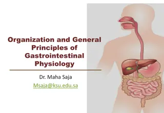
Overview of the Gastrointestinal System and Its Functions


The alimentary tract provides the body with a
continual supply of water, electrolytes, and
nutrients.To achieve this requires
(1) movement of food through
the alimentary tract; (2) secretion of digestive juices
and digestion of the food; (3) absorption of water,
various electrolytes, and digestive products; (4)
circulation of blood through the gastrointestinal
organs to carry away the absorbed substances; and
(5) control of all these functions by local, nervous,
and hormonal systems.
Introduction to the gastrointestinal system
The
gastrointestinal tract
(GIT) consists of a
hollow muscular tube starting from the oral cavity,
where food enters the mouth, continuing through
the pharynx, oesophagus, stomach and intestines
to the rectum and anus, where food is expelled.
There are various
accessory organs
that assist the
tract by secreting enzymes to help break down
food into its component nutrients. Thus the
salivary glands, liver, pancreas and gall bladder
have important functions in the
digestive system
.
Food is propelled along the length of the GIT by
peristaltic movements of the muscular walls.
functions of the digestive system include:
1
. Motility
. This refers to the movement of food through
the digestive tract through the processes of
a.
Ingestion: Taking food into the mouth.
b.
Mastication: Chewing the food and mixing it with
saliva.
c.
Deglutition: Swallowing food.
d.
Peristalsis: Rhythmic, wavelike contractions that
move food through the gastrointestinal tract.
2
. Secretion.
This includes both exocrine and endocrine
secretions.
a.
Exocrine secretions:Water, hydrochloric acid,
bicarbonate, and many digestive enzymes are secreted
into the lumen of the gastrointestinal tract. The
stomach alone, for example, secretes 2 to 3 liters of
gastric juice a day.
b.
Endocrine secretions:
The stomach and small
intestine secrete a number of hormones that help to
regulate the digestive system.
3
.Digestion.
This refers to the breakdown of food
molecules into their smaller subunits, which can be
absorbed.
4
. Absorption.
This refers to the passage of digested
end products into the blood or lymph.
5
. Storage and elimination.
This refers to the
temporary storage and subsequent elimination of
indigestible food molecules.
Layers of the Gastrointestinal Tract
The GI tract from the esophagus to the anal canal is
composed of four layers, or
tunics.
Each tunic contains a
dominant tissue type that performs specific functions in the
digestive process. The four tunics of the GI tract, from the
inside out, are the
mucosa, submucosa, muscularis, and
serosa
Mucosa
The mucosa, which lines the lumen of the GI tract, is the
absorptive and major secretory layer. It consists of a simple
columnar epithelium supported by the
lamina propria
, a thin
layer of areolar connective tissue containing numerous lymph
nodules, which are important in protecting against disease
External to the lamina propria is a thin layer of smooth muscle
called the
muscularis mucosae
.
This is the muscle layer
responsible for the numerous small folds in certain portions of
the GI tract. These folds greatly increase the absorptive
surface area. Specialized goblet cells in the mucosa secrete
mucus throughout most of the GI tract.
Submucosa
The relatively thick , submucosa is a highly vascular
layer of connective tissue that serves the mucosa.
Absorbed molecules that pass through the columnar
epithelial cells of the mucosa enter into blood and
lymphatic vessels of the submucosa. In addition to
blood vessels, the submucosa contains glands and
nerve plexuses. The
submucosal plexus (
Meissner’s
plexus)
provides an autonomic nerve supply to the
muscularis mucosae.
Muscularis
The
muscularis (also called the
muscularis externa)
is responsible for
segmental contractions and peristaltic movement through the GI tract.
The muscularis has an inner circular and an outer longitudinal layer of
smooth muscle. Contractions of these layers move the food through the
tract and physically pulverize and mix the food with digestive enzymes.
The
myenteric plexus
(
Auerbach’s plexus), located
between the two
muscle layers
,
provides the major nerve supply to the GI tract. It includes
fibers and ganglia from both the sympathetic and parasympathetic
divisions of the autonomic nervous system
Serosa
The outer serosa completes the wall of the GI tract. It is a binding and
protective layer consisting of areolar connective tissue covered with a
layer of simple squamous epithelium
.
The effects of the
sympathetic nerves
reduce
peristalsis and secretory activity and stimulate the contraction
of sphincter muscles along the GI tract; therefore, they are
antagonistic to the effects of parasympathetic nerve
stimulation.
The gut also contains the
enteric nervous
system
, a discrete web of 100 million neurons that can
control gut function without extrinsic control. It consists
of the myenteric plexus, located between longitudinal and
circular muscle layers, and the submucosal plexus.
Reflexes mediate
motility
,
secretions
,
hormone release
, and
vasculature.
components of the gastrointestinal system
Oral cavity
The oral cavity or mouth is responsible for the intake of
food.
Mastication
refers to the mechanical breakdown of food
by chewing and chopping actions of the teeth. The tongue,
a strong muscular organ, manipulates the food bolus to
come in contact with the teeth. It is also the sensing organ
of the mouth for touch, temperature and taste using its
specialised sensors known as papillae.
•
Insalivation
refers to the mixing of the oral cavity
contents with salivary gland secretions. The mucin (a
glycoprotein) in saliva acts as a lubricant. The oral cavity
also plays a limited role in the digestion of
carbohydrates. The enzyme serum amylase, a component
of saliva, starts the process of digestion of complex
carbohydrates. The final function of the oral cavity is
absorption of small molecules such as glucose and water,
across the mucosa. From the mouth, food passes through
the pharynx and oesophagus via the action of
swallowing.
Salivary glands
Three pairs of salivary glands communicate with the
oral cavity. Each is a complex gland with numerous
acini lined by secretory epithelium. The acini secrete
their contents into specialised ducts. Each gland is
divided into smaller segments called lobes. Salivation
occurs in response to the taste, smell or even
appearance of food. This occurs due to nerve signals
that tell the salivary glands to secrete saliva to prepare
and moisten the mouth. Each pair of salivary glands
secretes saliva with slightly different compositions
Parotids
:
The parotids produce a watery secretion
which is also rich in proteins. Immunoglobins are
secreted help to fight microorganisms and amylase
proteins start to break down complex carbohydrates.
Sublingual
:
They produce approximately 5% of the
saliva and their secretions are very sticky due to the
large concentration of mucin. The main functions are to
provide buffers and lubrication.
Submandibular
:These glands produce a more viscid
(thick) secretion, rich in mucin and with a smaller
amount of protein.
Mucin is a glycoprotein that acts as a lubricant
Esophagus
The
esophagus is that portion of the GI tract which
connects the pharynx to the stomach. It is a muscular
tube approximately 25 cm (10 in.) long, located
posterior to the trachea within the mediastinum of
the thorax. the esophagus passes through the
diaphragm by means of an opening called the
esophageal hiatus. The esophagus is lined
with a
nonkeratinized stratified squamous epithelium; its
walls contain either skeletal or smooth muscle,
depending on the location.
the middle third contains a mixture of skeletal and
smooth muscle, and the terminal portion contains
only smooth muscle
Swallowed food is pushed from the oral to the end of
the esophagus by a wavelike muscular contraction
called
peristalsis
the bolus along the digestive tract
occurs because the circular smooth muscle contracts
behind, and relaxes in front of, the bolus. This is
followed by shortening of the tube by longitudinal
muscle contraction. These contractions progress from
the superior end of the esophagus to the
astroesophageal junction at a
rate of 2 to 4 cm per
second as they empty the contents of the esophagus into
the cardiac region of the stomach.
The stomach:
•
the stomach
is a J shaped expanded bag, located just left of
the midline between the oesophagus and small intestine. The
stomach stores up to 2 liters of food
•
The functions of the stomach include
:
•
The short-term storage of ingested food.
•
Mechanical breakdown of food by churning and mixing
motions.
•
Chemical digestion of proteins by acids and enzymes.
•
Stomach acid kills germs.
•
Some absorption of substances such as alcohol.
•
Most of these functions are achieved by the secretion of
stomach juices by gastric glands in the body and fundus. Some
cells are responsible for secreting acid and others secrete
enzymes to break down proteins.
gastric juice:
thin, strongly acidic (
p
H varying from 1 to
3), almost colorless liquid secreted by the glands in the
lining of the stomach. Its essential constituents are the
digestive enzymes
pepsin
, hydrochloric acid, and mucus.
Pepsin converts proteins into simpler, more easily
absorbed substances; it is aided in this by hydrochloric
acid, which provides the acid environment in which pepsin
is most effective. Gastric secretion is stimulated by a
number of hormones and chemical substances, by the
presence of food in the stomach, and by a number of
psychological factors, such as the smell of a favorite food.
Certain cells of the stomach lining secrete a substance
known as intrinsic factor, which is necessary for the
absorption of vitamin B
12
; absence of this substance results
in pernicious anemia, or B
12
deficiency
Cell types in the stomach that help with digestion
There are four main types of cells for stomach secretions
spread all over the inner surface of the stomach:
Mucous cells
secrete the alkaline mucous for shielding
the epithelium from hydrochloric acid. These are found in
the fundic, cardiac, and pyloric region.
Parietal cells
, located in the fundic, cardiac, and pyloric
region, secrete hydrochloric acid; the acid activates release
of pepsin for protein digestion. The acid also kills micro-
organisms swallowed with the food.
Chief cells
secrete pepsin. These cells are located in the
fundic region.
G cells
are found in the fundic, pyloric, and gastric region.
These secrete gastrin which stimulates the secretion of
hydrochloric acid.
Motor Functions of the
Stomach
The motor functions of the stomach are threefold: (1)
storage of large quantities of food until the food can
be processed in the stomach, duodenum, and lower
intestinal tract; (2) mixing of this food with gastric
secretions until it forms a semifluid mixture called
chyme
; and (3) slow emptying of the chyme from the
stomach into the small intestine at a rate suitable for
proper digestion and absorption by the small intestine.
Figure 63–2 shows the basic anatomy of the
stomach. Anatomically, the stomach is usually divided
into two major parts: (1) the
body
and (2) the
antrum
.
Physiologically, it is more appropriately divided into
(1) the “orad” portion, comprising about the first two
thirds of the body, and (2) the “caudad” portion, comprising
the remainder of the body plus the antrum.
Storage Function of the Stomach
As food enters the stomach, it forms concentric circles
of the food in the orad portion of the stomach, the
newest food lying closest to the esophageal opening
and the oldest food lying nearest the outer wall of the
stomach. Normally, when food stretches the stomach,
a “vagovagal reflex” from the stomach to the brain
stem and then back to the stomach reduces the tone
in the muscular wall of the body of the stomach so that
the wall bulges progressively outward, accommodating
greater and greater quantities of food up to a limit in
the completely relaxed stomach of 0.8 to 1.5 liters.The
pressure in the stomach remains low until this limit is
approached.
The alimentary tract supplies the body with water, electrolytes, and nutrients through processes like movement of food, digestion, absorption, and circulation. The gastrointestinal tract, starting from the mouth to the anus, is aided by accessory organs for digestion. Functions include motility, secretion, digestion, absorption, and storage & elimination. This system ensures the body receives necessary components for sustenance.

Download Presentation

Please find below an Image/Link to download the presentation.
The content on the website is provided AS IS for your information and personal use only. It may not be sold, licensed, or shared on other websites without obtaining consent from the author. Download presentation by click this link. If you encounter any issues during the download, it is possible that the publisher has removed the file from their server.
E N D
Presentation Transcript
The alimentary tract provides the body with a continual supply of water, electrolytes, and nutrients.To achieve this requires (1) movement of food through the alimentary tract; (2) secretion of digestive juices and digestion of the food; (3) absorption of water, various electrolytes, and digestive products; (4) circulation of blood through the gastrointestinal organs to carry away the absorbed substances; and (5) control of all these functions by local, nervous, and hormonal systems.
Introduction to the gastrointestinal system The gastrointestinal tract (GIT) consists of a hollow muscular tube starting from the oral cavity, where food enters the mouth, continuing through the pharynx, oesophagus, stomach and intestines to the rectum and anus, where food is expelled. There are various accessory organs that assist the tract by secreting enzymes to help break down food into its component nutrients. Thus the salivary glands, liver, pancreas and gall bladder have important functions in the digestive system. Food is propelled along the length of the GIT by peristaltic movements of the muscular walls.
functions of the digestive system include: 1. Motility. This refers to the movement of food through the digestive tract through the processes of a. Ingestion: Taking food into the mouth. b. Mastication: Chewing the food and mixing it with saliva. c. Deglutition: Swallowing food. d. Peristalsis: Rhythmic, wavelike contractions that move food through the gastrointestinal tract.
2. Secretion. This includes both exocrine and endocrine secretions. a. Exocrine secretions:Water, hydrochloric acid, bicarbonate, and many digestive enzymes are secreted into the lumen of the gastrointestinal tract. The stomach alone, for example, secretes 2 to 3 liters of gastric juice a day. b. Endocrine secretions: The stomach and small intestine secrete a number of hormones that help to regulate the digestive system.
3.Digestion. This refers to the breakdown of food molecules into their smaller subunits, which can be absorbed. 4. Absorption. This refers to the passage of digested end products into the blood or lymph. 5. Storage and elimination. This refers to the temporary storage and subsequent elimination of indigestible food molecules.
Layers of the Gastrointestinal Tract The GI tract from the esophagus to the anal canal is composed of four layers, or tunics. Each tunic contains a dominant tissue type that performs specific functions in the digestive process. The four tunics of the GI tract, from the inside out, are the mucosa, submucosa, muscularis, and serosa
http://faculty.irsc.edu/FACULTY/TFischer/images/layers%20of%20dig%20tract.jpghttp://faculty.irsc.edu/FACULTY/TFischer/images/layers%20of%20dig%20tract.jpg
Mucosa The mucosa, which lines the lumen of the GI tract, is the absorptive and major secretory layer. It consists of a simple columnar epithelium supported by the lamina propria, a thin layer of areolar connective tissue containing numerous lymph nodules, which are important in protecting against disease External to the lamina propria is a thin layer of smooth muscle called the muscularis mucosae. This is the muscle layer responsible for the numerous small folds in certain portions of the GI tract. These folds greatly increase the absorptive surface area. Specialized goblet cells in the mucosa secrete mucus throughout most of the GI tract.
Submucosa The relatively thick , submucosa is a highly vascular layer of connective tissue that serves the mucosa. Absorbed molecules that pass through the columnar epithelial cells of the mucosa enter into blood and lymphatic vessels of the submucosa. In addition to blood vessels, the submucosa contains glands and nerve plexuses. The submucosal plexus (Meissner s plexus) provides an autonomic nerve supply to the muscularis mucosae.
http://humanphysiology2011.wikispaces.com/file/view/layers_of_the_digestive_tract.jpg/222326984/504x564/layers_of_the_digestive_tract.jpghttp://humanphysiology2011.wikispaces.com/file/view/layers_of_the_digestive_tract.jpg/222326984/504x564/layers_of_the_digestive_tract.jpg
Muscularis The muscularis (also called the muscularis externa) is responsible for segmental contractions and peristaltic movement through the GI tract. The muscularis has an inner circular and an outer longitudinal layer of smooth muscle. Contractions of these layers move the food through the tract and physically pulverize and mix the food with digestive enzymes. The myenteric plexus (Auerbach s plexus), located between the two muscle layers, provides the major nerve supply to the GI tract. It includes fibers and ganglia from both the sympathetic and parasympathetic divisions of the autonomic nervous system Serosa The outer serosa completes the wall of the GI tract. It is a binding and protective layer consisting of areolar connective tissue covered with a layer of simple squamous epithelium .
The effects of the sympathetic nerves reduce peristalsis and secretory activity and stimulate the contraction of sphincter muscles along the GI tract; therefore, they are antagonistic to the effects of parasympathetic nerve stimulation. The gut also contains the enteric nervous system, a discrete web of 100 million neurons that can control gut function without extrinsic control. It consists of the myenteric plexus, located between longitudinal and circular muscle layers, and the submucosal plexus. Reflexes mediate motility, secretions, hormone release, and vasculature.
components of the gastrointestinal system Oral cavity The oral cavity or mouth is responsible for the intake of food. Mastication refers to the mechanical breakdown of food by chewing and chopping actions of the teeth. The tongue, a strong muscular organ, manipulates the food bolus to come in contact with the teeth. It is also the sensing organ of the mouth for touch, temperature and taste using its specialised sensors known as papillae.
Insalivation refers to the mixing of the oral cavity contents with salivary gland secretions. The mucin (a glycoprotein) in saliva acts as a lubricant. The oral cavity also plays a limited role in the digestion of carbohydrates. The enzyme serum amylase, a component of saliva, starts the process of digestion of complex carbohydrates. The final function of the oral cavity is absorption of small molecules such as glucose and water, across the mucosa. From the mouth, food passes through the pharynx and oesophagus via the action of swallowing.
Salivary glands Three pairs of salivary glands communicate with the oral cavity. Each is a complex gland with numerous acini lined by secretory epithelium. The acini secrete their contents into specialised ducts. Each gland is divided into smaller segments called lobes. Salivation occurs in response to the taste, smell or even appearance of food. This occurs due to nerve signals that tell the salivary glands to secrete saliva to prepare and moisten the mouth. Each pair of salivary glands secretes saliva with slightly different compositions
Parotids :The parotids produce a watery secretion which is also rich in proteins. Immunoglobins are secreted help to fight microorganisms and amylase proteins start to break down complex carbohydrates. Sublingual :They produce approximately 5% of the saliva and their secretions are very sticky due to the large concentration of mucin. The main functions are to provide buffers and lubrication. Submandibular:These glands produce a more viscid (thick) secretion, rich in mucin and with a smaller amount of protein. Mucin is a glycoprotein that acts as a lubricant
Esophagus The esophagus is that portion of the GI tract which connects the pharynx to the stomach. It is a muscular tube approximately 25 cm (10 in.) long, located posterior to the trachea within the mediastinum of the thorax. the esophagus passes through the diaphragm by means of an opening called the esophageal hiatus. The esophagus is lined with a nonkeratinized stratified squamous epithelium; its walls contain either skeletal or smooth muscle, depending on the location. the middle third contains a mixture of skeletal and smooth muscle, and the terminal portion contains only smooth muscle
Swallowed food is pushed from the oral to the end of the esophagus by a wavelike muscular contraction called peristalsis the bolus along the digestive tract occurs because the circular smooth muscle contracts behind, and relaxes in front of, the bolus. This is followed by shortening of the tube by longitudinal muscle contraction. These contractions progress from the superior end of astroesophageal junction at a rate of 2 to 4 cm per second as they empty the contents of the esophagus into the cardiac region of the stomach. the esophagus to the
The stomach: the stomachis a J shaped expanded bag, located just left of the midline between the oesophagus and small intestine. The stomach stores up to 2 liters of food The functions of the stomach include: The short-term storage of ingested food. Mechanical breakdown of food by churning and mixing motions. Chemical digestion of proteins by acids and enzymes. Stomach acid kills germs. Some absorption of substances such as alcohol. Most of these functions are achieved by the secretion of stomach juices by gastric glands in the body and fundus. Some cells are responsible for secreting acid and others secrete enzymes to break down proteins.
http://humanphysiology2011.wikispaces.com/file/view/Primary_regions_and_structures_of_the_stomach.jpg/222328162/532x374/Primary_regions_and_structures_of_the_stomach.jpghttp://humanphysiology2011.wikispaces.com/file/view/Primary_regions_and_structures_of_the_stomach.jpg/222328162/532x374/Primary_regions_and_structures_of_the_stomach.jpg
gastric juice: thin, strongly acidic (pH varying from 1 to 3), almost colorless liquid secreted by the glands in the lining of the stomach. Its essential constituents are the digestive enzymes pepsin , hydrochloric acid, and mucus. Pepsin converts proteins into simpler, more easily absorbed substances; it is aided in this by hydrochloric acid, which provides the acid environment in which pepsin is most effective. Gastric secretion is stimulated by a number of hormones and chemical substances, by the presence of food in the stomach, and by a number of psychological factors, such as the smell of a favorite food. Certain cells of the stomach lining secrete a substance known as intrinsic factor, which is necessary for the absorption of vitamin B12; absence of this substance results in pernicious anemia, or B12 deficiency
Cell types in the stomach that help with digestion There are four main types of cells for stomach secretions spread all over the inner surface of the stomach: Mucous cells secrete the alkaline mucous for shielding the epithelium from hydrochloric acid. These are found in the fundic, cardiac, and pyloric region. Parietal cells, located in the fundic, cardiac, and pyloric region, secrete hydrochloric acid; the acid activates release of pepsin for protein digestion. The acid also kills micro- organisms swallowed with the food. Chief cells secrete pepsin. These cells are located in the fundic region. G cells are found in the fundic, pyloric, and gastric region. These secrete gastrin which stimulates the secretion of hydrochloric acid.
http://upload.wikimedia.org/wikipedia/commons/thumb/3/32/Gastric_gland.png/250px-Gastric_gland.pnghttp://upload.wikimedia.org/wikipedia/commons/thumb/3/32/Gastric_gland.png/250px-Gastric_gland.png
Motor Functions of the Stomach The motor functions of the stomach are threefold: (1) storage of large quantities of food until the food can be processed in the stomach, duodenum, and lower intestinal tract; (2) mixing of this food with gastric secretions until it forms a semifluid mixture called chyme; and (3) slow emptying of the chyme from the stomach into the small intestine at a rate suitable for proper digestion and absorption by the small intestine. Figure 63 2 shows the basic anatomy of the stomach. Anatomically, the stomach is usually divided into two major parts: (1) the body and (2) the antrum. Physiologically, it is more appropriately divided into (1) the orad portion, comprising about the first two thirds of the body, and (2) the caudad portion, comprising the remainder of the body plus the antrum.
Storage Function of the Stomach As food enters the stomach, it forms concentric circles of the food in the orad portion of the stomach, the newest food lying closest to the esophageal opening and the oldest food lying nearest the outer wall of the stomach. Normally, when food stretches the stomach, a vagovagal reflex from the stomach to the brain stem and then back to the stomach reduces the tone in the muscular wall of the body of the stomach so that the wall bulges progressively outward, accommodating greater and greater quantities of food up to a limit in the completely relaxed stomach of 0.8 to 1.5 liters.The pressure in the stomach remains low until this limit is approached.